. 2023 Dec;299(12):105371.
doi: 10.1016/j.jbc.2023.105371. Epub 2023 Oct 20.

Using CRISPR-Cas9/phosphoproteomics to identify substrates of calcium/calmodulin-dependent kinase 2δ

Affiliations

Using CRISPR-Cas9/phosphoproteomics to identify substrates of calcium/calmodulin-dependent kinase 2δ

Euijung Park et al. J Biol Chem. 2023 Dec.

Abstract

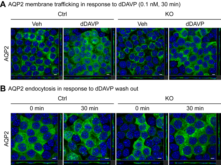
Ca2+/Calmodulin-dependent protein kinase 2 (CAMK2) family proteins are involved in the regulation of cellular processes in a variety of tissues including brain, heart, liver, and kidney. One member, CAMK2δ (CAMK2D), has been proposed to be involved in vasopressin signaling in the renal collecting duct, which controls water excretion through regulation of the water channel aquaporin-2 (AQP2). To identify CAMK2D target proteins in renal collecting duct cells (mpkCCD), we deleted Camk2d and carried out LC-MS/MS-based quantitative phosphoproteomics. Specifically, we used CRISPR/Cas9 with two different guide RNAs targeting the CAMK2D catalytic domain to create multiple CAMK2D KO cell lines. AQP2 protein abundance was lower in the CAMK2D KO cells than in CAMK2D-intact controls. AQP2 phosphorylation at Ser256 and Ser269 (normalized for total AQP2) was decreased. However, trafficking of AQP2 to and from the apical plasma membrane was sustained. Large-scale quantitative phosphoproteomic analysis (TMT-labeling) in the presence of the vasopressin analog dDAVP (0.1 nM, 30 min) allowed quantification of 11,570 phosphosites of which 169 were significantly decreased, while 206 were increased in abundance in CAMK2D KO clones. These data are available for browsing or download at https://esbl.nhlbi.nih.gov/Databases/CAMK2D-proteome/. Motif analysis of the decreased phosphorylation sites revealed a target preference of -(R/K)-X-X-p(S/T)-X-(D/E), matching the motif identified in previous in vitro phosphorylation studies using recombinant CAMK2D. Thirty five of the significantly downregulated phosphorylation sites in CAMK2D KO cells had exactly this motif and are judged to be likely direct CAMK2D targets. This adds to the list of known CAMK2D target proteins found in prior reductionist studies.

Keywords: aquaporin-2; mass spectrometry; vasopressin.


Conflict of interest statement

Conflict of interest The authors declare that they have no conflicts of interest with the contents of this article.

Figures

Figure 1
Figure 1
Generation of calcium/calmodulin-dependent kinase II δ KO mpkCCD cells.A, exons 8 and 9 which code for the catalytic domain of Camk2d were targeted by two separate gRNAs. B, actual sequences of the two gRNAs. C, semiquantitative immunoblots show responses to long-term dDAVP exposure. Clones that underwent the transfection procedure but lacked mutations in Camk2d were used as Camk2d-intact cells. Camk2d-KO clones maintained expression of AQP2 protein in the presence of dDAVP. Clones which are marked with ∗ below their respective immunoblot lanes were selected for the further experiments. AQP2, aquaporin-2; CAMK2D, CAMK2δ; dDAVP, 1-desamino-8-D-arginine-vasopressin; gRNA, guide RNA.
Figure 2
Figure 2
Phosphorylation of AQP2 in Camk2d KO cells.A, cell-culture strategy for short-term dDAVP (30 min, 0.1 nM) treatment. B, development of transepithelial resistance (TER). Cells developed high transepithelial resistance as a function of time but did not show differences between Camk2d-intact and Camk2d-KO cells. C and D, semiquantitative immunoblots and densitometry of CAMK2D, total AQP2, phosphorylated AQP2 (at Ser256 and Ser269) in response to dDAVP. The abundance of total AQP2 and ratios of pS256 and pS269 to total AQP2 were significantly decreased in Camk2d-KO clones compared to Camk2d intact clones. ∗p < 0.05. unpaired t test. AQP2, aquaporin-2; CAMK2D, CAMK2δ; dDAVP, 1-desamino-8-D-arginine-vasopressin.
Figure 3
Figure 3
Subcellular distribution of AQP2 in Camk2d KO cells.A, representative confocal images of Camk2d-intact and Camk2d-KO cells labeled with anti-AQP2 antibody in the presence of 0.1 nM dDAVP for 30 min (green). Apical distribution of AQP2 was detected both in Camk2d-intact and KO cells in response to dDAVP stimulation. B, representative confocal images of Camk2d intact and KO cells labeled with anti-AQP2 antibody with dDAVP washout for 30 min (green). Internalization of AQP2 was detected both in Camk2d intact and KO cells after dDAVP washout showing typical punctate labeling in cytoplasm. Confocal microscope magnification 63×. Scale bar represents 5 μm. DAPI labeling indicated in blue. AQP2, aquaporin-2; CAMK2D, CAMK2δ; dDAVP, 1-desamino-8-D-arginine-vasopressin.
Figure 4
Figure 4
Tight junction, distribution of actin cytoskeleton, and nuclear morphology in the presence of dDAVP.A and B, confocal images of Camk2d intact and KO cells labeled with antibody recognizing Zonula Occludens 1 (ZO-1) protein. Epithelial polarization and the cell cross sectional area was unaffected by Camk2d deletion. The ZO-1 intensity and cross sectional area were calculated from the cell shown entire image field using ImageJ. Nuclear counts per unit area were used to estimate cell density. Maximum intensity Z-projection of ZO-1 was divided by the number of cells. Three randomly chosen areas from three intact and five KO cell lines were used for analysis. C and D, nuclei volume and surface areas in intact and KO cells. Nuclei volume and nuclear surface area were calculated with Imaris software. The calculation showed no significant changes in Camk2d KO cells compared to the intact cells. Nuclei volume and surface area were calculated using the Surfaces tool of Imaris. A total of 240 nuclei from intact cells (three random area per clone, three clones) and total 468 nuclei from KO cells (three random area per clone, five clones) were analyzed. E and F, confocal images of Camk2d intact and KO cells labeled with phalloidin. Subcellular distribution of F-actin was not demonstrably changed with Camk2d deletion. The sum of phalloidin intensities and the sum of DAPI intensities were obtained from Imaris. The nuclei were stained by 4,6-diamidino-2-phenylindole (DAPI). Morphometry was carried out in three randomly selected areas for both intact and KO clones. Confocal microscope magnification 63×. Scale bar without a number represents 5 μm. Three randomly chosen areas from three intact and five KO cell lines were used for analysis. p values are from unpaired t-tests throughout. CAMK2D, CAMK2δ; dDAVP, 1-desamino-8-D-arginine-vasopressin.
Figure 5
Figure 5
Total and phospho-proteomics analysis of Camk2d KO versus Camk2d intact cells.A, the strategy for the sample preparation of total and phospho-proteomics by mass spectrometry. Cells were treated with dDAVP for short-term dDAVP (30 min, 0.1 nM). B, volcano plot of total proteome. One hundred forty-two proteins (60 with Log2(KO/Ctrl) >0 and 82 with Log2(KO/Ctrl) <0) underwent significant changes in response to dDAVP stimulation (pjoint < 0.0005). C, volcano plot of phospho-proteome. Three hundred seventy-five phosphosites (206 with Log2(KO/Ctrl) >0 and 169 with Log2(KO/Ctrl) <0) underwent significant changes in response to dDAVP stimulation (pjoint < 0.0005, see Experimental procedures). From the 11,570 identified phosphosites, phosphosites which underwent significant changes in total proteomics data were not included for pjoint calculation. CAMK2D, CAMK2δ; dDAVP, 1-desamino-8-D-arginine-vasopressin.
Figure 6
Figure 6
Motif analysis based on the increased and decreased phosphosites of Camk2d KO cells.A, motif preference for the decreased phosphosites in Camk2d KO cells. Putative CAMK2D target site has R/K at position −3 and D/E at position +2. B, motif preference for the increased phosphosites in Camk2d KO cells shows preference for P at position +1. C, motif analysis from two previous in vitro phosphorylation studies (see text for references). In both studies, motif analysis showed that CAMK2 prefers R/K at position −3 and D/E at position +2. D, seven significantly decreased phosphosites with K/R at position −3 and D/E at position +2 were also identified in previous studies (see text for references). CAMK2, Ca2+/Calmodulin-dependent protein kinase 2; CAMK2D, CAMK2δ.
Figure 7
Figure 7
In vitro phosphorylation.A, dot blot of Syntide2. Syntide2 which is a known Camk2d target peptide was phosphorylated in the presence of CAMK2D. BG, the ratio of phospho to total intensity obtained from the proteomics data. CDK7tide (negative control) was not phosphorylated, and syntide2 (positive control) was significantly phosphorylated by CAMK2D. Tjp1 (S1051), Pea15 (S116), Synpo (S258), and Lad (T19) were significantly phosphorylated in the presence of CAMK2D while those peptides were not phosphorylated in the absence of CAMK2D. ∗ indicates significance (p < 0.05), one-way ANOVA test. Samples were triplicated. Intensity values calculated in MaxQuant. CAMK2D, CAMK2δ.
Figure 8
Figure 8
RNA-seq–based transcriptomic analysis of Camk2d KO versus Camk2d intact cells.A, cell culture strategy for long-term dDAVP (7 days, 0.1 nM) stimulation. B, plot for the cut-off threshold to determine the optimal false discovery rate (FDR) correction. The cut-off threshold was calculated, and it was determined by the value which assigned the most significant genes. C, volcano plot of RNA-seq. Only 19 transcripts underwent significant changes (14 decreased genes and 5 increased genes). D, distribution of RNA-seq reads across gene bodies of selected genes: Camk2d (decreased), Gsdmc2 (decreased), Gsdmc3 (decreased), Gsdmc4 (decreased), AQP2 (unchanged). AQP2, aquaporin-2; CAMK2D, CAMK2δ; dDAVP, 1-desamino-8-D-arginine-vasopressin.

References

    1. Soderling T.R., Chang B., Brickey D. Cellular signaling through multifunctional Ca2+/calmodulin-dependent protein kinase II. J. Biol. Chem. 2001;276:3719–3722. - PubMed
    1. Braun A.P., Schulman H. The multifunctional calcium/calmodulin-dependent protein kinase: from form to function. Annu. Rev. Physiol. 1995;57:417–445. - PubMed
    1. Colbran R.J., Schworer C.M., Hashimoto Y., Fong Y.L., Rich D.P., Smith M.K., et al. Calcium/calmodulin-dependent protein kinase II. Biochem. J. 1989;258:313–325. - PMC - PubMed
    1. Bradford D., Raghuram V., Wilson J.L., Chou C.L., Hoffert J.D., Knepper M.A., et al. Use of LC-MS/MS and Bayes' theorem to identify protein kinases that phosphorylate aquaporin-2 at Ser256. Am. J. Physiol. Cell Physiol. 2014;307:C123–C139. - PMC - PubMed
    1. Chen L., Chou C.L., Knepper M.A. A comprehensive map of mRNAs and their isoforms across all 14 renal tubule segments of mouse. J. Am. Soc. Nephrol. 2021;32:897–912. - PMC - PubMed

Publication types

Substances