. 2023 Dec;299(12):105365.
doi: 10.1016/j.jbc.2023.105365. Epub 2023 Oct 20.

A highly specific antibody against the core fucose of the N-glycan in IgG identifies the pulmonary diseases and its regulation by CCL2

Affiliations

A highly specific antibody against the core fucose of the N-glycan in IgG identifies the pulmonary diseases and its regulation by CCL2

Noriko Kanto et al. J Biol Chem. 2023 Dec.

Abstract

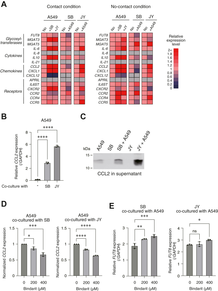
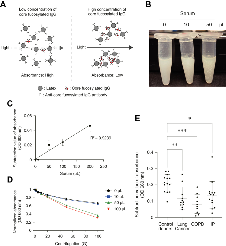
Glycan structure is often modulated in disease or predisease states, suggesting that such changes might serve as biomarkers. Here, we generated a monoclonal antibody (mAb) against the core fucose of the N-glycan in human IgG. Notably, this mAb can be used in Western blotting and ELISA. ELISA using this mAb revealed a low level of the core fucose of the N-glycan in IgG, suggesting that the level of acore fucosylated (noncore fucosylated) IgG was increased in the sera of the patients with lung cancer, chronic obstructive pulmonary disease, and interstitial pneumonia compared to healthy subjects. In a coculture analysis using human lung adenocarcinoma A549 cells and antibody-secreting B cells, the downregulation of the FUT8 (α1,6 fucosyltransferase) gene and a low level of core fucose of the N-glycan in IgG in antibody-secreting B cells were observed after coculture. A dramatic alteration in gene expression profiles for cytokines, chemokines, and their receptors were also observed after coculturing, and we found that the identified C-C motif chemokine 2 was partially involved in the downregulation of the FUT8 gene and the low level of core fucose of the N-glycan in IgG in antibody-secreting B cells. We also developed a latex turbidimetric immunoassay using this mAb. These results suggest that communication with C-C motif chemokine 2 between lung cells and antibody-secreting B cells downregulate the level of core fucose of the N-glycan in IgG, i.e., the increased level of acore fucosylated (noncore fucosylated) IgG, which would be a novel biomarker for the diagnosis of patients with pulmonary diseases.

Keywords: N-linked glycosylation; biomarker; chemokine; immunoglobulin G (IgG); lung.


Conflict of interest statement

Conflict of interest The authors declare no conflicts of interest with the contents of this article.

Figures

Figure 1
Figure 1
Knockout of the Fut8 gene disrupts the reactivity of the antibody against core fucose of N-glycan in IgG.A, representative N-glycan structure in human IgG. FUT8 (α1,6 fucosyltransferase) is an enzyme to generate the core fucose of N-glycan. B, PhoSL and LCA lectin blotting in Fut8-deficient HEK293 cells (Fut8−/−) and wildtype cells (WT). Total cell lysates (20 μg) were analyzed, and GAPDH was used as a loading control. The protein expression of FUT8 was also analyzed with an anti-FUT8 antibody. C, lectin blotting of IgGs derived from Fut8-deficient HEK293 cells (Fut8−/−) and wildtype cells (WT). After transfection with an IgG expression vector (human IgG1 CH1/CH2/CH3 portion), the secreted IgGs in the culture media were purified and enriched with ProteinG/Sepharose. A part of those samples was analyzed with lectins (AAL, ConA, PHA-E4, PHA-L4, SSA, MAM, PhoSL, and LCA). Arrows indicate the CH1/CH2/CH3 portion of human IgG1. D, Western blotting of IgGs. Enriched IgGs used in C were also analyzed with an antibody against the core fucose of N-glycan in IgG and an anti-human IgG antibody. AAL, Aleuria aurantia lectin; CH2, constant heavy 2; FUT8, α1,6 fucosyltransferase; IgG, immunoglobulin G; LCA, Lens culinaris agglutinin lectin; mAb, monoclonal antibody; MAM, Maackia amurensis lectin; PhoSL, Pholiota squarrosa lectin; SSA, Sambucus sieboldiana lectin.
Figure 2
Figure 2
Low level of core fucosylation of IgG in sera of the patients with pulmonary diseases.A, a typical standard curve for ELISA for purified normal human IgG using the antibody against core fucose of N-glycan in IgG. B, ELISA using the antibody against the core fucose of the N-glycan in IgG to measure the core fucosylation level of IgGs in sera of lung cancer, chronic obstructive pulmonary disease (COPD), interstitial pneumonia (IP) patients, and control healthy donors. The ratios of core fucosylated IgG against total IgG were plotted. Lung cancer: N = 29, COPD: N = 31, IP: N = 15, healthy donors: N = 18. ∗∗∗∗p < 0.0001. IgG, immunoglobulin G.
Figure 3
Figure 3
Coculturing with lung cancer cells downregulates the Fut8 gene and the level of core fucose of N-glycan in IgG in antibody-secreting B cells.A, scheme for the coculture analyses under conditions of contact or no-contact using human lung adenocarcinoma A549 cells and antibody-secreting B cells, SB and JY. A549 is an adherent cell, while SB and JY cells are nonadherent cells. B, FUT8 gene expression was examined by RT-qPCR in antibody-secreting SB and JY cells after coculture with A549 cells on the condition of contact or no-contact for 3 days. Expressions were normalized to the GAPDH gene. ∗∗∗p < 0.005, ∗∗∗∗p < 0.001. C, the level of core fucose of N-glycan in IgGs in SB and JY cells after coculturing with A549 cells. Secreted IgGs into the culture media were enriched with ProteinG/Sepharose and examined by Western blotting using an antibody against the core fucose of N-glycan in IgG and an anti-human IgG antibody. The values under the images indicate the normalized band intensities of the level of core fucose of N-glycan in IgG in SB and JY cells after coculture with A549 cells relative to those without coculture. D, the band intensities in C. Three independent experiments were performed, and the band intensities normalized to the condition without coculture were measured. ∗p < 0.05, ∗∗p < 0.01. FUT8, α1,6 fucosyltransferase; IgG, immunoglobulin G; ns, not significant; RT-qPCR, reverse transcription-quantitative polymerase chain reaction.
Figure 4
Figure 4
Gene expression profiles in A549 and antibody-secreting B cells after coculturing.A, heatmaps of gene expression examined by RT-qPCR in A549, SB, and JY cells after coculture for 3 days under conditions of contact or no-contact. Gene expression levels in the cells after coculturing relative to the cells without co∗-culture are shown. Gray color indicates no expression. B, CCL2 gene expression in A549 cells after coculture with SB or JB cells on the condition of contact examined by RT-qPCR. Expressions were normalized to the GAPDH gene. C, CCL2 protein expression in supernatant examined by Western blotting. After coculture SB and JB cells with or without A549 cells on the condition of contact, culture supernatant was collected and concentrated. An equally part of resulting samples was analyzed. D, CCL2 gene expression in A549 cells under the presence of Bindarit, an inhibitor of CCL2 expression, was examined by RT-qPCR. Expressions were normalized to the condition without Bindarit treatment (0 μM). E, FUT8 gene expression in SB and JY cells after coculture with A549 under the presence or absence of Bindarit. Expressions were examined by RT-qPCR and normalized to the GAPDH gene. ∗p < 0.05, ∗∗p < 0.01, ∗∗∗p < 0.005, ∗∗∗∗p < 0.001. CCL2, C-C motif chemokine 2; FUT8, α1,6 fucosyltransferase; ns: not significant; RT-qPCR, reverse transcription-quantitative polymerase chain reaction.
Figure 5
Figure 5
The latex turbidimetric immunoassay can be used to identify patients with pulmonary diseases.A, scheme for the latex turbidimetric immunoassay using the antibody against core fucose of N-glycan in IgG. The aggregation of latex beads coated with the antibody can be observed in a dose-dependent manner. A lower absorbance value indicates a higher concentration of core fucose of N-glycan in IgG in the subjects. B, an image of aggregated latex beads. Human serum from a control healthy donor (0, 10, or 50 μl) was incubated with the solution of latex beads coated with the antibody against core fucose of N-glycan in IgG. C, a typical standard curve for the latex turbidimetric immunoassay. The human pooled serum (0, 10, 50, 100, or 200 μl) was analyzed, and the absorbance values (600 nm) were subtracted from those just after the addition of sera (time = 0). D, the absorbance of the solution of latex beads after centrifugation. After incubation with human pooled serum (0, 10, 50, or 100 μl), the latex solution was centrifugated at 2, 5, 10, 20, 50, or 100 G for 20 s, then, the absorbance of the solution was analyzed. The absorbance values (600 nm) were normalized to those without centrifugation. E, the absorbance of the solution of latex beads after incubation with the sera of the patients with lung cancer (N = 11), chronic obstructive pulmonary disease (COPD) (N = 10), interstitial pneumonia (IP) (N = 14), and control healthy donors (N = 14). The absorbance values (660 nm) were subtracted from those just after the addition of sera (time = 0). ∗p < 0.05, ∗∗p < 0.01, ∗∗∗p < 0.001. IgG, immunoglobulin G.

References

    1. Varki A. Biological roles of glycans. Glycobiology. 2017;27:3–49. - PMC - PubMed
    1. Moremen K.W., Tiemeyer M., Nairn A.V. Vertebrate protein glycosylation: diversity, synthesis and function. Nat. Rev. Mol. Cell Biol. 2012;13:448–462. - PMC - PubMed
    1. Freeze H.H., Kinoshita T., Varki A. In: Essentials of Glycobiology. 3rd Ed. Varki A., Cummings R.D., Esko J.D., Stanley P., Hart G.W., Aebi M., et al., editors. Cold Spring Harbor Laboratory Press; Cold Spring Harbor, NY: 2015. Glycans in acquired human diseases.
    1. Sackstein R., Hoffmeister K.M., Stowell S.R., Kinoshita T., Varki A., Freeze H.H. In: Essentials of Glycobiology. 4th Ed. Varki A., Cummings R.D., Esko J.D., Stanley P., Hart G.W., Aebi M., et al., editors. Cold Spring Harbor Laboratory Press; Cold Spring Harbor, NY: 2022. Glycans in acquired human diseases.
    1. Wang M., Zhu J., Lubman D.M., Gao C. Aberrant glycosylation and cancer biomarker discovery: a promising and thorny journey. Clin. Chem. Lab. Med. 2019;57:407–416. - PMC - PubMed

Publication types

MeSH terms