. 2023 Dec;299(12):105382.
doi: 10.1016/j.jbc.2023.105382. Epub 2023 Oct 21.

Discovery of FERM domain protein-protein interaction inhibitors for MSN and CD44 as a potential therapeutic approach for Alzheimer's disease

Affiliations

Discovery of FERM domain protein-protein interaction inhibitors for MSN and CD44 as a potential therapeutic approach for Alzheimer's disease

Yuhong Du et al. J Biol Chem. 2023 Dec.

Abstract

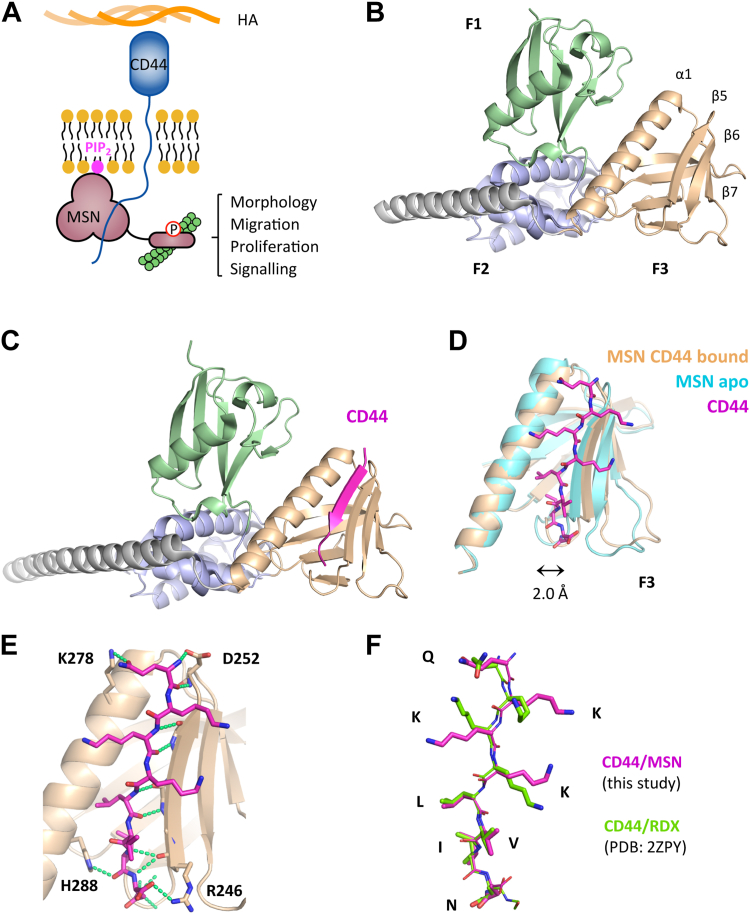
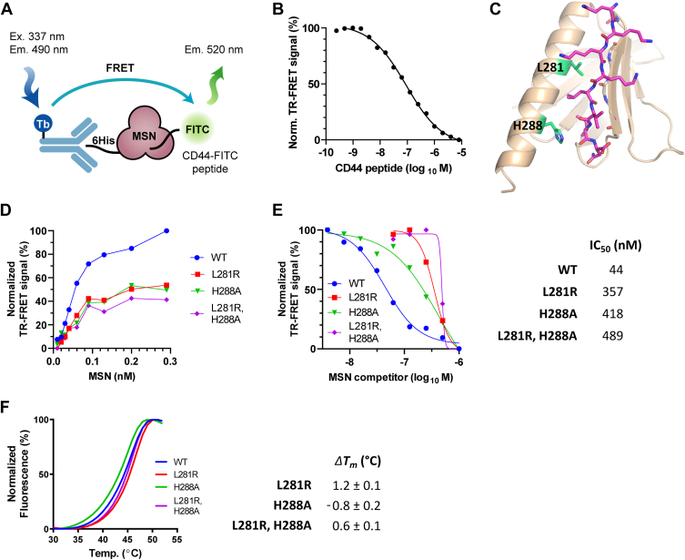
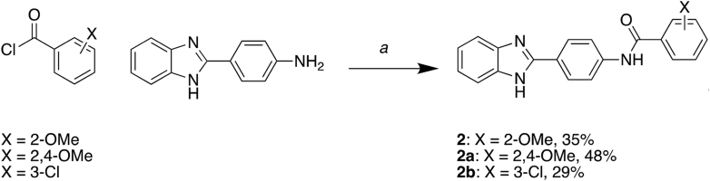
Proteomic studies have identified moesin (MSN), a protein containing a four-point-one, ezrin, radixin, moesin (FERM) domain, and the receptor CD44 as hub proteins found within a coexpression module strongly linked to Alzheimer's disease (AD) traits and microglia. These proteins are more abundant in Alzheimer's patient brains, and their levels are positively correlated with cognitive decline, amyloid plaque deposition, and neurofibrillary tangle burden. The MSN FERM domain interacts with the phospholipid phosphatidylinositol 4,5-bisphosphate (PIP2) and the cytoplasmic tail of CD44. Inhibiting the MSN-CD44 interaction may help limit AD-associated neuronal damage. Here, we investigated the feasibility of developing inhibitors that target this protein-protein interaction. We have employed structural, mutational, and phage-display studies to examine how CD44 binds to the FERM domain of MSN. Interestingly, we have identified an allosteric site located close to the PIP2 binding pocket that influences CD44 binding. These findings suggest a mechanism in which PIP2 binding to the FERM domain stimulates CD44 binding through an allosteric effect, leading to the formation of a neighboring pocket capable of accommodating a receptor tail. Furthermore, high-throughput screening of a chemical library identified two compounds that disrupt the MSN-CD44 interaction. One compound series was further optimized for biochemical activity, specificity, and solubility. Our results suggest that the FERM domain holds potential as a drug development target. Small molecule preliminary leads generated from this study could serve as a foundation for additional medicinal chemistry efforts with the goal of controlling microglial activity in AD by modifying the MSN-CD44 interaction.

Keywords: Alzheimer disease; CD44; FERM domain; MSN; crystal structure; fluorescence resonance energy transfer; high-throughput screening; inhibitor; microglia; moesin; structure-activity relationship.


Conflict of interest statement

Conflict of interest The authors declare that they have no conflicts of interest with the contents of this article.

Figures

Figure 1
Figure 1
Crystal structure of MSN-FERM, either ligand free, or bound to CD44 cytoplasmic tail.A, model showing MSN activation through binding the C-terminal cytoplasmic tail of the CD44 receptor and PIP2. B, crystal structure of ligand-free FERM domain of human MSN (PDB: 6TXQ). The three FERM subdomains of MSN, shown as ribbons, are colored in green (F1), metallic blue (F2), and wheat color (F3), while the long C-terminal alpha helix is colored in gray. C, crystal structure of human MSN bound to a short synthetic CD44 peptide (678-QKKKLVIN-685). CD44 (magenta, ribbon) binds within the F3 domain, forming part of the beta-sheet. D, CD44 within the F3 lobe (magenta, stick model) is accommodated by a displacement of the beta-sheet away from the alpha helix by 2.0 Å, measured from H288 and R246 carbonyl carbon atoms. MSN bound to CD44 is shown in wheat colour, while unbound MSN is shown in cyan. E, interactions between MSN and CD44, showing hydrogen bonding (green dashed lines). The majority of bonding is backbone–backbone, although a few MSN side chain residue interactions (H288, D252, R246) were identified. The MSN–K278 interaction is most likely an artefact of crystal packing. F, overlay of the FERM domain–bound CD44 to either MSN (this work) or radixin (PDB: 2ZPY). FREM, four-point-one ezrin radixin moesin; MSN, moesin; PIP2, phosphatidylinositol 4,5-bisphosphate.
Figure 2
Figure 2
Structure-guided mutagenesis reveals MSN residues involved in CD44 binding.A, schematic representation of the in vitro TR-FRET assay for MSN–CD44 interaction using purified components. Signal generated from Tb-linked anti-6HIS antibody bound to MSN is transferred to the FITC-conjugated CD44 peptide. B, inhibition of the TR-FRET signal using unlabeled CD44 peptide as a competitor (IC50 = 66 nM). C, side chains of mutated MSN residues (L281 and H288) within the MSN–CD44 binding pocket are shown (green). D, comparison of MSN–CD44 TR-FRET response using varying levels of 6His-tagged WT or MSN mutant protein (0.01–0.29 nM). CD44 peptide concentration was kept constant (8 nM). E, competition of mutant MSN protein in MSN–CD44 TR-FRET assay. Dose response of untagged WT or mutant MSN protein in TR-FRET assay containing WT 6His-MSN (2 nM) and CD44 peptide (8 nM). IC50 values of competitor proteins are shown on the right (n = 3). F, thermal shift assay, showing melting temperature (Tm) curves of WT, and mutant proteins (left panel). The ΔTm, compared to WT protein, is shown in the right panel (n = 3; ±SD). MSN, moesin; PPI, protein–protein interaction; TR-FRET, time-resolved FRET.
Figure 3
Figure 3
Phage display screening identifies peptides that bind MSN at distinct sites.A, sequences of phage display peptides. Underlined region corresponds to the variable region within the phage-derived sequence. B, crystal structure of FERM domain of MSN bound to the C3P-pd peptide. FERM subdomains are displayed as in 1B. C3P-pd peptide (magenta) binds in a pocket between the F1 and F3 subdomains. C, close-up view of C3P-pd peptide bound to MSN. Peptide intramolecular H-bonds are shown (purple dashed lines). Side chains of MSN residues interacting with the peptide are displayed, together with their bonding to the peptide (gold dashed lines). D, superimposed F3 subdomains of MSN from apo (cyan) and C3P-pd bound (light yellow) structures. C3P-pd peptide binding causes a 2.3 Å movement of the beta-sheet away from the alpha helix in the MSN F3 lobe, measured from H288 and R246 carbonyl carbon atoms. E, crystal structure of FERM domain of MSN bound to both C3P-pd and C3S1-pd peptides. F, close-up of C3S1-pd binding to F3 lobe of MSN. H-bond contacts are shown (green dashed lines). G, space-filling model of MSN FERM domain, showing C3P-pd (magenta) and CD44 (cyan) binding relative to proposed PIP2 binding pocket (PIP2-BP; blue positively charged surface). HJ, MSN TR-FRET inhibition assays, with acceptor fluorophore conjugated to either (H) CD44, (I) C3P-pd, or (J) C3S1-pd peptides. Unlabeled peptides were used as competitors (C3S1-pd, blue circle; C3P-pd, red square; CD44, green triangle). K, IC50 values derived from (HJ). Asterisk denotes stimulatory rather than inhibitory values. L, binding properties of MSN associated to C3P-pd and C3S1-pd peptides were measured by ITC. See Fig. S3 for thermograms and corresponding fitted curves. ΔH, enthalpy; FERM, four-point-one ezrin radixin moesin; ITC, isothermal titration calorimetry; KD, dissociation constant; MSN, moesin; N, stoichiometry; PPI, protein–protein interaction; TR-FRET, time-resolved FRET.
Figure 4
Figure 4
uHTS of a 138k compound library identifies MSN–CD44 small-molecule PPI inhibitors.A, scatter plot showing percentage inhibition with 138,214 compound library in the MSN–CD44 TR-FRET assay. Primary hit rate is 0.2%, with 271 compounds giving >50% inhibition. B, screening flow chart showing hit-to-lead identification strategy. C, structures of lead compounds based upon different chemical scaffolds. D, confirmatory TR-FRET dose-response curves of compound 1 (IC50 = 8.1 ±0.8 μM; h = 1.7) and compound 2 (IC50 = 8.1 ±0.8 μM; h = 15.8) E, dose response of compounds in GST pull-down assay. Cell lysates were obtained from cells transfected with full-length constructs of GST-tagged MSN and Flag-tagged CD44 and were incubated with the compounds. Immunoblotting was performed to detect GST and Flag. Presented are representative immunoblots among replicates, demonstrating that compounds 1 and 2 induce the dissociation of CD44 from MSN. F, thermal shift assay, showing melting temperature (Tm) curves of MSN with compounds. The average change in Tm (av. ΔTm), compared to DMSO alone, was determined from two experiments (1: 1.1 °C at 20 μM; 2: 1.9 °C at 20 μM). G, dose response of compound binding to MSN FERM domain, measured by SPR. KD measurements were determined for compounds 1 (4.2 μM) and 2 (0.7 μM). RU = response units. H, dose response of compounds in cell-based NanoBiT bioluminescence assay, with HEK293 cells expressing LgBiT-MSN(T588D) and SmBit-CD44 fusions. Luminescence from reconstituted split luciferase was measured after substrate addition. Activities were determined from two independent experiments (1: 0.57 ± 0.17 and 0.69 ± 0.12 μM; 2: 0.18 ± 0.04 and 0.19 ± 0.06 μM). I, solubility of compounds (PBS buffer, 1% DMSO), as measured by nephelometry. DMSO, dimethyl sulfoxide; FERM, four-point-one ezrin radixin moesin; GST, glutathione-S-transferase; LgBiT, large binary technology; MSN, moesin; PPI, protein–protein interaction; RNU, relative nephelometric unit; SmBiT, small binary technology; TR-FRET, time-resolved FRET; uHTS, ultrahigh-throughput screening.
Figure 5
Figure 5
Compound 2 and analogs disrupt the MSN–CD44 PPI.A, structures of compound 2 and analogs, with associated solubility data (PBS, 1% DMSO), and activity in both the MSN–CD44 TR-FRET assay and an unrelated PPI (SYK-FCER1G) TR-FRET assay. The bottom panel shows the MSN–CD44 TR-FRET dose response for compound 2a (h = 2.4) and compound 2b (h = 2.3). Solubility was measured by nephelometry. B, Dose response of compounds in GST pull-down assay. Cell lysates were obtained from cells transfected with full-length constructs of GST-tagged MSN and Flag-tagged CD44 and were incubated with the compounds. Immunoblotting was performed to detect GST and Flag, and a representative immunoblot from two experiments is shown. C, thermal shift assay, showing representative melting temperature (Tm) curves of MSN with compounds. The average change in Tm (av. ΔTm), compared to DMSO alone, was determined from two experiments (2a: 1.3 °C at 50 μM; 2b: 1.2 °C at 50 μM). DMSO, dimethyl sulfoxide; GST, glutathione-S-transferase; MSN, moesin; PPI, protein–protein interaction; TR-FRET, time-resolved FRET.
Figure 6
Figure 6
Synthesis of compound 1.a, 4-((tert-butoxycarbonyl)amino)benzoic acid, CH2Cl2, SOCl2, 0 to 40 °C; (b) tert-butyl (4-(chlorocarbonyl)phenyl)carbamate, diisopropylethylamine, tetrahydrofolic acid, 65 °C; and (c) TFA, CH2Cl2.
Figure 7
Figure 7
Synthesis of compounds 2, 2a, and 2b.a, acyl chloride, diisopropylethylamine, tetrahydrofolic acid, 0 to 65 °C.

Update of

References

    1. Patterson C. The State of the Art of Dementia Research: New Frontiers. An Analysis of Prevalence, Incidence, Cost and Trends. Alzheimer’s Disease International (ADI); London: 2018.
    1. Haass C., Selkoe D. If amyloid drives Alzheimer disease, why have anti-amyloid therapies not yet slowed cognitive decline? PLoS Biol. 2022;20 - PMC - PubMed
    1. The Lancet Lecanemab for Alzheimer's disease: tempering hype and hope. Lancet. 2022;400:1899. - PubMed
    1. van Dyck C.H., Sabbagh M., Cohen S. Lecanemab in early Alzheimer's disease. N. Engl. J. Med. 2023;388:1631–1632. - PubMed
    1. Wang C., Zong S., Cui X., Wang X., Wu S., Wang L., et al. The effects of microglia-associated neuroinflammation on Alzheimer's disease. Front. Immunol. 2023;14 - PMC - PubMed

Publication types