. 2023 Oct 23;8(1):404.
doi: 10.1038/s41392-023-01657-4.

Fe65-engineered neuronal exosomes encapsulating corynoxine-B ameliorate cognition and pathology of Alzheimer's disease

Affiliations

Fe65-engineered neuronal exosomes encapsulating corynoxine-B ameliorate cognition and pathology of Alzheimer's disease

Ashok Iyaswamy et al. Signal Transduct Target Ther. .

Abstract

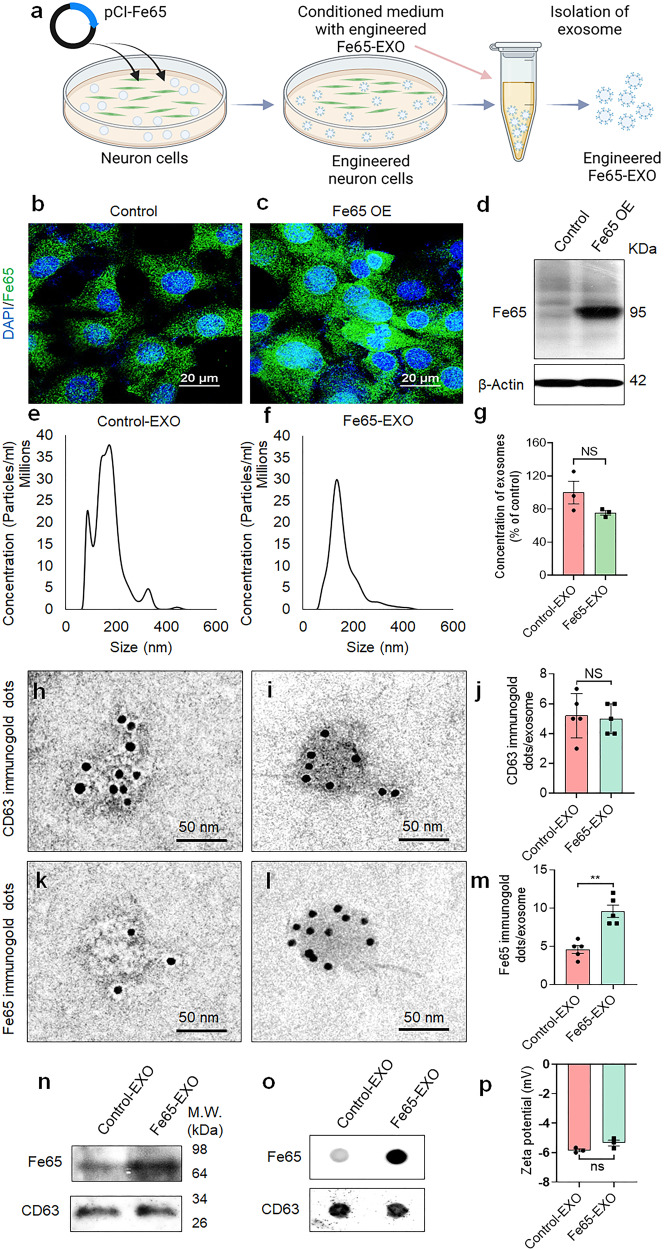
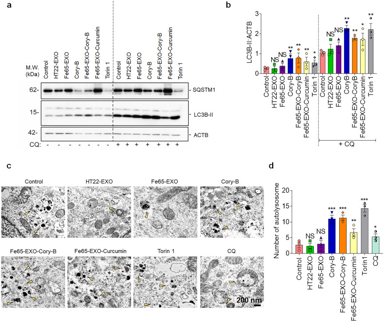
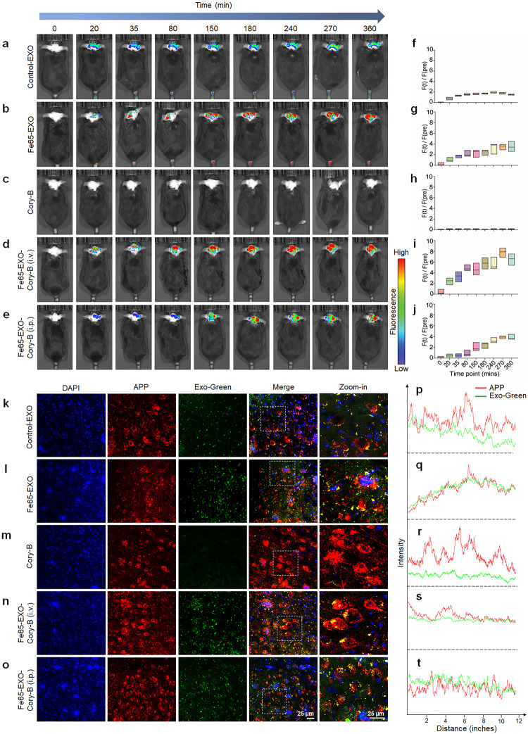
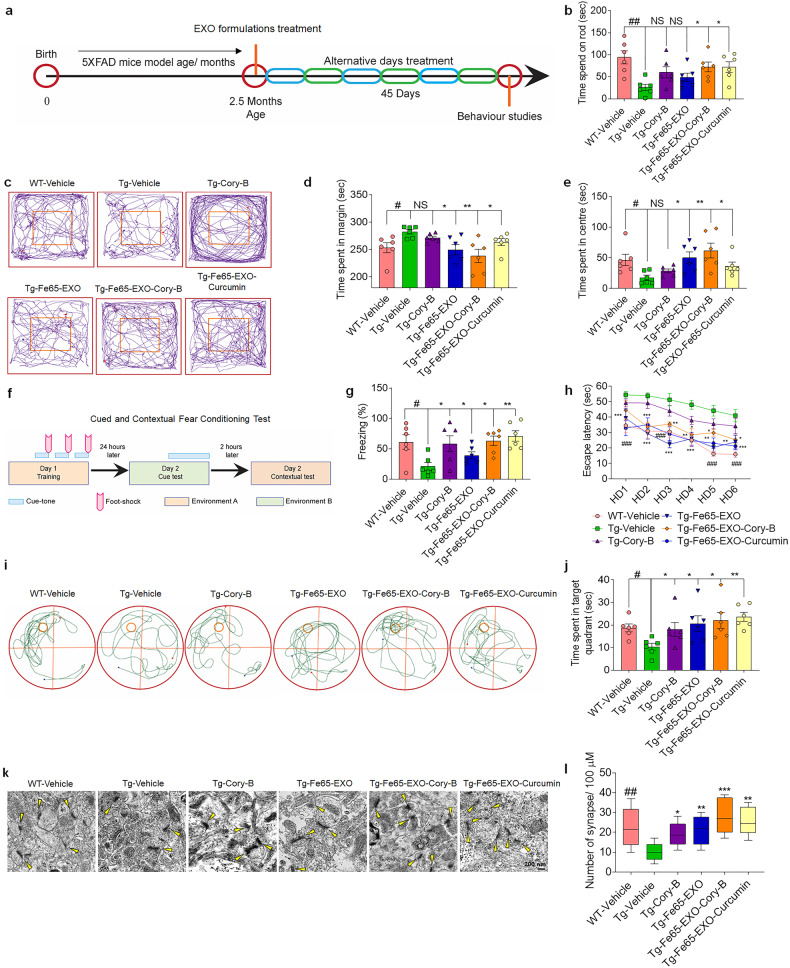
Alzheimer's disease (AD) is a neurodegenerative disorder characterized by the predominant impairment of neurons in the hippocampus and the formation of amyloid plaques, hyperphosphorylated tau protein, and neurofibrillary tangles in the brain. The overexpression of amyloid-β precursor protein (APP) in an AD brain results in the binding of APP intracellular domain (AICD) to Fe65 protein via the C-terminal Fe65-PTB2 interaction, which then triggers the secretion of amyloid-β and the consequent pathogenesis of AD. Apparently, targeting the interaction between APP and Fe65 can offer a promising therapeutic approach for AD. Recently, exosome, a type of extracellular vesicle with diameter around 30-200 nm, has gained much attention as a potential delivery tool for brain diseases, including AD, due to their ability to cross the blood-brain barrier, their efficient uptake by autologous cells, and their ability to be surface-modified with target-specific receptor ligands. Here, the engineering of hippocampus neuron cell-derived exosomes to overexpress Fe65, enabled the development of a novel exosome-based targeted drug delivery system, which carried Corynoxine-B (Cory-B, an autophagy inducer) to the APP overexpressed-neuron cells in the brain of AD mice. The Fe65-engineered HT22 hippocampus neuron cell-derived exosomes (Fe65-EXO) loaded with Cory-B (Fe65-EXO-Cory-B) hijacked the signaling and blocked the natural interaction between Fe65 and APP, enabling APP-targeted delivery of Cory-B. Notably, Fe65-EXO-Cory-B induced autophagy in APP-expressing neuronal cells, leading to amelioration of the cognitive decline and pathogenesis in AD mice, demonstrating the potential of Fe65-EXO-Cory-B as an effective therapeutic intervention for AD.


Conflict of interest statement

A.I., A.T., M.L., and H.J.C. are inventors on a patent filed with the United States Patent and Trademark Office (USPTO) about “Exosomal compositions and methods for treating neurodegenerative diseases (Application number: 63/479,758. Attorney Docket Number: 22-1827-US-PRO)”. All other authors have no competing interests.

Figures

Fig. 1
Fig. 1
Development and characterization of Fe65-engineered exosomes. a Schematic diagram showing the steps involved in the production of engineered Fe65-EXO. Representative b, c immunofluorescence images showing expression of Fe65, d protein level of Fe65 in control- and Fe65 OE- HT22 hippocampus neuron cells. Representative eg size distribution, hj, km CD63-, and Fe65-positive immunogold dots, and their relative quantification, n, o protein level of Fe65 by n WB, and o dot-blot, and p zeta potential of exosomes isolated from control- and Fe65 OE- HT22 hippocampus neuron cells. In g, j, m, and p data are shown as mean ± standard error mean (SEM). Statistical analysis was performed by Student’s t-test to compare Control-EXO vs. Fe65-EXO. Significance level: **P < 0.01, NS not significant
Fig. 2
Fig. 2
Higher autologous uptake of Fe65-engineered exosomes by APP overexpressed hippocampus neuron cells. ah Enhanced autologous uptake of Fe65-EXO by APP OE-HT22 cells compared with HT22 cells, and ip enhanced heterologous uptake of Fe65-EXO by APP OE-N2a cells compared with N2a cells after 24 h. Nuclei: DAPI (blue), cell membrane: APP (red), Fe65-EXO: Green (green). EXO-APP (yellow). In ap; scale bar = 10 µm. q, r Effect of Fe65-EXO on the viability of HT22- and N2a- neuron cells, respectively. HT22- and N2a- neuron cells were treated with cell culture medium (control), control EXO, or Fe65-EXO for 24 h, followed by the measurement of viability using MTT assay protocol. In q and r, data are shown as mean ± S.E.M (N = 6). Statistical analysis was performed by one-way ANOVA for multiple comparison of Vehicle Control vs. Control EXO or Fe65-EXO, Significance level: **P < 0.01, NS not significant
Fig. 3
Fig. 3
Delivery of Cory-B by Fe65-EXO caused BECN1-dependent augmented autophagy in neuronal cells. a, b Representative immunoblots depicting the protein level of autophagy markers (SQSTM1 and LC3B-II) in N2a cells, without and with pre-treatment of chloroquine (CQ, 100 μM), and the corresponding bar graphs. c, d Representative electron micrograph of N2a cells showing the formation of autolysosome and the corresponding bar graphs, respectively. Scale bar: 200 nm. In b and d, data are presented as mean ± SEM (N = 3). Statistical analysis was performed by one-way ANOVA with Dunnett’s multiple comparison test to compare Control vs. treatment groups. Significance level at *P < 0.05, **P < 0.01, and ***P < 0.001
Fig. 4
Fig. 4
Biodistribution of Fe65-EXO-Cory-B in the brain of AD mouse. Intravenously injected a control exosomes, b Fe65-EXO, c Cory-B, and d Fe65-EXO-Cory-B in 5xFAD mouse model. e Intraperitoneally injected Fe65-EXO-Cory-B in 5xFAD mouse model. The exosomes are labeled with Exo-Glow dye, and after its administration, AD mice were imaged at various times using the IVIS In Vivo Imaging System. fj The quantitative time course bar graphs show the biodistribution of control-EXO, Fe65-EXO, Cory-B, and Fe65-EXO-Cory-B (i.v. or i.p.) in the brain of a 5xFAD mouse model. ko Immunofluorescence staining showing colocalization of APP (shown in red) and exosomes (labeled with Exo-Green; shown in green) in the hippocampus region of brain of 5xFAD mouse model treated with control-EXO, Fe65-EXO, Cory-B, and Fe65-EXO-Cory-B (i.v. or i.p.), and pt the corresponding fluorescence intensity of APP and Exo-Green, depicting the strength of their colocalization. In ko, scale bar = 25 µm
Fig. 5
Fig. 5
Fe65-EXO-Cory-B improves cognitive behavior in AD mice. a A schematic plan of the experiment showing the treatment schedule, followed by behavior experiments in 5xFAD mice. b Representative bar graph shows the time spent by the AD mice on the accelerating rotarod platform under various treatment conditions, as analyzed by rotarod test. ce Analysis of general activity level, gross locomotor activity, and exploration habits of AD mice by open field test in a white Plexiglas box under various treatment conditions. Bar graphs d and e show the time spent by the AD mice in the margin and center of the Plexiglas box, based on the recorded tracks of the mice as shown in c. f Diagrammatic illustration of cued and contextual fear conditioning. g Representative graph showing freezing time of AD mice under various treatment conditions after the cue tone, as analyzed from the contextual fear conditioning. h Representative graph showing the escape latency of AD mice. i, j Analysis of spatial learning and memory in AD mice by Morris’s water maize (MWM) test under various treatment conditions. Representative i graph showing time spent by AD mice in target quadrant based on their swim paths or mouse trajectory j recorded in MWM. k, l Representative k electron microscopy images showing the formation of synapses (scale bar: 200 nm), and l the corresponding bar graph depicting the number of synapses formed in the hippocampus region of AD mouse brain under various treatment conditions. Data are presented as mean ± SEM (N = 6 mice/group). Statistical analysis was performed by one-way ANOVA with Dunnett’s multiple comparison test to compare Tg-Vehicle vs. treatment groups. Significance level at *P < 0.05, **P < 0.01, ***P < 0.001. Comparison of Tg-Vehicle vs. WT-Vehicle group. Significance level at #P < 0.05, ##P < 0.01, ###P < 0.001

References

    1. Ballard C, et al. Drug repositioning and repurposing for Alzheimer disease. Nat. Rev. Neurol. 2020;16:661–673. - PMC - PubMed
    1. Yee A, et al. Alzheimer’s disease: insights for risk evaluation and prevention in the Chinese population and the need for a comprehensive programme in Hong Kong/China. Hong Kong Med. J. 2018;24:492. - PubMed
    1. Mattson MP. Pathways towards and away from Alzheimer’s disease. Nature. 2004;430:631–639. - PMC - PubMed
    1. Selkoe DJ, Schenk D. Alzheimer’s disease: molecular understanding predicts amyloid-based therapeutics. Annu. Rev. Pharmacol. Toxicol. 2003;43:545–584. - PubMed
    1. Jack CR, et al. Tracking pathophysiological processes in Alzheimer’s disease: an updated hypothetical model of dynamic biomarkers. Lancet Neurol. 2013;12:207–216. - PMC - PubMed

Publication types

LinkOut - more resources