. 2023 Oct 23;21(1):231.
doi: 10.1186/s12915-023-01736-6.

SRSF2 is required for mRNA splicing during spermatogenesis

Affiliations

SRSF2 is required for mRNA splicing during spermatogenesis

Wen-Long Lei et al. BMC Biol. .

Abstract

Background: RNA splicing plays significant roles in fundamental biological activities. However, our knowledge about the roles of alternative splicing and underlying mechanisms during spermatogenesis is limited.

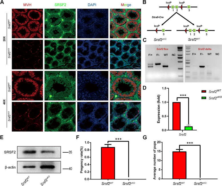
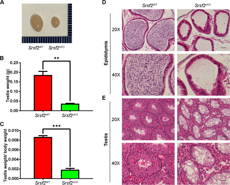
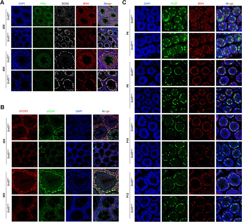
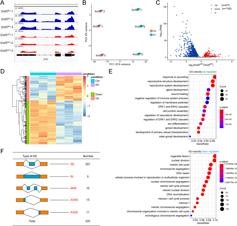
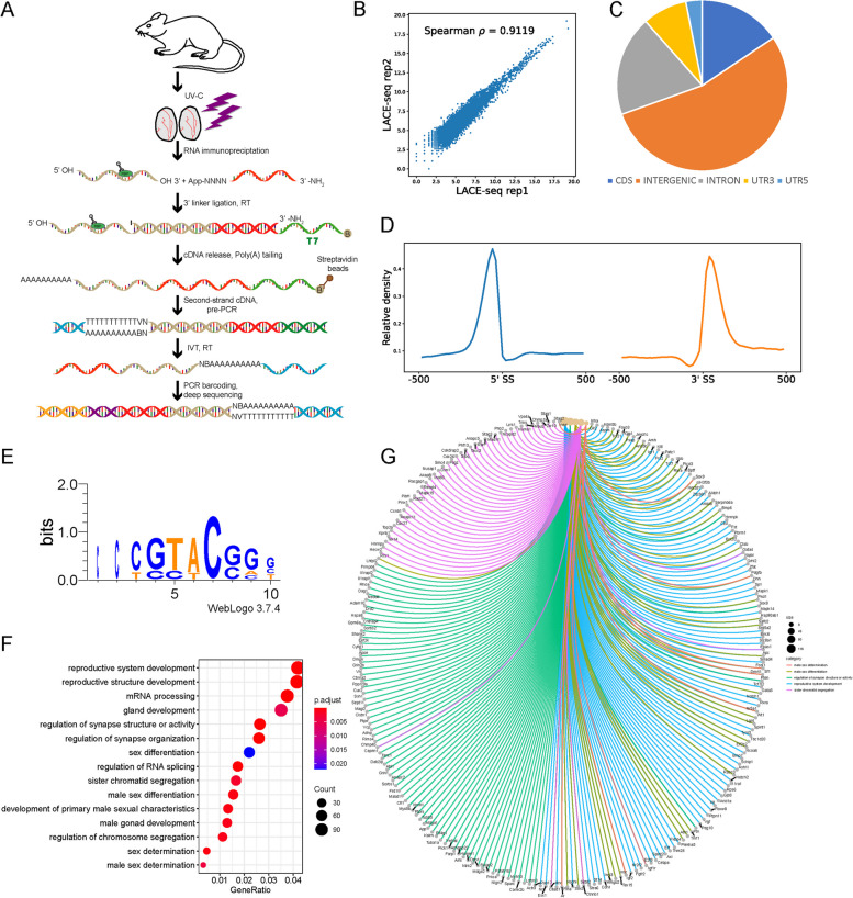
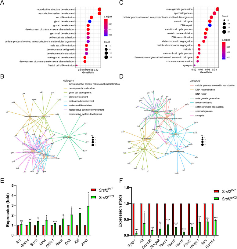
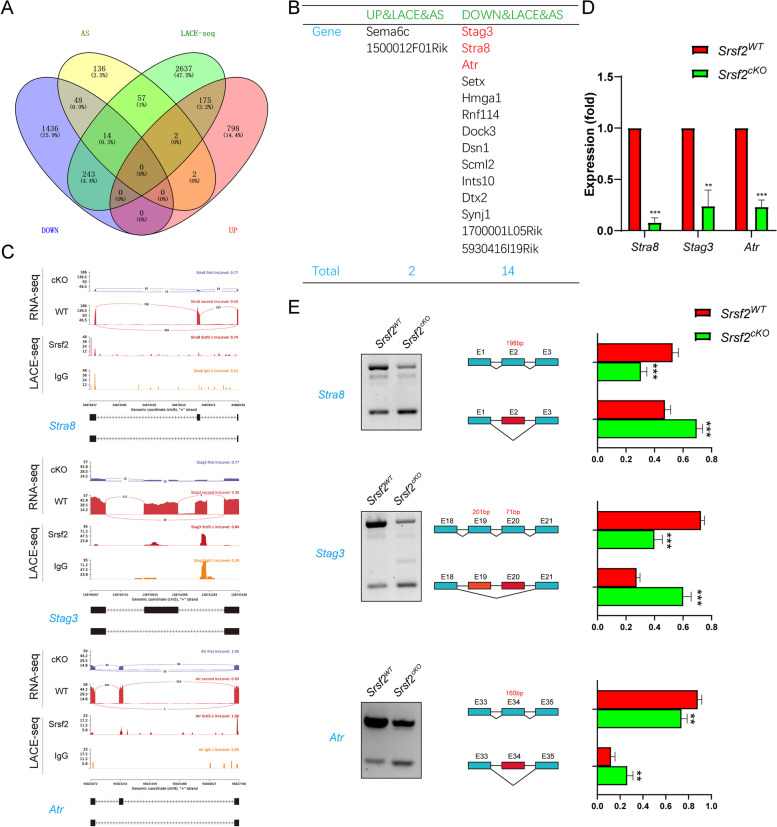
Results: Here, we report that Serine/arginine-rich splicing factor 2 (SRSF2), also known as SC35, plays critical roles in alternative splicing and male reproduction. Male germ cell-specific deletion of Srsf2 by Stra8-Cre caused complete infertility and defective spermatogenesis. Further analyses revealed that deletion of Srsf2 disrupted differentiation and meiosis initiation of spermatogonia. Mechanistically, by combining RNA-seq data with LACE-seq data, we showed that SRSF2 regulatory networks play critical roles in several major events including reproductive development, spermatogenesis, meiotic cell cycle, synapse organization, DNA recombination, chromosome segregation, and male sex differentiation. Furthermore, SRSF2 affected expression and alternative splicing of Stra8, Stag3 and Atr encoding critical factors for spermatogenesis in a direct manner.

Conclusions: Taken together, our results demonstrate that SRSF2 has important functions in spermatogenesis and male fertility by regulating alternative splicing.

Keywords: Alternative splicing; LACE-seq; Male infertility; SRSF2; Spermatogenesis.


Conflict of interest statement

The authors declare that they have no competing interests.

Figures

Fig. 1
Fig. 1
SRSF2 is essential for male fertility. A Representative images of localization of SRSF2 (green) and MVH (red) in the control and Srsf2cKO testes of 8-week-old mice. The DNA was stained with DAPI. Scale bar: (top) 50 μm; (bottom) 20 μm. B Schematic diagram of deletion of Srsf2 exons 1 and 2 and generation of Srsf2 Δ allele by Stra8 Cre-mediated recombination in male germ cells. C Genotyping PCRs were performed using Srsf2 flox and Srsf2 delta primers. D Quantitative RT-PCR analyses showing Srsf2 mRNA level was decreased. β-actin was used as the internal control. E Western blotting analysis of SRSF2 protein in Srsf2WT and Srsf2cKO total testes of 8-week-old mice. β-actin was detected as an internal control. F Pregnancy rates (%) of plugged wild-type females after mating with Srsf2WT and Srsf2cKO 8-week-old males. G Average litter size of plugged wild-type females after mating with Srsf2WT and Srsf2.cKO 8-week-old males. For this part, 3 mice (8-week-old) of each genotype were used for the analysis. Data are presented as the mean ± SEM. P < 0.05(*), 0.01(**) or 0.001(***)
Fig. 2
Fig. 2
SRSF2 is required for spermatogenesis. A The testes of Srsf2cKO were smaller than those of the control (8-week-old, the same as below). B Testis weight of Srsf2WT and Srsf2cKO 8-week-old male mice (n = 3). C Testis weight to body weight ratio of Srsf2WT and Srsf2cKO 8-week-old male mice (n = 3). Data are presented as the mean ± SEM. P < 0.05(*), 0.01(**) or 0.001(***). D Histological analysis of the caudal epididymes of the Srsf2WT and Srsf2cKO mice. (Scale bar: 50 μm) (E) Histological analysis of the seminiferous tubules of the Srsf2WT and Srsf2.cKO mice. Scale bar: (top) 100 μm; (bottom) 50 μm. For this part, 3 mice (8-week-old) of each genotype were used for the analysis. Data are presented as the mean ± SEM. P < 0.05(*), 0.01(**) or 0.001(***)
Fig. 3
Fig. 3
Srsf2 deficient germ cells fail to progress into meiosis. A PNA-lectin histochemistry (green), SOX9 (a marker of Sertoli cells, white) and MVH (a marker of germ cells, red) immunofluorescence analysis of the Srsf2WT and Srsf2cKO 8-week-old male mice. Scale bar: (top) 50 μm; (bottom) 20 μm. B γH2AX (green) and SYCP3 (red) immunofluorescence analysis of the Srsf2WT and Srsf2cKO 8-week-old male mice. Scale bar: (top) 50 μm; (bottom) 20 μm. C PLZF (green) and MVH (red) immunofluorescence analysis of the Srsf2WT and Srsf2cKO male mice at P6, P8, P10 and P12. Scale bar, 20 μm. In this part, 3 mice of each genotype were used for the analysis
Fig. 4
Fig. 4
Transcriptome and splicing of transcripts changes in SRSF2-null testes. A RNA-seq results showing the reduction of Srsf2 RNA in Srsf2cKO mice testes. Three independent RNA-seq experiments are shown. B Srsf2cKO groups rather than to Srsf2WT groups are clustered together by PCA. C Volcano plot showing transcriptome changes between Srsf2WT and Srsf2cKO testes. D Heatmap showing hierarchical clustering of differential expression genes of Srsf2WT and Srsf2cKO male mice testes. E GO term enrichment analysis of upregulated genes and downregulated genes. F The five different types of alternative splicing (AS) events. The numbers of abnormal AS events were counted between Srsf2WT and Srsf2cKO testes by rMATS software. In this part, 3 mice of each genotype were used for the analysis
Fig. 5
Fig. 5
Global landscape of SRSF2-binding sites in mouse testes as revealed by using LACE-seq. A Flowchart of the LACE-seq method. RBP, represents RNA-binding protein. A circled B represents biotin modification. N, represents random nucleotide; V represents A, G or C. IVT, represents in vitro transcription. B Spearman correlation plot between SRSF2 LACE-seq replicates in total testes for assessing the reproducibility of the data. Spearman correlation for the reads counts of each sample was calculated from two replicates. C Genomic distribution of SRSF2 binding sites in testes. CDS, coding sequence. UTR3, 3′ untranslated region. UTR5, 5′ untranslated region. D Schematic analysis showing the distribution of SRSF2-binding sites in the vicinity of the 5′ exon–intron and the 3′ intron–exon boundaries (500 nt upstream and 500 nt downstream of 3′SS; 500 nt upstream and 500 nt downstream of 5′SS). E SRSF2-binding motifs identified by LACE-seq in mouse testes. F GO enrichment map of SRSF2-binding genes. G Network analysis of the enriched GO terms of SRSF2-specific targets
Fig. 6
Fig. 6
The expressions of key SRSF2-binding genes involved in the spermatogenesis change after Srsf2 KO. A Correlation analysis between the RNA-seq and LACE-seq. GO analysis of the significantly upregulated genes and SRSF2-binding genes. B Network analysis of the enriched GO terms of the significantly upregulated genes and SRSF2-specific targets. C Correlation analysis between the RNA-seq and LACE-seq. GO analysis of the significantly downregulated genes and SRSF2-binding genes. D Network analysis of the enriched GO terms of the significantly downregulated genes and SRSF2-specific targets. E Quantitative RT-PCR validation of the expression of genes involved in (B). β-actin was used as the internal control. Data are presented as the mean ± SEM. P < 0.05(*), 0.01(**) or 0.001(***). F Quantitative RT-PCR validation of the expression of genes involved in (D). β-actin was used as the internal control. Data are presented as the mean ± SEM. P < 0.05(*), 0.01(**) or 0.001(***)
Fig. 7
Fig. 7
SRSF2 affects expression and alternative splicing of Stra8, Stag3 and Atr in a direct manner. A Venn diagram shows the correlation among SRSF2-binding genes, DEGs, and AS genes. B The detailed genes of SRSF2-binding, differentially expressed, and AS. C A magnified view showing RNA-seq and LACE-seq signals of the selected candidate genes. IgG, immunoglobulin G. D Quantitative RT-PCR validation of the expression of Stra8, Stag3, and Atr. E Semiquantitative RT-PCR analysis of AS patterns of the changed spliced genes in Srsf2WT and Srsf2cKO testes at P10 (n = 4 per group). PCR primers are listed in Additional file 5: Table S1. The scheme and cumulative data on percentage of the indicated fragments are shown accordingly

References

    1. Oatley JM, Brinster RL. Regulation of spermatogonial stem cell self-renewal in mammals. Annu Rev Cell Dev Biol. 2008;24:263–286. - PMC - PubMed
    1. Yang QE, Oatley JM. Spermatogonial stem cell functions in physiological and pathological conditions. Curr Top Dev Biol. 2014;107:235–267. - PubMed
    1. Kanatsu-Shinohara M, Shinohara T. Spermatogonial stem cell self-renewal and development. Annu Rev Cell Dev Biol. 2013;29:163–187. - PubMed
    1. Song HW, Wilkinson MF. Transcriptional control of spermatogonial maintenance and differentiation. Semin Cell Dev Biol. 2014;30:14–26. - PMC - PubMed
    1. Hogarth CA, Griswold MD. The key role of vitamin A in spermatogenesis. J Clin Invest. 2010;120(4):956–962. - PMC - PubMed

Publication types