. 2023 Oct 23;21(1):229.
doi: 10.1186/s12915-023-01656-5.

Prevalent bee venom genes evolved before the aculeate stinger and eusociality

Affiliations

Prevalent bee venom genes evolved before the aculeate stinger and eusociality

Ivan Koludarov et al. BMC Biol. .

Abstract

Background: Venoms, which have evolved numerous times in animals, are ideal models of convergent trait evolution. However, detailed genomic studies of toxin-encoding genes exist for only a few animal groups. The hyper-diverse hymenopteran insects are the most speciose venomous clade, but investigation of the origin of their venom genes has been largely neglected.

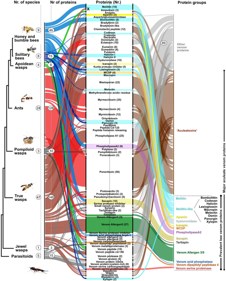
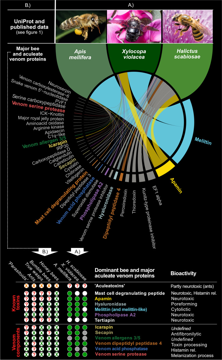
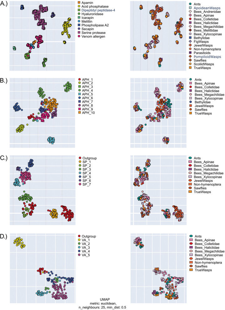
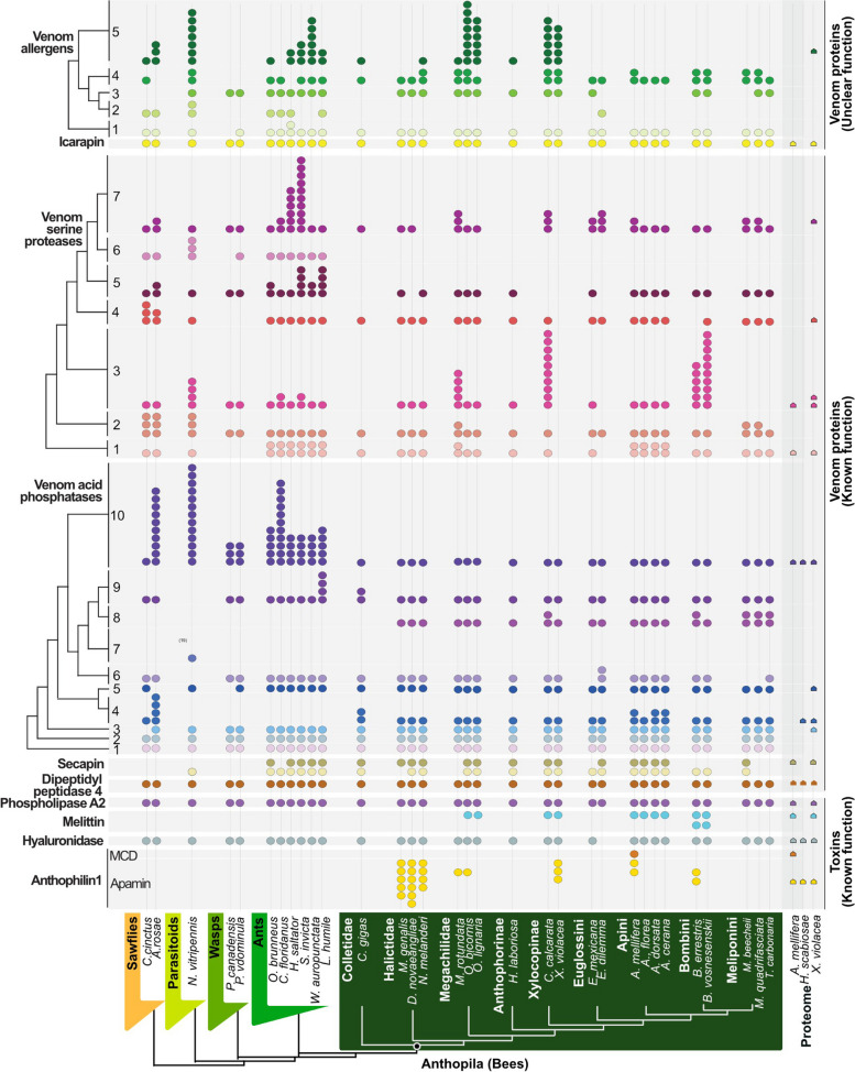
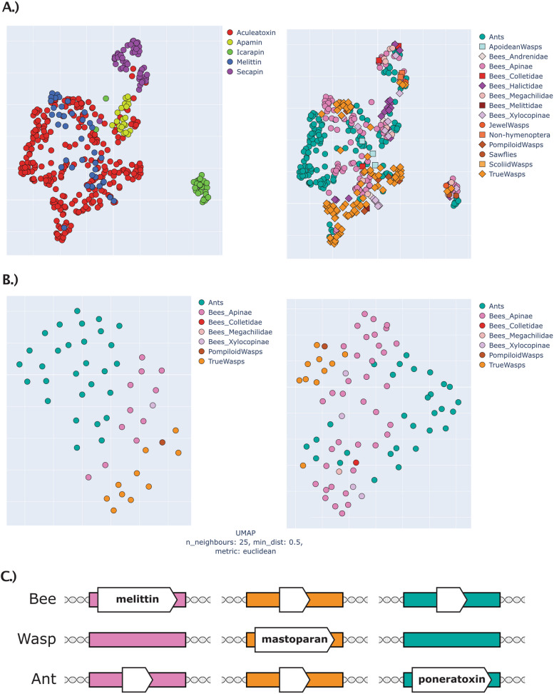
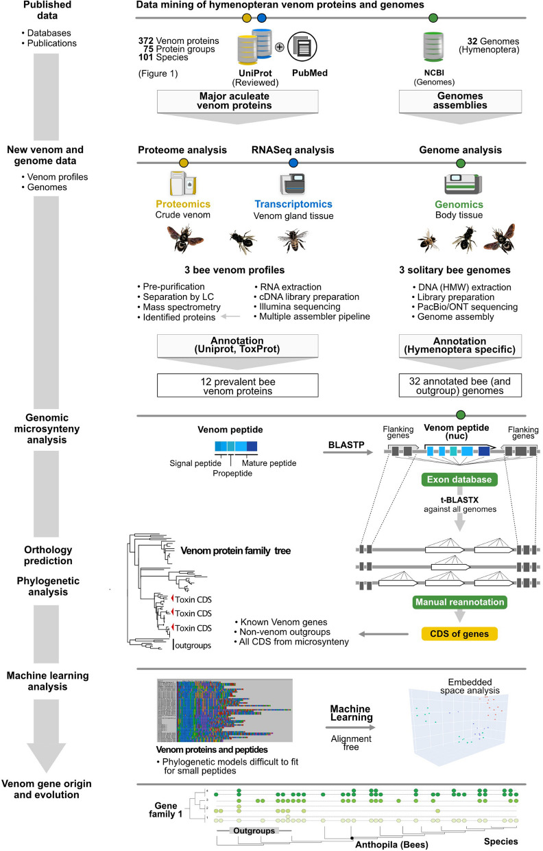
Results: Utilizing a combination of genomic and proteo-transcriptomic data, we investigated the origin of 11 toxin genes in 29 published and 3 new hymenopteran genomes and compiled an up-to-date list of prevalent bee venom proteins. Observed patterns indicate that bee venom genes predominantly originate through single gene co-option with gene duplication contributing to subsequent diversification.

Conclusions: Most Hymenoptera venom genes are shared by all members of the clade and only melittin and the new venom protein family anthophilin1 appear unique to the bee lineage. Most venom proteins thus predate the mega-radiation of hymenopterans and the evolution of the aculeate stinger.

Keywords: Aculeatoxins; Apamin; Bee toxins; Genomics; Hymenoptera venom; Machine learning; Melittin; Proteo-transcriptomics; Solitary bee venom; Venom gene evolution.


Conflict of interest statement

The authors declare they have no competing interests.

Figures

Fig. 1
Fig. 1
Reviewed venom proteins for hymenopteran taxa in respect to protein and species numbers from UniProt. Major hymenopteran clades are shown on the left (species numbers in circles). The second numbers in circles within the colour-coded lines indicate venom proteins (grouped according to their names). The twelve herein proposed prevalent bee venom protein families (PBVP) are illustrated on the right, together with the toxins proposed as “Aculeatoxins” (brown) according to Robinson et al. [29]. Novel, and further undescribed peptides and proteins are shown in grey. The hymenopteran groups are based on the recent phylogeny according to Peters et al. [33]. Please note that three melittin sequences from wasps are falsely annotated in UniProt as wasp melittins (marked by a black X). Our analyses clearly show that genes encoding peptides highly similar to honey bee melittin are not present in wasps, see also von Reumont et al. [22]. The phylogeny is pruned to the groups for which data is available based on Peters et al. [33]
Fig. 2
Fig. 2
The most prevalent bee venom proteins. Components selected from our own data (A.) A. mellifera, H. scabiosae and X. violacea profiles, and (B.) published bee and aculeate venom components. In (A.) only venom protein transcripts validated by the proteome data are listed. Transcript expression is shown as thickness of the Circos plot lines and based on the percentage of scaled transcript per million (TPM) values including only proteome-validated sequences. The twelve selected venom proteins that we discuss herein further as dominant bee venom proteins are printed in bold in the colour code used for these proteins in this manuscript. Peptide names in white were not identified by our proteo-transcriptome data but are present in published data. For our new proteo-transcriptome data (A.), the green circles indicate venom proteins identified by proteo-transcriptomics, grey circles indicate transcriptome-only hits. White circles illustrate missing data. For published data the green X indicate major components identified in literature, red questions marks highlight missing/unclear data. Orange X highlight the “aculeatoxin” peptides (According to Robison et al. [29], melittin is also a member of the proposed aculeatoxin family, which is separately shown as part of the PBVPs)
Fig. 3
Fig. 3
Machine learning generated protein space representations of hymenopteran venoms corresponds with gene phylogeny-based clustering. In each case, the left panel shows the breakdown of subgroups revealed in this study (protein families for the entire dataset, subfamilies for each of the protein families) while the right panel shows the same space coloured by taxa, clearly highlighting that each protein group is a gene clade, not a species clade. A Representation of the entire non-redundant dataset. B Acid phosphatase family. C Serine protease family. D Venom allergens family
Fig. 4
Fig. 4
Overview of prevalent bee venom genes. The presence of venom gene orthologs and copy number variation is mapped onto the phylogenetic relationship between the species we surveyed according to Peters et al. [33]. Coloured circles represent genes with identical microsynteny in the genomes of the surveyed species. Please note that tertiapin is now included within anthophilin1 as variant of apamin
Fig. 5
Fig. 5
Microsyntenic pattern for the apamin family (Anthophilin1). Question marks indicate coding sequences with products of unknown functions. Pseudogenes are symbolized by ψ. The arrows reflect gene orientation. We show here only species for which the genomic sequence in the region with apamin genes is contiguous. Note that “apamin-like” genes are also known as “tertiapin”. Apis lineages are in dark green, other non-Apini bees in grass green and ants and wasps in light green
Fig. 6
Fig. 6
Microsynteny around the melittin sequence. All species for which the genome data allowed for microsyntenic analysis are shown. Vollenhovia emeryi was not included in other genomic analyses due to its relatively low genome quality. However, it is shown because it was the only one of the eight analysed ant species that features a seemingly related gene in the correct position but with a very different mature sequence. Genes labelled with ψ in ants and wasps bear little similarity with melittin genes; however, they might be sister genes to the melittin group that underwent severe pseudogenization. Note that Osmia melittin is also called “osmin”, Colletes—collectin, Bombus—bombolittin, and Xylocopa—xylopin. Apis lineages are in dark green, other non-Apini bees in grass green and ants and wasps in light green
Fig. 7
Fig. 7
“Protein space” of small peptidic aculeatan toxins as revealed by machine learning analysis and their genomic position in respect to each other. A Combined data of available verified toxin sequences from Robinson et al., and the present study (including ToxProt part of the UniProt) for all peptidic toxins, on the left coloured by protein family, on the right—coloured by taxa. B Data from Robinson et al., on the left sequences with signal peptide included, on the right only mature peptides. For the interactive plots see Additional file 11. C Schematic of genomic position of the three groups of hymenopteran toxins. Coloured rectangles represent regions of microsynteny: pink for melittin, orange for mastoparan and green for poneratoxins. See text for details
Fig. 8
Fig. 8
Simplified visualization of the prevalent bee venom proteins and their representation in outgroup taxa. The numbers of genomes are shown in brackets after the family names. Genes are colour-coded and feature a colour range for duplicates. Duplications are summarized by numbers. Phylogeny and divergence times are shown as previously described in Peters et al. [33]. The nodes for monophyletic aculeates and bees are highlighted in green. The red lined circle indicates the secondary loss of the stinger in sweat bees (Meliponini)
Fig. 9
Fig. 9
Description of the proteo-transcriptomic and genomic workflow applied in this study. Details of each step are given in material and methods

References

    1. Oeyen JP, Baa-Puyoulet P, Benoit JB, Beukeboom LW, Bornberg-Bauer E, Buttstedt A, et al. Sawfly genomes reveal evolutionary acquisitions that fostered the mega-radiation of parasitoid and eusocial Hymenoptera. Lavrov D, editor. Genome Biol Evol. 2020;12(7):1099–188. - PMC - PubMed
    1. Casewell NR, Wüster W, Vonk FJ, Harrison RA, Fry BG. Complex cocktails: the evolutionary novelty of venoms. Trends Ecol Evol. 2013;28(4):219–229. - PubMed
    1. von Reumont BM, Anderluh G, Antunes A, Ayvazyan N, Beis D, Caliskan F, et al. Modern venomics—current insights, novel methods, and future perspectives in biological and applied animal venom research. GigaScience. 2022;11:giac048. - PMC - PubMed
    1. Wang T, Zhao M, Rotgans BA, Ni G, Dean JFD, Nahrung HF, et al. Proteomic analysis of the venom and venom sac of the woodwasp, Sirex noctilio - towards understanding its biological impact. J Proteomics. 2016;146:195–206. - PubMed
    1. Piek T. Venoms of the Hymenoptera. London: Academic Press Inc. (London) Ltd.; 1986.

Publication types

LinkOut - more resources