. 2023 Nov;10(33):e2303369.
doi: 10.1002/advs.202303369. Epub 2023 Oct 22.

Actomyosin Activity and Piezo1 Activity Synergistically Drive Urinary System Fibroblast Activation

Affiliations

Actomyosin Activity and Piezo1 Activity Synergistically Drive Urinary System Fibroblast Activation

Guo Chen et al. Adv Sci (Weinh). 2023 Nov.

Abstract

Mechanical cues play a crucial role in activating myofibroblasts from quiescent fibroblasts during fibrosis, and the stiffness of the extracellular matrix is of significant importance in this process. While intracellular force mediated by myosin II and calcium influx regulated by Piezo1 are the primary mechanisms by which cells sense and respond to mechanical forces, their intercellular mechanical interaction remains to be elucidated. Here, hydrogels with tunable substrate are used to systematically investigate the crosstalk of myosin II and Piezo1 in fibroblast to myofibroblast transition (FMT). The findings reveal that the two distinct signaling pathways are integrated to convert mechanical stiffness signals into biochemical signals during bladder-specific FMT. Moreover, it is demonstrated that the crosstalk between myosin II and Piezo1 sensing mechanisms synergistically establishes a sustained feed-forward loop that contributes to chromatin remodeling, induces the expression of downstream target genes, and ultimately exacerbates FMT, in which the intracellular force activates Piezo1 by PI3K/PIP3 pathway-mediated membrane tension and the Piezo1-regulated calcium influx enhances intracellular force by the classical FAK/RhoA/ROCK pathway. Finally, the multifunctional Piezo1 in the complex feedback circuit of FMT drives to further identify that targeting Piezo1 as a therapeutic option for ameliorating bladder fibrosis and dysfunction.

Keywords: Piezo1; actomyosin; fibroblasts; fibrosis; urinary Systems.


Conflict of interest statement

The authors declare no conflict of interest.

Figures
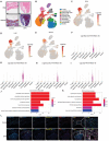
Figure 1
Figure 1
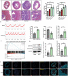
Bladder fibroblasts are the predominant cell type involved in ECM remodeling. A) Images of HE and Masson staining in BPH bladder and control bladder. Scale bar = 500 µm. B) Single‐cell of human bladders: Cell clusters found therein representing ten cell types (n = 5). C) Fibroblasts identified using canonical marker DCN. D) Fibroblasts identified using canonical marker LUM. E) Fibroblasts identified using canonical marker ACTA2. F) Expression of COL1A1 in each of the ten cell types, with the highest expression observed in fibroblasts. G) Expression of COL1A2 in each of the ten cell types, with the highest expression observed in fibroblasts. H) Expression of COL3A1 in each of the ten cell types, with the highest expression observed in fibroblasts. I) Expression of FN1 in each of the ten cell types, with the highest expression observed in fibroblasts. J) GO enrichment analysis of DEGs between BPH bladder and control bladder, showing that DEGs of fibroblasts were primarily involved in extracellular matrix remodeling. K) KEGG enrichment analysis of DEGs between BPH bladder and control bladder. L) Immunofluorescence was performed to co‐localize cellular traction force‐related markers and fibroblast activation markers (n = 3). Scale bar = 200 µm. Shown is the mean ± SD.
Figure 2
Figure 2
Cellular traction force and FMT were positively correlated in mice BOO bladder development. A) Masson of bladder tissues in sham‐operated group and BOO group; Scale bar = 200 µm. Bladder weight and collagen ration of sham‐operated group and BOO group (n = 4). B) Urodynamic curve(left) and quantification(right) of micturition intervals, baseline pressure, and micturition pressure of sham‐operated group and BOO group (n = 4). C) GSEA data showed that focal adhesion pathway significantly increased in BOO bladder. D) WB (left) and quantification(right) of p‐NMIIA and Lamin A/C in sham‐operated group and BOO group. E) Cellular traction force‐related markers are positively correlated with BOO‐induced FMT (n = 3); Scale bar = 500 µm.*p < 0.05, **p < 0.01; Shown is the mean ± SD.
Figure 3
Figure 3
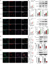
Stiffness regulates bladder FMT via NMIIA‐mediated cellular traction force. A–C) Increased stiffness enhanced the p‐NMIIA and Lamin A/C labeling intensity (n = 50). D–F) WB analysis shows the increased stiffness induced the expression of p‐NMIIA and Lamin A/C. G–I) Increased stiffness enhanced the Col1 and α‐SMA labeling intensity (n = 50). J–L) WB analysis shows the increased stiffness induced Col1 and α‐SMA expression. M–O) Blebb decreased the p‐NMIIA and Lamin A/C labeling intensity. P–R) WB analysis shows that Blebb decreased the p‐NMIIA and Lamin A/C expression. S–U) Blebb decreased the Col1 and α‐SMA labeling intensity. V–X) WB analysis shows that Blebb decreased the Col1 and α‐SMA expression. Scale bar = 20 µm; *p < 0.05, **p < 0.01; Shown is the mean ± SD.
Figure 4
Figure 4
Piezo1 mediates stiff ECM‐induced bladder FMT. A) KEGG enrichment analysis of the DEGs between the sham‐operated mice bladder and BOO mice bladder indicated that the calcium ion pathway was chiefly responsible for BOO‐induced bladder remodeling (n = 3). B) Cluster analysis of all ion channels in the transcriptome sequencing results. C) Cluster analysis of all ion channels in the transcriptome sequencing results show that Piezo1 messenger RNA levels were the most abundantly expressed channel and it was prominently upregulated in the BOO bladder. D) Piezo subtypes expression in sham‐operated mice bladder and BOO mice bladder were detected by WB and quantification(blow) shown in (E) (n = 3). F) Immunofluorescence was performed to co‐localize cellular traction force markers and activated fibroblast markers (n = 3). Scale bar = 200 µm. G–I) Inhibition of Piezo1 by siRNA was confirmed at functional (Ca2+ influx) and biochemical (IF) levels; Scale bar = 20 µm. J–L) Inhibition of Piezo1 by siRNA decreased the Col1 and α‐SMA intensity (n = 50); Scale bar = 20 µm. *p < 0.05, **p < 0.01;Shown is the mean ± SD.
Figure 5
Figure 5
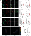
The crosstalk between Piezo1 and NMIIA in the stiffness‐induced FMT. A–C) Stiffen substrate‐activated Piezo1 was confirmed at functional (Ca2+ influx) and biochemical (IF) levels. D–F) Blebb decreased Piezo1 was confirmed at functional (Ca2+ influx) and biochemical (IF) levels. G–I) Inhibition of Piezo1 by siRNA decreased the p‐NMIIA and Lamin A/C labeling intensity (n = 50). J,K) TFM results show that inhibition of Piezo1 decreased the traction forces (n = 25). Scale bar = 20 µm; *p < 0.05, **p < 0.01; Shown is the mean ± SD.
Figure 6
Figure 6
Piezo1 and NMIIA could directly and indirectly influence the stiffness‐induced FMT. A–C) After the treatment with Blebb, concertation of Yoda1 to adjust the intracellular Ca2+ to a baseline level as the control group. D–F) Yoda1 attenuated the Blebb‐decreased fibroblast activated markers, while still lower than the control group (n = 50). G–I) Concertation of Calyc to adjust the p‐NMIIA expression to baseline level of the control group (n = 50). J–L) Blebb attenuated the siRNA Piezo1‐decreased fibroblast activation markers, while still lower than the control group (n = 50). Scale bar = 20 µm; *p < 0.05, **p < 0.01; n.s, not significant. The data are expressed as the mean±SD of three independent experiments.
Figure 7
Figure 7
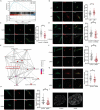
The underlying mechanism of the crosstalk between Myosin II and Piezo1. A) GSEA data analysis shows PI3K and focal adhesion pathway were significantly altered in BOO bladder. B) Stiffen substrate activated p‐PI3K labeling intensity (n = 50). C) PI3K inhibitor LY29004 decreased the p‐PI3K labeling intensity (n = 50). D) LY294002 decreased the Piezo1 labeling intensity (n = 50). E) Co‐immunoprecipitation coupled to mass spectrometry assay enriched in actin‐filament and plasma membrane; F) Inhibition of Piezo1 by siRNA decreased the p‐FAK labeling intensity (n = 50). G) Inhibition of Piezo1 by siRNA decreased the RhoA labeling intensity (n = 50). H) Inhibition of Piezo1 by siRNA decreased the ROCK labeling intensity. I) Inhibition of Piezo1 by siRNA results in condensation of chromatin (n = 15, two technical replicates), Scale bar = 20 µm; *p < 0.05, **p < 0.01; Shown is the mean ± SD.
Figure 8
Figure 8
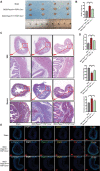
Deletion of fibroblast Piezo1 attenuates BOO remodeling and bladder dysfunction. A) Bladder harvested from sham‐operated, BOO, and deletion of fibroblast in BOO group (n = 4). B) Bladder weight in sham‐operated, BOO, and deletion of fibroblast in BOO group (n = 4). C) HE and Masson staining of bladder in sham‐operated, BOO, and deletion of fibroblast in BOO group (n = 3). D) Urodynamic parameters analysis in sham‐operated, BOO, and deletion of fibroblast in BOO group (n = 3). E) Immunofluorescence was performed to co‐localize Piezo1 and activated fibroblast markers of bladder in sham‐operated, BOO, and deletion of fibroblast in BOO group (n = 3); *p < 0.05, **p < 0.01. Shown is the mean ± SD.
Figure 9
Figure 9
Schematic illustration of mechanotransduction circuit in fibroblasts responding to stiffness: Myosin II and Piezo1 have a feed‐forward crosstalk through the PI3K/PIP3 pathway‐mediated membrane tension and the FAK/RhoA/ROCK pathway, which synergistically contribute to stiffness‐induced fibroblast activation.

References

    1. Friedman S. L., Sheppard D., Duffield J. S., Violette S., Sci. Transl. Med. 2013, 5, 167sr1. - PubMed
    1. Rockey D. C., Bell P. D., Hill J. A., N. Engl. J. Med. 2015, 372, 1138. - PubMed
    1. Wang W., Ai J., Liao B., Xiao K., Lin L., Chen H., Zhou L., Int. Immunopharmacol. 2021, 99, 107947. - PubMed
    1. Bushman W. A., Jerde T. J., Am. J. Physiol. Renal. Physiol. 2016, 311, F817. - PubMed
    1. Akino H., Gobara M., Okada K., Int. J. Urol. 1996, 3, 441. - PubMed

Publication types

LinkOut - more resources