

A case report on rare finding of microfilaria in pus sample of an ulcer over elephantiasis leg
- PMID: 37867533
- PMCID: PMC10589395
- DOI: 10.1002/ccr3.8102
A case report on rare finding of microfilaria in pus sample of an ulcer over elephantiasis leg
Abstract
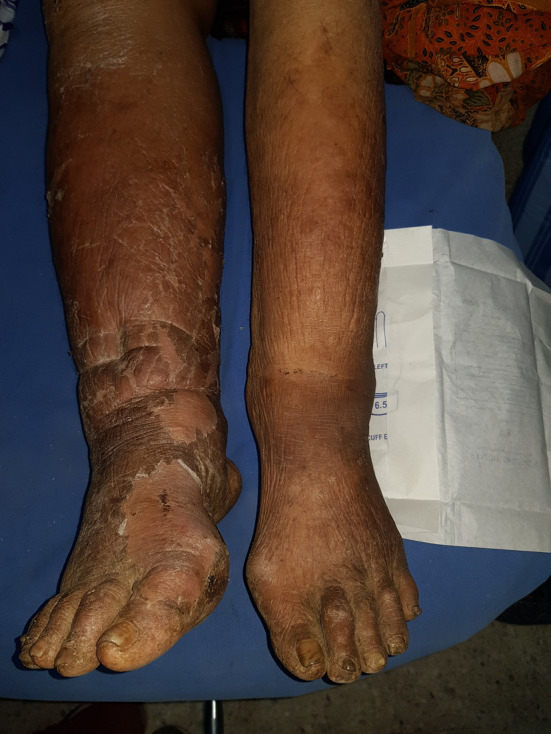
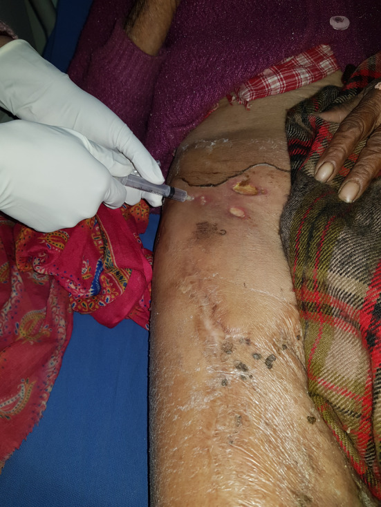
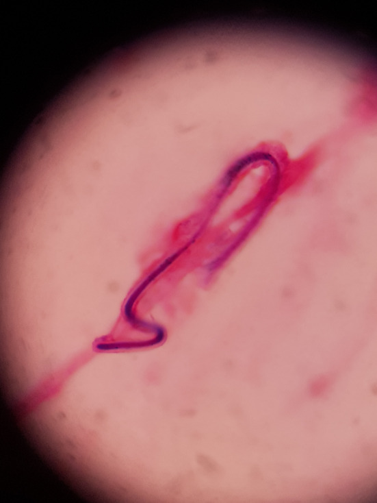
Skin ulcerations are a significant cause of morbidity and can be challenging to manage. Among the various causes of chronic non-healing ulcers, lymphedema is also considered a possible diagnosis in countries such as Nepal. Lymphatic filariasis has been a significant public health issue in endemic areas. Wuchereria bancrofti is a common nematode parasite that causes lymphatic filariasis. Excessive retention of lymphatic fluid in the interstitial compartment can cause localized tissue swelling, known as lymphedema, which is caused by impaired lymphatic drainage. Microfilariae can be detected in peripheral blood, body fluids, and needle aspirates. Microfilaria is not commonly found in ulcers on elephantiasis legs. We discuss here a case of 73-year-old women with elephantiasis legs with pus discharging ulcers in the thighs. Microscopic examination of pus discharge revealed microfilaria which highlights the importance of pus examination as diagnostic modality in endemic countries.
Keywords: chronic ulcer; elephantiasis; filariasis; microfilaria.
© 2023 The Authors. Clinical Case Reports published by John Wiley & Sons Ltd.
Conflict of interest statement
The author(s) declared no potential conflicts of interest with respect to the research, authorship, and/or publication of this article.
Figures
References
-
- Kar H, Singh G, Urhekar AD. (n.d.) Microfilaria in pus sample of an ulcer over elephantiasis leg: an unusual case presentation . Ijcmas.com. Retrieved July 17, 2023, from https://www.ijcmas.com/vol‐2‐7/Harapriya%20Kar,%20et%20al.pdf
Publication types
LinkOut - more resources
Full Text Sources
