Review
. 2023 Oct 6:10:1252529.
doi: 10.3389/fmolb.2023.1252529. eCollection 2023.

Cryo-electron microscopy in the fight against COVID-19-mechanism of virus entry

Affiliations
Review

Cryo-electron microscopy in the fight against COVID-19-mechanism of virus entry

Satish Bodakuntla et al. Front Mol Biosci. .

Abstract

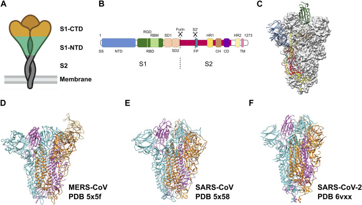
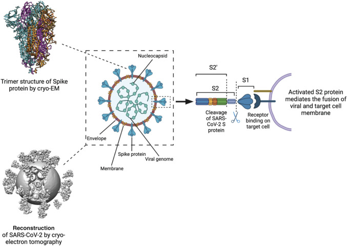
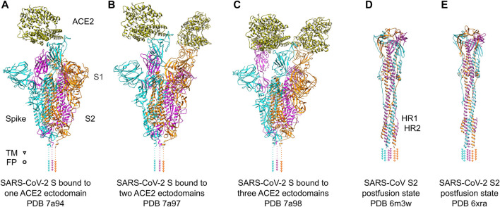
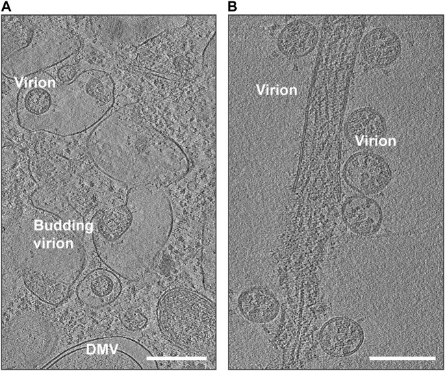
Cryogenic electron microscopy (cryo-EM) and electron tomography (cryo-ET) have become a critical tool for studying viral particles. Cryo-EM has enhanced our understanding of viral assembly and replication processes at a molecular resolution. Meanwhile, in situ cryo-ET has been used to investigate how viruses attach to and invade host cells. These advances have significantly contributed to our knowledge of viral biology. Particularly, prompt elucidations of structures of the SARS-CoV-2 spike protein and its variants have directly impacted the development of vaccines and therapeutic measures. This review discusses the progress made by cryo-EM based technologies in comprehending the severe acute respiratory syndrome coronavirus-2 (SARS-Cov-2), the virus responsible for the devastating global COVID-19 pandemic in 2020 with focus on the SARS-CoV-2 spike protein and the mechanisms of the virus entry and replication.

Keywords: ACE2; COVID-19; SARS-CoV-2; cryo-EM; cryo-ET; spike protein.


Conflict of interest statement

The authors declare that the research was conducted in the absence of any commercial or financial relationships that could be construed as a potential conflict of interest.

Figures

FIGURE 1
FIGURE 1
Pre-fusion structure of the S protein. (A) Schematic of the pre-fusion state of the trimeric spike. (B) Domain architecture of the S protein: N-terminal domain (NTD), receptor binding domain (RBD) with RGD motif and receptor binding motif (RBM), spike subdomain 1 (SD1), spike subdomain 2 (SD2), fusion peptide (FP), heptad repeats (HR1, HR2), central helix region (CH), connector domain (CD), transmembrane helix (TM), cytoplasmic tail (CT). (C) Pre-fusion structure of the ectodomain of the SARS-CoV-2 S protein. Adapted from Wrapp et al. (2020). Domains are colored as in (B). Adapted from Wrapp et al. (2020). (D–F) Ectodomain timers of S proteins from related viruses MERS-CoV, SARS-CoV and SARS-CoV-2, respectively. Individual monomers are colored in cyan, magenta and orange, respectively.
FIGURE 2
FIGURE 2
Processing and activation of SARS-CoV-2 spike before fusion. Left Structure of the S protein ectodomain (top) and a virus capsid (bottom, Yao et al., 2020). Center Schematic of the SARS-CoV2 virion. Spike in blue, membrane in brown, M protein in yellow, E protein in purple, RNP in green. Right the S protein is cut by host cell proteases at 2 positions after docking to a receptor. The process releases the S1 domain and part of the S2 domain and results in conformational change of the S2 domain and the M protein.
FIGURE 3
FIGURE 3
SARS-Cov-2 S protein recognizing ACE2 receptors and conformational change after fusion. (A–C) SARS-Cov-2 S protein trimer ectodomains bound to one, two or three ACE2 receptor ectodomains. Individual spike monomers are colored in cyan, magenta and orange, respectively. The positions of the transmembrane (TM) helices are indicated as triangles. The ACE2 ectodomain is shown in gold. (D,E) Postfusion state of the S2 domain of SARS-CoV and SARS-CoV-2, respectively. After a “jackknife”-motion, the TM helices and the fusion peptides (FP) are transposed to the same topological side (indicated as triangles and rings, respectively). The HR1 and HR2 domains form a characteristic six-helix-bundle. The coloring is the same as in (A–C).
FIGURE 4
FIGURE 4
Snapshots of cryo-EM reconstructions of SARS-CoV-2 virions inside VeroE6 cells. (A) Budding SARS-CoV-2 virions (EMD-11863). The location of a double-membrane vesicle (DMV) is indicated. Scale Bar: 300 nm. (B) Release of SARS-CoV-2 virions (EMD-11867). Scale Bar: 200 nm.

Similar articles

Cited by

References

    1. Aboudounya M. M., Heads R. J. (2021). COVID-19 and toll-like receptor 4 (TLR4): SARS-CoV-2 may bind and activate TLR4 to increase ACE2 expression, facilitating entry and causing hyperinflammation. Mediat. Inflamm. 2021, 8874339. 10.1155/2021/8874339 - DOI - PMC - PubMed
    1. Arya R., Kumari S., Pandey B., Mistry H., Bihani S. C., Das A., et al. (2021). Structural insights into SARS-CoV-2 proteins. J. Mol. Biol. 433, 166725. 10.1016/j.jmb.2020.11.024 - DOI - PMC - PubMed
    1. Astuti I., Ysrafil (2020). Severe acute respiratory syndrome coronavirus 2 (SARS-CoV-2): an overview of viral structure and host response. Diabetes Metab. Syndr. 14, 407–412. 10.1016/j.dsx.2020.04.020 - DOI - PMC - PubMed
    1. Baindara P., Roy D., Mandal S. M., Schrum A. G. (2022). Conservation and enhanced binding of SARS-CoV-2 Omicron spike Protein to coreceptor neuropilin-1 Predicted by docking analysis. Infect. Dis. Rep. 14, 243–249. 10.3390/idr14020029 - DOI - PMC - PubMed
    1. Barcena M., Barnes C. O., Beck M., Bjorkman P. J., Canard B., Gao G. F., et al. (2021). Structural biology in the fight against COVID-19. Nat. Struct. Mol. Biol. 28, 2–7. 10.1038/s41594-020-00544-8 - DOI - PubMed