

Exploring Spinal Subarachnoid Hemorrhage: A Neurosurgical Case Series
- PMID: 37868412
- PMCID: PMC10588961
- DOI: 10.7759/cureus.45627
Exploring Spinal Subarachnoid Hemorrhage: A Neurosurgical Case Series
Abstract
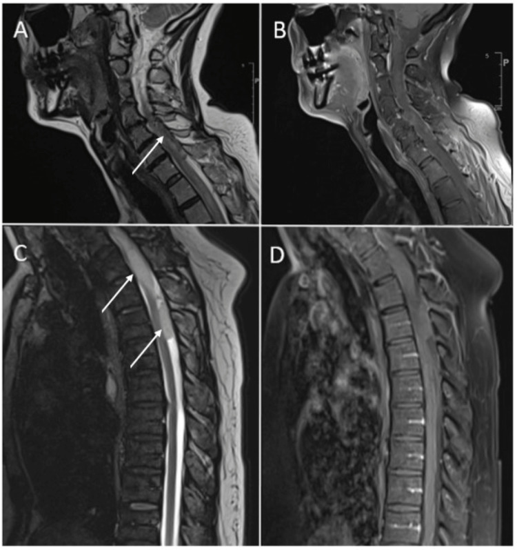
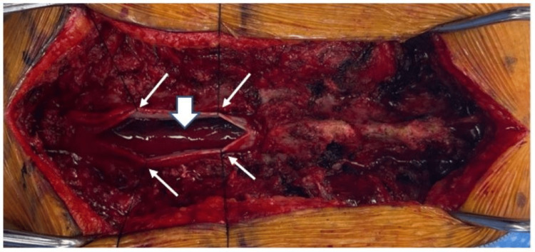
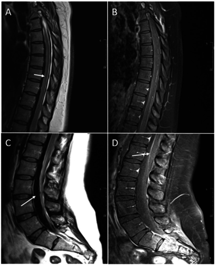
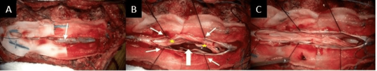
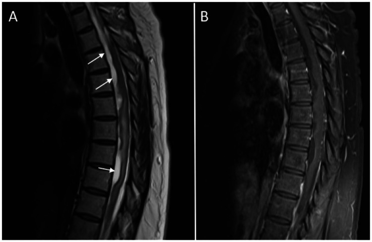
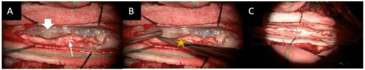
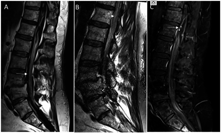
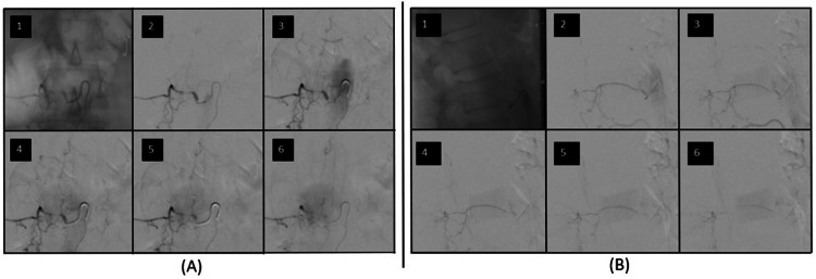
Spinal subarachnoid hemorrhage (SSAH) is a rare condition that can cause spinal cord or nerve root compression and permanent neurologic damage. The reported etiologies include trauma, vascular malformations or aneurysms, coagulopathies, neoplasms, autoimmune disease, and spontaneous hemorrhage. If there is evidence of neurologic deterioration, it is commonly managed as a surgical emergency, but cases of conservative management have also been reported. In this case series, we present three patients who suffered from SSAH. The first was a spontaneous cervical SSAH that occurred following cardiac catheterization, the second was a spontaneous thoracolumbar SSAH in a patient with a known history of coagulopathy, and the third was a thoracolumbar SSAH that was caused by a dural arteriovenous fistula (dAVF). All three patients exhibited neurologic deficits and thus underwent emergent decompression and hematoma evacuation. The patient with the dAVF also required open ligation of the fistula. Following surgical intervention, all three patients regained at least partial neurologic function, but one patient developed symptomatic arachnoid cysts that required further intervention. The presented case series highlights the importance and time-sensitivity of surgical decompression in patients experiencing neurologic deficits from SSAH. These cases underscore the urgency of timely neurosurgical intervention to mitigate neurologic impairment and add insights to the existing literature on this rare condition.
Keywords: dural arteriovenous fistula (davf); intraoperative neurologic monitoring; neurologic exam; spinal angiogram; spinal decompression; spinal subarachnoid cyst; spinal subarachnoid hemorrhage.
Copyright © 2023, Sankarappan et al.
Conflict of interest statement
The authors have declared that no competing interests exist.
Figures
References
-
- Spinal hematoma: a literature survey with meta-analysis of 613 patients. Kreppel D, Antoniadis G, Seeling W. Neurosurg Rev. 2003;26:1–49. - PubMed
-
- Spinal subarachnoid hematomas: our experience and literature review. Domenicucci M, Ramieri A, Paolini S, Russo N, Occhiogrosso G, Di Biasi C, Delfini R. Acta Neurochir (Wien) 2005;147:741–750. - PubMed
-
- Spinal subarachnoid hematoma. Frager D, Zimmerman RD, Wisoff HS, Leeds NE. https://pubmed.ncbi.nlm.nih.gov/6800242/ AJNR Am J Neuroradiol. 1982;3:77–79. - PMC - PubMed
-
- Spinal subarachnoid hemorrhage and aneurysms. Maiti TK, Bir SC, Nanda A. Handb Clin Neurol. 2017;143:215–223. - PubMed
-
- Outcome and prognostic factors in the surgical treatment of spinal dural arteriovenous fistulas. A long-term study. Tacconi L, Lopez Izquierdo BC, Symon L. Br J Neurosurg. 1997;11:298–305. - PubMed
Publication types
LinkOut - more resources
Full Text Sources