

Dual-layer spectral computed tomography aortography using a seventy-five-percent-reduced iodine dose protocol and multiparameter spectral imaging: comparison with conventional computed tomography imaging
- PMID: 37869326
- PMCID: PMC10585532
- DOI: 10.21037/qims-23-101
Dual-layer spectral computed tomography aortography using a seventy-five-percent-reduced iodine dose protocol and multiparameter spectral imaging: comparison with conventional computed tomography imaging
Abstract
Background: Computed tomography angiography (CTA) is the recommended diagnostic and follow-up imaging modality for acute aortic dissection (AD). However, the high-contrast medium burden associated with repeated CT aortography follow-ups remains a significant concern. This prospective study aimed to assess whether an ultra-low contrast dose (75% cutoff) aortic CTA protocol on dual-layer spectral CT could achieve comparable image quality with the full dose protocol. We also investigated the image quality of the virtual noncontrast (VNC) images derived from the ultra-low dose protocol.
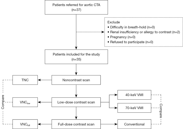
Methods: This study included 37 consecutive patients who were referred to aortic CTA from May 2022 to August 2022. The enrolled patients underwent full-dose contrast CTA and ultra-low dose (reduced to 25% of conventional) contrast CTA on dual-layer spectral CT in 1 day. Virtual monochromatic images (VMIs) were reconstructed with 40 and 70 keV. The VNC images were reconstructed for both protocols. Objective image quality evaluation, recorded as signal-to-noise ratios (SNRs) and contrast-to-noise ratios (CNRs), was compared between the groups using 1-way analysis of variance and post hoc analysis with Bonferroni correction. Subjective image quality was also compared between the groups. Finally, VNC images derived from the low-dose (VNClow) and full-dose (VNCfull) protocols were compared to the true noncontrast (TNC) images.
Results: Neither CNR nor SNR was lower for the 40-keV images reconstructed from the ultra-low dose group compared to the conventional images. Both were significantly higher than those of the 70-keV images. Regarding subjective image quality, vessel enhancement was not significantly different between the 40-keV VMI and full-dose images [ascending aorta (AAO): 4.37±0.46 vs. 4.57±0.48, P=0.096; brachiocephalic arteries: 4.34±0.45 vs. 4.51±0.49, P=0.152; abdominal aortic side branch: 4.42±0.48 vs. 4.51±0.49, P=0.480]. The VNClow images were similar to the TNC images but significantly different from the VNCfull images (P<0.001).
Conclusions: Ultra-low contrast aortic CTA with a 75%-reduced iodine dose using dual-layer spectral CT and the derived VNC achieved image quality comparable to that of conventional CTA and TNC images.
Keywords: Aortic computed tomography aortography (aortic CTA); aortic dissection (AD); dual-layer spectral CT (DLCT); virtual noncontrast images (VNC images).
2023 Quantitative Imaging in Medicine and Surgery. All rights reserved.
Conflict of interest statement
Conflicts of Interest: All authors have completed the ICMJE uniform disclosure form (available at https://qims.amegroups.com/article/view/10.21037/qims-23-101/coif). SD and HL, employees of Philips Healthcare, contributed to the description of technical principles and image post-processing without impacting the research results. The other authors have no conflicts of interest to declare.
Figures


References
-
- Raff GL, Chinnaiyan KM, Cury RC, Garcia MT, Hecht HS, Hollander JE, O'Neil B, Taylor AJ, Hoffmann U, Society of Cardiovascular Computed Tomography Guidelines Committee . SCCT guidelines on the use of coronary computed tomographic angiography for patients presenting with acute chest pain to the emergency department: a report of the Society of Cardiovascular Computed Tomography Guidelines Committee. J Cardiovasc Comput Tomogr 2014;8:254-71. 10.1016/j.jcct.2014.06.002 - DOI - PubMed
-
- MacGillivray TE, Gleason TG, Patel HJ, Aldea GS, Bavaria JE, Beaver TM, Chen EP, Czerny M, Estrera AL, Firestone S, Fischbein MP, Hughes GC, Hui DS, Kissoon K, Lawton JS, Pacini D, Reece TB, Roselli EE, Stulak J. The Society of Thoracic Surgeons/American Association for Thoracic Surgery Clinical Practice Guidelines on the Management of Type B Aortic Dissection. Ann Thorac Surg 2022;113:1073-92. 10.1016/j.athoracsur.2021.11.002 - DOI - PubMed
LinkOut - more resources
Full Text Sources
Miscellaneous