. 2024 Mar 4;26(3):458-472.
doi: 10.1093/neuonc/noad205.

Systematic characterization of antibody-drug conjugate targets in central nervous system tumors

Affiliations

Systematic characterization of antibody-drug conjugate targets in central nervous system tumors

Shannon Coy et al. Neuro Oncol. .

Abstract

Background: Antibody-drug conjugates (ADCs) enhance the specificity of cytotoxic drugs by directing them to cells expressing target antigens. Multiple ADCs are FDA-approved for solid and hematologic malignancies, including those expressing HER2, TROP2, and NECTIN4. Recently, an ADC targeting HER2 (Trastuzumab-Deruxtecan) increased survival and reduced growth of brain metastases in treatment-refractory metastatic breast cancer, even in tumors with low HER2 expression. Thus, low-level expression of ADC targets may be sufficient for treatment responsiveness. However, ADC target expression is poorly characterized in many central nervous system (CNS) tumors.

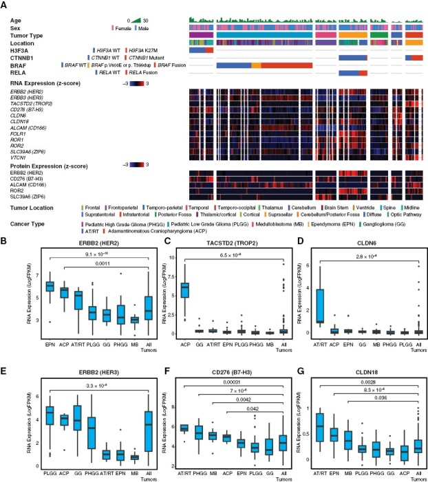
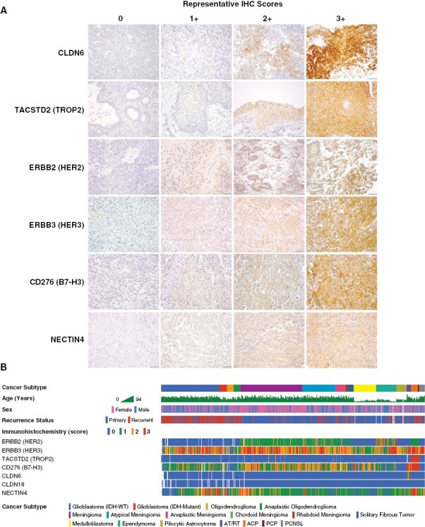
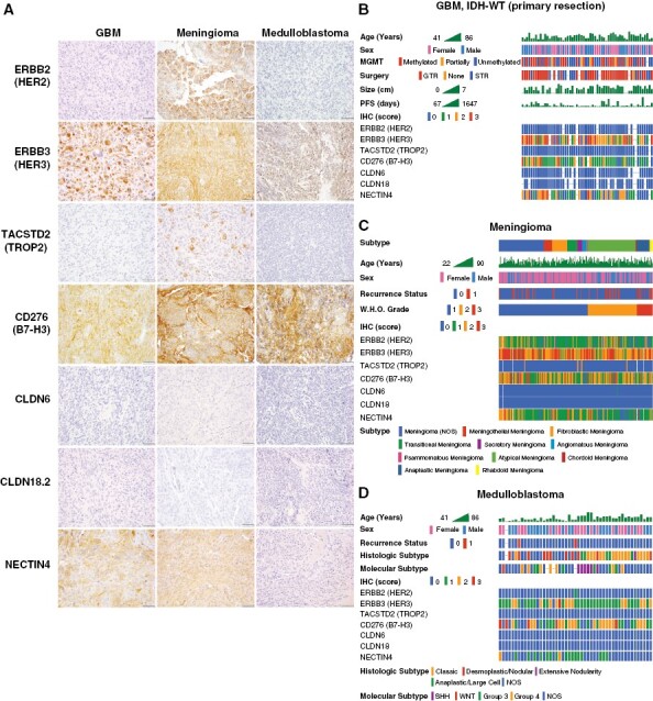
Methods: We analyzed publicly available RNA-sequencing and proteomic data from the children's brain tumor network (N = 188 tumors) and gene-expression-omnibus RNA-expression datasets (N = 356) to evaluate expression of 14 potential ADC targets that are FDA-approved or under investigation in solid cancers. We also used immunohistochemistry to measure the levels of HER2, HER3, NECTIN4, TROP2, CLDN6, CLDN18.2, and CD276/B7-H3 protein in glioblastoma, oligodendroglioma, meningioma, ependymoma, pilocytic astrocytoma, medulloblastoma, atypical teratoid/rhabdoid tumor (AT/RT), adamantinomatous craniopharyngioma (ACP), papillary craniopharyngioma (PCP), and primary CNS lymphoma (N = 575).

Results: Pan-CNS analysis showed subtype-specific expression of ADC target proteins. Most tumors expressed HER3, B7-H3, and NECTIN4. Ependymomas strongly expressed HER2, while meningiomas showed weak-moderate HER2 expression. ACP and PCP strongly expressed B7-H3, with TROP2 expression in whorled ACP epithelium. AT/RT strongly expressed CLDN6. Glioblastoma showed little subtype-specific marker expression, suggesting a need for further target development.

Conclusions: CNS tumors exhibit subtype-specific expression of ADC targets including several FDA-approved for other indications. Clinical trials of ADCs in CNS tumors may therefore be warranted.

Keywords: craniopharyngioma; ependymoma; glioma; immunoconjugates; meningioma.


Conflict of interest statement

K.L.L. has received research support from Dana-Farber Cancer Institute from Rarecyte and Bristol Myers Squibb, consulting fees from BMS, Integragen, Blaze Biosciences, and Travera Inc., and is an equity holder and founder of Travera Inc. P.K.S. is a co-founder and member of the Board of Directors of Glencoe Software, a member of the Board of Directors for Applied Biomath, and a member of the Scientific Advisory Board for RareCyte, NanoString, and Montai Health; he holds equity in Glencoe, Applied Biomath, and RareCyte. P.K.S. is a consultant for Merck and the Sorger lab has received research funding from Novartis and Merck in the past five years. P.Y.W. has received honoraria for consulting from Astra Zeneca, Bayer, Black Diamond, Boehringer Ingelheim, Boston Pharmaceuticals, Celularity, Chimerix, Day One Bio, Genenta, Glaxo Smith Kline, Karyopharm, Merck, Mundipharma, Novartis, Novocure, Nuvation Bio, Prelude Therapeutics, Sapience, Servier, Sagimet, Vascular Biogenics, and VBI Vaccines.

Figures

Figure 1.
Figure 1.
Molecular profiling of ADC targets in pediatric CNS tumors. To characterize expression of potential ADC targets in pediatric CNS tumors, we analyzed publicly available bulk-RNA sequencing, selected whole-genome sequencing (WGS), and proteomic (mass spectrometry) data from the CPTAC database. This dataset included 188 tumors from seven major subtypes, including pediatric high-grade glioma (PHGG), pediatric low-grade glioma (PLGG), medulloblastoma (MB), ependymoma (EPN), ganglioglioma (GG), atypical teratoid/rhabdoid tumor (AT/RT), and adamantinomatous craniopharyngioma (ACP). Relevant oncogenic drivers were analyzed for each histologic subtype, including H3F3A in PHGG, BRAF in PLGG, CTNNB1 in CP, and RELA in ependymoma (A). ERBB2/HER2 RNA expression was enriched relative to other tumors in EPN (P = 9.1 × 10−10) and ACP (P = .001) (B), though only EPN showed enrichment in proteomic data (A). TACSTD2/TROP2 RNA was strongly enriched in ACP (P = 6.5 × 10−8) and very low in other subtypes (C). CLDN6 RNA was strongly enriched in AT/RT (P = 2.8 × 10−6), with low expression in other subtypes (D). ERBB3/HER3 RNA showed moderate expression in multiple subtypes, with slight enrichment in PLGG (P = 3.3 × 10−6) (E). CD276/B7-H3 RNA was moderately expressed by all tumors, with relative enrichment in ACP (P = .042), AT/RT (P = .0003), MB (P = .004), and PHGG (P = 7 × 10−5) (F). CLDN18 RNA showed very low expression in all subtypes, with slight relative enrichment in AT/RT (P = .0028), EPN (P = 8.3 × 10−5), and MB (P = .036) (G). All statistical comparisons performed using a Student’s t-test, unpaired, 2-sided with α = .05.
Figure 2.
Figure 2.
Molecular profiling of ADC targets in adult and pediatric CNS tumors. To broaden and validate our analysis of ADC targets in CNS tumors, we analyzed RNA microarray expression data from publicly available CNS tumor GEO datasets (n = 356 total tumors). We again found that ERBB/HER2 was selectively enriched in EPN (P = 2.6 × 10−10), and also found significant enrichment in meningioma (MEN, P = 2.22 × 10−16) and AT/RT (P = .009) (A) TACSTD2/TROP2 was modest in most tumors with relative enrichment in primary central nervous system lymphoma (PCNSL) (P = 1.5 × 10−6); craniopharyngiomas were not present for comparison (B). CLDN6 was strongly enriched in AT/RT (P = 4.9 × 10−6), with slight enrichment in PCNSL (P = .0003) and a single PB (C). FOLR1 was enriched in EPN (P = 4.2 × 10−9) and MEN (2.1 × 10−8) (D). HER3/ERBB3 was broadly expressed with enrichment in pilocytic astrocytoma (PA, P = 2.22 × 10−16) and to a lesser extent adult GBM (P = .0076) (E). CD276/B7-H3 showed broad expression, with relative enrichment in AT/RT (P = 1.7 × 10−10), GBM (P = .0004), and MB (.0002) (F). NECTIN4 showed broad expression with relative enrichment in EPN (P = .008) and PCNSL (P = .002) (G). CLDN18 showed low expression in all subtypes, with slight relative enrichment in PCNSL (P = .0008) (H). All statistical comparisons performed using a Student’s t-test, unpaired, 2-sided with α = .05.
Figure 3.
Figure 3.
Immunohistochemical analysis of ADC target protein expression in human tumor tissue. To directly analyze ADC target antigen expression in human tumor tissues and explore the feasibility of antibody markers for clinical screening, we developed immunohistochemistry (IHC) assays for a selected group of ADC targets, including HER2, HER3, TROP2, B7-H3, NECTIN4, CLDN6, and CLDN18.2. Target marker expression was semi-quantitatively (0–3+) scored by a pathologist, with the same scoring system and relative intensity for any tumor type. Representative staining for each marker is shown in samples from AT/RT (CLDN6), craniopharyngioma (TROP2), ependymoma (HER2), and meningioma (HER3, NECTIN4, B7-H3) specimens (A). A cohort of 575 human CNS tumors from diverse histologic subtypes were stained by IHC using tissue microarrays (TMA) and whole-slide sections (WSS). These data were correlated with clinicopathologic data for each case, showing distinctive patterns of ADC target marker expression in each subtype (B). Scale bars 50 μm.
Figure 4.
Figure 4.
Characterization of ADC target proteins in pediatric and rare brain tumors. Immunohistochemistry for ADC targets was performed in a broad panel of pediatric CNS tumors, including pilocytic astrocytoma (PA), ependymoma (EPN), atypical teratoid/rhabdoid tumor (AT/RT), adamantinomatous craniopharyngioma (ACP), and papillary craniopharyngioma (PCP). HER2 showed moderate-strong expression in ependymoma, as well as region-specific expression in whorled epithelial cells in ACP (arrows). HER3 was expressed by all tumor subtypes, with frequent strong expression in ependymoma, AT/RT, and PCP, as well as expression in whorled cells in ACP. TROP2 showed strong/diffuse expression in all PCP examined, and exceptionally strong expression in whorled epithelial cells in ACP. B7-H3 was moderately to strongly expressed by all tumors except PA. In contrast to prior markers, B7-H3 was expressed by basaloid rather than whorled cells in ACP. CLDN6 was uniquely and strongly expressed by AT/RT. No expression of CLDN18.2 was identified in any tumor subtype. NECTIN4 typically showed weak-moderate expression in each tumor subtype.
Figure 5.
Figure 5.
Characterization of ADC target protein expression in brain tumors by histologic and molecular subtype. Immunohistochemistry for ADC target antigens were analyzed in common adult and pediatric CNS tumors by histologic and molecular subtype, including GBM, meningioma, and medulloblastoma. GBM, meningioma, and medulloblastoma frequently show moderate-strong expression of HER3, B7-H3, and NECTIN4. There was no expression of CLDN6 or CLDN18.2 in GBM, meningioma, or medulloblastoma. Meningiomas frequently exhibited weak-moderate expression of HER2, while this antigen target was absent in GBM and medulloblastoma. While TROP2 was absent in GBM and medulloblastoma, meningiomas rarely showed expression of TROP2 in scattered tumor cells (A). Sub-group analysis of primary IDH-WT GBM cases (n = 58) showed a typical distribution of clinicopathologic characteristics but no definite correlations with ADC marker expression (B). Meningiomas (n = 234) were stratified according to WHO histologic group, revealing that rare cases with TROP2 expression are predominantly secretory meningiomas, while chordoid meningiomas show lower expression of HER2 and B7-H3 (C). Analysis of medulloblastoma cases (n = 49) by molecular and histologic subtype revealed lower expression of HER3 and B7-H3 in SHH-group tumors, with no definite correlation with histologic subtype (D).
Figure 6.
Figure 6.
Semi-quantitative evaluation of ADC target expression in CNS tumors. ADC marker expression was compared in each subtype via semi-quantitative index scoring by a pathologist. HER2 expression was enriched in ependymoma (P = 3 × 10−15), with moderate expression in all meningioma subtypes and craniopharyngioma (A). TROP2 was strongly enriched in ACP and PCP (P < 1 × 10−15), with rare expression in meningioma (B). CLDN6 was strongly enriched in AT/RT (P < 1 × 10−15) with minimal expression in other tumors (C). HER3 was broadly expressed by all subtypes, with relatively lower expression levels in oligodendroglioma (D). B7-H3 was broadly expressed with enrichment in AT/RT (P = 5 × 10−6), ACP (P = 1 × 10−10), and PCP (P = .002) (E). NECTIN4 showed weak-moderate expression in all subtypes, with slight enrichment in AT/RT (P = 2 × 10−13) (F). Schematic representation of ADC target expression in CNS tumors. Colors indicate typical intensity of staining (IHC index score), while percentages indicate the overall number of cases with any detectable expression (IHC score = 1–3) (G). All statistical comparisons were performed using a Student’s t-test, unpaired, 2-sided with α = .05.

References

    1. Louis DN, Ohgaki H, Wiestler OD, et al. . WHO Classification of Tumours of the Central Nervous System. 6th ed. Lyon: International Agency for Research on Cancer; 2021.
    1. Dias-Santagata D, Lam Q, Vernovsky K, et al. . BRAF V600E mutations are common in pleomorphic xanthoastrocytoma: diagnostic and therapeutic implications. PLoS One. 2011;6(3):e17948. - PMC - PubMed
    1. Brastianos PK, Taylor-Weiner A, Manley PE, et al. . Exome sequencing identifies BRAF mutations in papillary craniopharyngiomas. Nat Genet. 2014;46(2):161–165. - PMC - PubMed
    1. Brastianos PK, Shankar GM, Gill CM, et al. . Dramatic response of BRAF V600E mutant papillary craniopharyngioma to targeted therapy. J Natl Cancer Inst. 2015;108(2):djv310. - PMC - PubMed
    1. Brastianos PK, Twohy E, Geyer S, et al. . BRAF-MEK inhibition in newly diagnosed papillary craniopharyngiomas. N Engl J Med. 2023;389(2):118–126. - PMC - PubMed

Publication types