. 2023 Nov 27;51(21):11732-11747.
doi: 10.1093/nar/gkad863.

Structural and functional basis of inositol hexaphosphate stimulation of NHEJ through stabilization of Ku-XLF interaction

Affiliations

Structural and functional basis of inositol hexaphosphate stimulation of NHEJ through stabilization of Ku-XLF interaction

Antonia Kefala Stavridi et al. Nucleic Acids Res. .

Abstract

The classical Non-Homologous End Joining (c-NHEJ) pathway is the predominant process in mammals for repairing endogenous, accidental or programmed DNA Double-Strand Breaks. c-NHEJ is regulated by several accessory factors, post-translational modifications, endogenous chemical agents and metabolites. The metabolite inositol-hexaphosphate (IP6) stimulates c-NHEJ by interacting with the Ku70-Ku80 heterodimer (Ku). We report cryo-EM structures of apo- and DNA-bound Ku in complex with IP6, at 3.5 Å and 2.74 Å resolutions respectively, and an X-ray crystallography structure of a Ku in complex with DNA and IP6 at 3.7 Å. The Ku-IP6 interaction is mediated predominantly via salt bridges at the interface of the Ku70 and Ku80 subunits. This interaction is distant from the DNA, DNA-PKcs, APLF and PAXX binding sites and in close proximity to XLF binding site. Biophysical experiments show that IP6 binding increases the thermal stability of Ku by 2°C in a DNA-dependent manner, stabilizes Ku on DNA and enhances XLF affinity for Ku. In cells, selected mutagenesis of the IP6 binding pocket reduces both Ku accrual at damaged sites and XLF enrolment in the NHEJ complex, which translate into a lower end-joining efficiency. Thus, this study defines the molecular bases of the IP6 metabolite stimulatory effect on the c-NHEJ repair activity.


Figures

Graphical Abstract
Graphical Abstract
Figure 1.
Figure 1.
Cryo-EM and crystal structures of the human Ku heterodimers bound to inositol hexaphosphate. (A) Domains of Ku70 and Ku80. The C-terminal regions (dash area) are not visible in the cryo-EM structures and are deleted for crystallization of the KuΔC construct. The boundaries of the domains are indicated. (B) Structure of the inositol hexaphosphate (IP6) with the position and orientation of the phosphates. (C, D) Cryo-EM structure of (C) full-length Ku alone and (D) Ku bound to a 15 bp DNA with a 15mer 5′ overhang at 3.5 and 2.74 Å resolution, respectively. IP6 co-purified with Ku expressed in Sf9 insect cells. Ku70 is shown in orange, Ku80 in forest green, DNA in yellow and IP6 as a red surface. (E) Crystal structure at 3.7 Å resolution of KuΔC with a 21–34 bp hairpin DNA (hDNA) and IP6. (F) Distance distribution, P(r), from SAXS analysis of full-length Ku with or without IP6.
Figure 2.
Figure 2.
Structure of the IP6 binding pocket of Ku (A) IP6 (coloured by heteroatom, carbons: magenta, phosphorus: orange, oxygens: red) binds to a solvent exposed pocket at the interface between Ku70 (orange) and Ku80 (green). (B) Ku is shown as a surface and coloured according to its electrostatic potential. IP6 binds to a highly positively charged pocket at the surface of Ku. (C) Detailed visualisation of the IP6 binding pocket. The enumeration of the carbons is reported. Salt bridges and hydrogen bond are respectively in magenta dashed and plain lines (distances are presented in Supplementary Table 3). (D) Visualisation of the IP6 pocket coloured according to the conservation rate of the amino acids deduced from multiple sequence alignments of Ku70 or Ku80 eukaryotes sequences (Supplementary Figure 7). (Figure 2a,b and c, d are made with the cryoEM and X-ray structures of Ku-DNA-IP6, respectively).
Figure 3.
Figure 3.
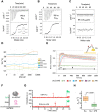
Impacts of IP6 on Ku stabilization and interaction with DNA. (A–C) Isothermal titration calorimetry (ITC) analyses of the interaction between (A) KuFL and IP6, (B) KuΔC and IP6 and (C) the mutant Ku-2E (Ku70-K357E, Ku80-K481E) and IP6. All ITC experiments were done in duplicate. (D) Analysis of the thermal stability (Ti) by nano-differential scanning fluorimetry (nanoDSF) of KuFL, Ku-2E and Ku-4E (Ku70-H359E and H360E)/Ku80-K413E and H414E), in presence or absence of a hairpin DNA as indicated and with IP6 concentration ranging from 0 to 10 μM. All nanoDSF experiments were done in triplicate. (E) switchSENSE kinetics analysis of the Ku interaction with a dsDNA (48 bp) bound to a chip. The experiments were performed in the presence of 3 μM IP6, with 3 min association time ‘a’ and 15 min dissociation time ‘d’, based on a duplicate experiment. Interactions were performed with Ku concentration ranging from 15 nM to 240 nM (see legend below x-axis). The same experiment was performed in parallel without IP6 (Supplementary Figure 10a). The fits of the association steps are superimposed on the raw data. (F) Principle of the single-molecule colocalization experiment. (G) Representative single-molecule time traces for Ku-AF647 binding on 18 bp DNA-Cy3 without (top) and with (bottom) 10 μM IP6. Red arrows indicate the binding of Ku-AF647 protein with DNA. The green laser is turned on at the end of the experiment, indicated by a green arrow, to confirm the presence of DNA. (H) Comparative kOFF box plot of binding of Ku on DNA without (orange, n > 10 000) and with (green, n > 10 000) IP6, based on four independent experiments. P = 0.0081 indicates a significant difference between kOFF without and with IP6 based on the Student's t-test. Error bars represent standard deviation.
Figure 4.
Figure 4.
Effects of IP6 on Ku-XLF interaction and of combined Ku70 and Ku80 mutations on Ku and XLF recruitment to laser-induced DSBs in cells. (A, B) Position of IP6 (magenta) binding site relative to the binding sites on Ku of DNA-PKcs (grey). Interaction sites of Ku with XLF KBM (Ku site in yellow, XLF KBM in brown), APLF KBM (Ku site in dark blue, APLF KBM in light blue) and PAXX KBM (Ku site in cyan, PAXX KBM in violet) (KBM peptides are represented in stick). The XLF and PAXX KBMs bind respectively to Ku80 and Ku70 vWA internal position upon an opening of the vWA domains of each subunit. (The figure is made with the X-ray structure Ku-DNA-IP6 and the position of vWA domain of Ku70 observed in the X-ray structure with PAXX KBM (8BHY).) (C) switchSENSE analysis of Ku-DNA interaction with XLF (8 μM) in presence or not of 20 μM IP6 during both Ku association (step 1) and XLF titration (step 2). Results with additional concentrations of XLF are shown in Supplementary Figure 12. Binding curves in presence or absence of IP6 were fitted with a two steps binding mode (thin lines). Kinetics data are reported in a table in Supplementary Figure 12. (D) Schematic diagram illustrating the steps of cell lines construction. U2OS cells expressing Tet-On inducible shRNAs against Ku80 and Ku70 were complemented with lentiviral constructs allowing the expression of both Ku80-K481E mutant and GFP-tagged Ku70-K357E mutant (Ku-2E) or both wild-type Ku80 and wild-type GFP-tagged Ku70 (wt) as a control. Rescued cell populations were selected by doxycycline treatment (+doxy) and further transduced to express mCherry-tagged XLF (mCh-XLF). (E) Expression levels of Ku80 and Ku70 at each step of cell lines construction were assessed by western blot. (F) GFP and mCherry-fluorescent cells expressing either Ku-wt or Ku-2E (as in D) and mCh-XLF were then micro-irradiated. GFP-Ku and mCh-XLF recruitment at irradiated areas was measured. Typical images of fluorescent nuclei before and 60 s post-irradiation. The irradiated areas are indicated by arrows. (G) Quantification of fluorescence accumulation at laser-damaged sites over 1 min for U2OS cells expressing GFP-Ku-wt (green) or GFP-Ku-2E (orange) and mCh-XLF. Results were plotted as mean values ± standard error of the mean (SEM) from 19 (Ku-wt) and 20 (Ku-2E) individual nuclei. P values at various time points were calculated using unpaired two-tailed t-test. P values for GFP-Ku recruitment (Ku-wt vs Ku-2E) at 10, 20, 30, 40, 50 and 60 s were 0.1741 (ns), 0.0482 (*), 0.0319 (*), 0.0318 (*), 0.0396 (*) and 0.1073 (ns), respectively. P values for mCherry-XLF recruitment (Ku-wt versus Ku-2E) were < 0.001 (***) at 10 s and <0.0001 (****) at 30 and 60 s.
Figure 5.
Figure 5.
Impact of combined Ku70 and Ku80 mutations on c-NHEJ complex assembly in cells. (A) Western blot of cell extracts from HEK-293T cells showing mAID-Ku70/Ku80 expression levels before and after overnight treatment with auxin (IAA). (B) Schematic diagram illustrating the steps of cell lines construction and dual DSB repair (DSBR) reporter assay. In HEK-293T cells expressing OsTIR1, both Ku subunits were silenced (by shRNA expression for Ku70 and knock-out for Ku80, respectively) in order to replace endogenous Ku70 and Ku80 expression by degron-tagged constructs, mAID-Ku70 (D70) and mAID-Ku80 (D80), respectively. These cells are further transduced to express Ku-2E mutant (Ku70-K357E, Ku80-K481E) or wild-type forms as a control. Degron-tagged Ku subunits are then degraded upon addition of auxin (+IAA) leaving only Ku-2E or Ku-wt dimers. (C) Expression levels of Ku80 and Ku70 at the different steps of the cell lines construction. Asterisks in the Ku80 western blot indicate residual Ku70 signals from previous hybridization that resisted membrane stripping. (D) Western blot of chromatin and soluble fractions as described in Materials and Methods section, from HEK-293T cells expressing Ku WT or Ku-2E (as in B) or mAID-Ku70 (as in A), treated with IAA for 18 h and then treated or not with calicheamicin doses as stated for 1 h.
Figure 6.
Figure 6.
Impact of combined Ku70 and Ku80 mutations on End-Joining activity in cells. (A) The dual reporter substrate for both End-Joining and Homologous Recombination repair activities consists of two non-functional copies of mTagBFP2 cDNA (BFP) separated by an intervening EGFP cDNA (GFP). The first copy of BFP is interrupted near the chromophore coding sequence (between codons 67 and 68) by a cassette containing the HSV-TK polyadenylation sequence flanked by two inverted copies of a Cas9 target sequence (bold characters; the PAM AGG sequence is underlined). The downstream copy of BFP is truncated and stops at codon Y201. Following Cas9-mediated double cleavage, the HSV-TK polyadenylation sequence is deleted and the resulting DNA blunt-ends can be rejoined directly through NHEJ. This direct end-joining (dEJ) does not restore the BFP coding sequence because of a frameshift insertion (red asterisk and subsequent frameshifted BFP sequence (fs-BFP), but enables the expression of the downstream EGFP cDNA which lies in a different reading frame. In order to allow EGFP expression, all stop codons downstream the DNA cutting sites have been removed from BFP cDNA by silent mutations. Alternatively, the DSB can be repaired by gene conversion using the second BFP copy. A stuffer coding DNA fragment (StD) together with a nuclear localization sequence (NLS) have been fused upstream both BFP cDNA copies to provide 400 bp homology on both sides of the DSB, as indicated by grey and black lines, respectively. The homologous recombination event (HR) restores a full-length BFP cDNA whereas the EGFP cDNA lies in a different reading frame (fs-GFP) and is not expressed. (B) The dual DSBR reporter assay is performed by transfecting the dual reporter substrate together with a Cas9/gRNA-expressing vector to cleave the substrate and a mCherry-expressing vector to normalize for transfection efficiency. Fluorescence expression is analysed 48 h later by flow cytometry. Green (GFP) and blue (BFP) fluorescence represent direct end-joining (dEJ) and homologous recombination (HR), respectively. (C and D) Validation of the dual DSBR reporter assay in HEK-293T cells upon treatment with 3 μM DNA-PK inhibitor NU7441 (PKi) or 10 μM Rad51 inhibitor B02 (RAD51i). Results are presented as GFP versus BFP scatter plots of a representative experiment (C), or as a histogram with mean values ± SEM from 4–7 experiments (D). The control condition corresponds to a complete repair reaction without inhibitors. The P-values are as follows: < 0.0001 (****, dEJ, +PKi vs control), 0.1190 (ns, dEJ, + RAD51i versus control), 0.0089 (**, HR, + PKi versus control), 0.0332 (*, HR, + RAD51i versus control), 0.0076 (**, HR, + PKi/+ RAD51i versus + PKi). (E) Simultaneous measurement of direct end-joining (dEJ) and homologous recombination (HR) activities in HEK-293T cells depleted (+IAA) or not of Ku (Figure 5A) and in of HEK-293T cells expressing Ku-wt or Ku-2E (see Figure 5B) following two days post-transfection with the dual DSB repair reporter substrate. Results are normalized to 100% for the untreated respective control cells and presented as mean values ± SEM from X experiments. The P-values are <0.0001 (****, dEJ, +IAA versus –IAA, HR, +IAA versus –IAA and dEJ, 2E versus wt) and 0.0036 (**, HR, 2E versus wt).

References

    1. Lieber M.R. The mechanism of double-strand DNA break repair by the nonhomologous DNA end-joining pathway. Annu. Rev. Biochem. 2010; 79:181–211. - PMC - PubMed
    1. Downs J.A., Jackson S.P.. A means to a DNA end: the many roles of Ku. Nat. Rev. Mol. Cell Biol. 2004; 5:367–378. - PubMed
    1. Zahid S., Seif El Dahan M., Iehl F., Fernandez-Varela P., Le Du M.H., Ropars V., Charbonnier J.B. The multifaceted roles of Ku70/80. Int. J. Mol. Sci. 2021; 22:4134–4155. - PMC - PubMed
    1. Frit P., Ropars V., Modesti M., Charbonnier J.B., Calsou P.. Plugged into the Ku-DNA hub: the NHEJ network. Prog. Biophys. Mol. Biol. 2019; 147:62–76. - PubMed
    1. Chaplin A.K., Hardwick S.W., Liang S., Kefala Stavridi A., Hnizda A., Cooper L.R., De Oliveira T.M., Chirgadze D.Y., Blundell T.L.. Dimers of DNA-PK create a stage for DNA double-strand break repair. Nat. Struct. Mol. Biol. 2020; 28:13–19. - PubMed

Publication types

LinkOut - more resources