. 2023 Nov 7;23(22):4876-4887.
doi: 10.1039/d3lc00543g.

Nano-injection molding with resin mold inserts for prototyping of nanofluidic devices for single molecular detection

Affiliations

Nano-injection molding with resin mold inserts for prototyping of nanofluidic devices for single molecular detection

Farhad Shiri et al. Lab Chip. .

Abstract

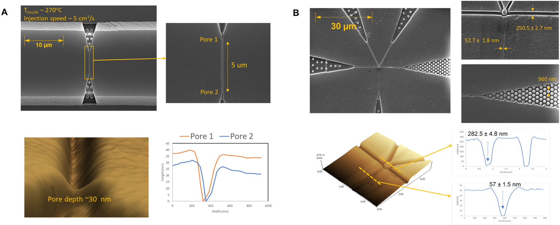
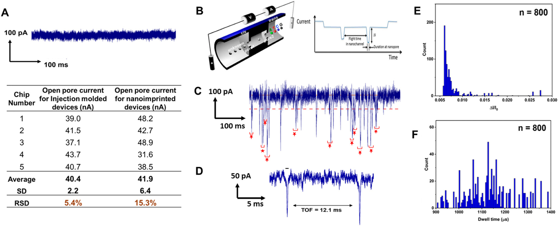
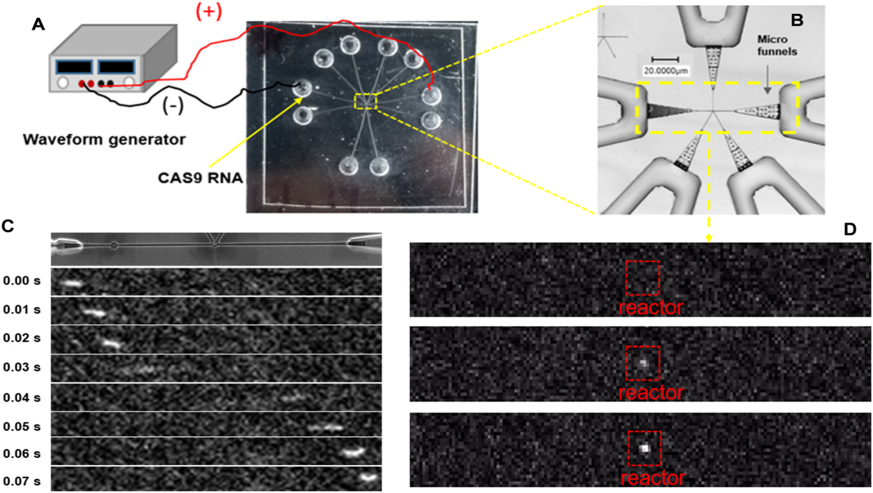
While injection molding is becoming the fabrication modality of choice for high-scale production of microfluidic devices, especially those used for in vitro diagnostics, its translation into the growing area of nanofluidics (structures with at least one dimension <100 nm) has not been well established. Another prevailing issue with injection molding is the high startup costs and the relatively long time between device iterations making it in many cases impractical for device prototyping. We report, for the first time, functional nanofluidic devices with dimensions of critical structures below 30 nm fabricated by injection molding for the manipulation, identification, and detection of single molecules. UV-resin molds replicated from Si masters served as mold inserts, negating the need for generating Ni-mold inserts via electroplating. Using assembled devices with a cover plate via hybrid thermal fusion bonding, we demonstrated two functional thermoplastic nanofluidic devices. The first device consisted of dual in-plane nanopores placed at either end of a nanochannel and was used to detect and identify single ribonucleotide monophosphate molecules via resistive pulse sensing and obtain the effective mobility of the molecule through nanoscale electrophoresis to allow its identification. The second device demonstrated selective binding of a single RNA molecule to a solid phase bioreactor decorated with a processive exoribonuclease, XRN1. Our results provide a simple path towards the use of injection molding for device prototyping in the development stage of any nanofluidic or even microfluidic application, through which rapid scale-up is made possible by transitioning from prototyping to high throughput production using conventional Ni mold inserts.


Conflict of interest statement

Conflicts of interest

The authors have declared no conflict of interest.

Figures

Fig. 1
Fig. 1
(A) Images of the injection molding machine equipped with a Master Unit Die (MUD) consisting of a movable and a stationary platens. A blank mold insert was placed on the surface of the moving platen while a resin mold insert was fixed to the stationary platen. (B) SEM images of the Si master used for nanoscale electrophoresis, which consist of dual in-plane nanopores flanked into both ends of a nanochannel. (C) SEM images of a nanofluidic structure used for selective binding of a single RNA molecule to a solid phase bioreactor. The structure consist of a nanopillar in the center which is connected to four input/output nanochannel of ∼250 nm × 250 nm in width × depth and one nanochannel of ∼50 nm × 50 nm in width × depth to capture the reaction products from the solid phase reactor. (D) and (E) AFM images of replicated MD700 molds with positive and negative toned structures fabricated by repetitive replication from Si master with the dual in-plane nanopore Time-of-Flight (ToF) sensor structure.
Fig. 2
Fig. 2
Production of nanofluidic structures by injection molding with COP from MD700 resin molds. (A) Top: Scanning electron microscopy (SEM) images of dual in-plane nanopore time-of-flight (ToF) sensor that consists of dual in-plane nanopores flanked into both ends of a nanochannel. Bottom: Atomic force microscopy (AFM) image of an in-plane nanopore (from resin mold 2, see Fig. 1B) with a ultrasharp AFM tip and cross-sectional profiles of both in-plane nanopores. (B) Top: SEM images of the nanofluidic structure used for selective binding of a single RNA molecule. The structure consists of a solid phase bioreactor located in the center of the image, four input/output nanochannels, and a nanochannel for capturing the reaction products from the solid phase reactor. The input/output channels also contain in-plane sensing pores and entropic trap. Bottom: AFM image near the solid phase bioreactor and cross sectional profiles of the nanochannels at two different locations. Both input/output channels were ∼280 nm in width and depth.
Fig. 3
Fig. 3
Dual in-plane nanopore sensor for measuring the Time-of-Flight (ToF) of single molecules, such as ribonucleotide monophosphates. (A) A 0.5 s current transient trace obtained prior to introducing rCMPs. The inset table shows open pore currents for five randomly picked assembled nanosensors produced by injection molding and nanoimprint lithography. (B) Diagram illustrating the dual-nanopore ToF sensor configuration, featuring a pair of in-plane nanopores positioned on opposite sides of a nanochannel serving as the nanochannel column for nanoscale electrophoresis. The schematic was taken from ref. 54. (C) A 0.5 s current transient trace of signal amplitudes obtained after injecting rCMPs (10 nM) in 1× NEBuffer 3 at pH.7.9 into the nanosensor. (D) Example current transient data showing a peak pair from the two in-plane nanopores for translocation of a single rCMP molecule. (E) Histogram of the normalized peak amplitude (ΔI/Io) data from the current transient data shown in Fig. 3C. (F) Histogram of the dwell time data from the current transient data shown in Fig. 3C.
Fig. 4
Fig. 4
RNA translocation through an injection molded single-molecule sequencing device. (A) Experimental setup showing the electrical connections to the chip with a waveform generator for supplying the electrical field for driving the RNA (CAS9) through the chip. (B) Rapid scanning confocal image of the single-molecule sequencing device with the yellow box showing the area that is imaged with the single-molecule laser-induced fluorescence tracking microscope. (C) Fluorescence image Syto 82 labeled RNA electrically translocating through the input/output channels of the mixed-scale sequencing device. In this case, there was no ribo-exonuclease covalently attached to the solid-phase bioreactor portion of the device. Also, this device did not contain the in-plane nanopores within the input/output channel network. (D) Same conditions as shown and discussed in (C), but in this case, there was XRN1 ribo-exonuclease attached to the solid-phase bioreactor, which associates to the translocating RNA molecule causing it to remain stationary.

References

    1. Haywood DG, Saha-Shah A, Baker LA and Jacobson SC, Fundamental Studies of Nanofluidics: Nanopores, Nanochannels, and Nanopipets, Anal. Chem., 2015, 87, 172–187. - PMC - PubMed
    1. Mawatari K, Kazoe Y, Shimizu H, Pihosh Y and Kitamori T, Extended-Nanofluidics: Fundamental Technologies, Unique Liquid Properties, and Application in Chemical and Bio Analysis Methods and Devices, Anal. Chem., 2014, 86, 4068–4077. - PubMed
    1. Chen J, Yu H, Fan J, Wang F, Lu D, Liu H and Wu H, Channel-width dependent pressure-driven flow characteristics of shale gas in nanopores, AIP Adv., 2017, 7, 045217.
    1. Nanofluidics is on the rise, Nat. Mater., 2020, 19, 253. - PubMed
    1. Riehn R, Lu M, Wang Y-M, Lim SF, Cox EC and Austin RH, Restriction mapping in nanofluidic devices, Proc. Natl. Acad. Sci. U. S. A., 2005, 102, 10012–10016. - PMC - PubMed

Publication types