. 2023 Oct 23;17(10):e0011693.
doi: 10.1371/journal.pntd.0011693. eCollection 2023 Oct.

Trichomonas vaginalis adherence phenotypes and extracellular vesicles impact parasite survival in a novel in vivo model of pathogenesis

Affiliations

Trichomonas vaginalis adherence phenotypes and extracellular vesicles impact parasite survival in a novel in vivo model of pathogenesis

Brenda M Molgora et al. PLoS Negl Trop Dis. .

Abstract

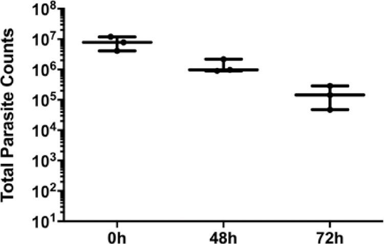
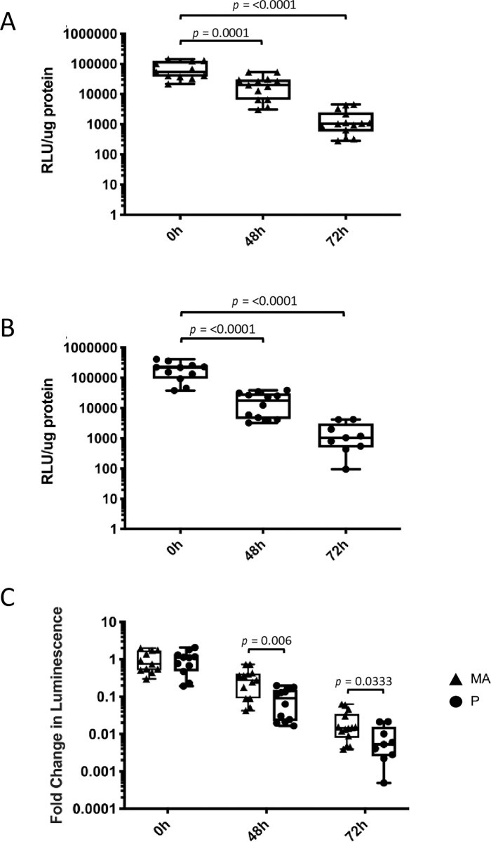
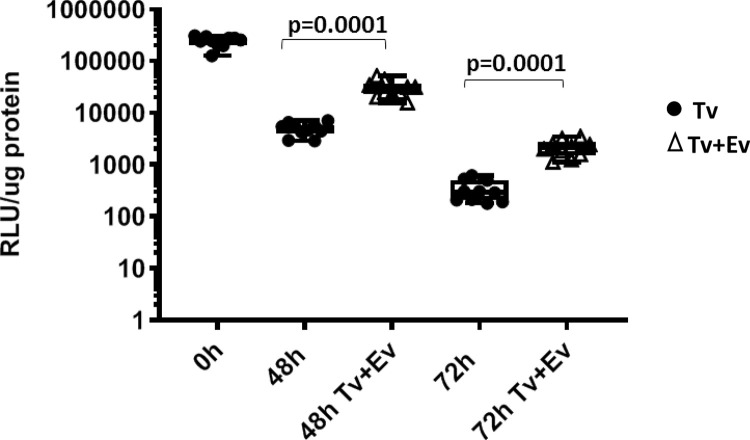
Trichomonas vaginalis is a human infective parasite responsible for trichomoniasis-the most common, non-viral, sexually transmitted infection worldwide. T. vaginalis resides exclusively in the urogenital tract of both men and women. In women, T. vaginalis has been found colonizing the cervix and vaginal tract while in men it has been identified in the upper and lower urogenital tract and in secreted fluids such as semen, urethral discharge, urine, and prostatic fluid. Despite the over 270 million cases of trichomoniasis annually worldwide, T. vaginalis continues to be a highly neglected organism and thus poorly studied. Here we have developed a male mouse model for studying T. vaginalis pathogenesis in vivo by delivering parasites into the murine urogenital tract (MUT) via transurethral catheterization. Parasite burden was assessed ex-vivo using a nanoluciferase-based gene expression assay which allowed quantification of parasites pre- and post-inoculation. Using this model and read-out approach, we show that T. vaginalis can be found within MUT tissue up to 72 hrs post-inoculation. Furthermore, we also demonstrate that parasites that exhibit increased parasite adherence in vitro also have higher parasite burden in mice in vivo. These data provide evidence that parasite adherence to host cells aids in parasite persistence in vivo and molecular determinants found to correlate with host cell adherence in vitro are applicable to infection in vivo. Finally, we show that co-inoculation of T. vaginalis extracellular vesicles (TvEVs) and parasites results in higher parasite burden in vivo. These findings confirm our previous in vitro-based predictions that TvEVs assist the parasite in colonizing the host. The establishment of this pathogenesis model for T. vaginalis sets the stage for identifying and examining parasite factors that contribute to and influence infection outcomes.


Conflict of interest statement

The authors disclose no competing interests that could be perceived to bias this work.

Figures

Fig 1
Fig 1. Tv is delivered into the mouse male urogenital tract (MUT) using urethral catheterization.
(A) Diagram of catheterization and Tv delivery into the mouse MUT. 108 Nluc expressing parasites suspended in 100 μl RPMI-1640 medium were introduced through the urethral meatus into the urethra via a syringe and polyethylene catheter. Additional mouse penile features such as the glans, penile bone, and the male urogenital mating protuberance (MUMP) are depicted. Mouse penis sketch adapted from a mid-sagittal sectioning of the adult mouse penis [74]. (B) A simplified diagram of a lateral view of the male urogenital system in adult mice modified from Cunha et. al. [75]. Parasites introduced into the mouse MUT via urethral catheterization would be delivered through the urethra (U) to the anterior (AP), dorsal (DP), lateral (LP), and ventral (VP) prostatic lobes. The bladder and seminal vesicles (SV) are also depicted. Histological validation of Tv delivery into the mouse MUT (C) versus the vehicle control. (E) The right panels are a 20X magnification of the left anterior prostate lobe (marked by a circle in the left panels). T. vaginalis parasites are indicated by a blue arrow. (D) 100X magnification of Tv parasite found in infected mouse MUT tissue. Blue arrow indicates the presence of flagella.
Fig 2
Fig 2. Linearity of luminescence in different Nluc-expressing Tv strains.
T. vaginalis strains LSU 160 MA and P (A) and MSA 1103 (B) nucleofected with pMasterNeo-Nluc plasmid were measured for luminescence at varying amounts of parasites from 0 to 106 parasites (as indicated on the X axis) to determine the linear range of the assay for each strain. Data shown are averages of luminescence signal from 3 technical replicates with standard deviation for each strain.
Fig 3
Fig 3. Quantifying parasite burden using nanoluciferase-expressing Tv.
(A) Schematic of mouse MUT tissue sample preparation and quantification of Tv parasite burden. Excised mouse MUT tissue is finely minced and suspended in prostate lysis buffer (2 mL/mg tissue). MUT tissue then undergoes additional homogenization via probe sonication to ensure complete tissue dissociation. Insoluble material is pelleted via centrifugation and the supernatant is transferred to a new tube to further quantify luciferase activity and protein concentration using the Nano-Glo luciferase and Bradford assays, respectively. Detailed description found in materials and methods. (B) Mice were inoculated with 108 parasites (n = 3) or RPMI vehicle control (n = 3) and promptly euthanized. Samples were processed and analyzed by Nluc and Bradford assays to quantify parasite load. Data is shown as average luminescence signal/μg protein of the sample.
Fig 4
Fig 4. Tv found within the murine MUT tissue 72 hours post-inoculation.
Quantification of Nluc-expressing MSA 1103 parasites in infected mouse MUT tissues 48 hrs or 72 hrs post-inoculation (as indicated on the X axis). Data are based on 12 infected mice per time point per strain and are averages of luminescence/μg protein of the sample with standard deviation. Data are representative of 3 independent experiments.
Fig 5
Fig 5. Viable parasites are recovered from mouse MUT tissue 72 hours post-inoculation.
108 Tv parasites were introduced into the mouse urogenital tract and the infected tissue was then excised at 0 hr, 48 hrs, and 72 hrs post-inoculation (as indicated on the X axis). Tissue was minced, dissociated using Accutase cell detachment solution, resuspended in completed modified TYM media and counted via hemocytometer. Data shown are the total number of parasites per sample with standard deviation and were carried out using 3 mice per timepoint.
Fig 6
Fig 6. Tv adherence to host cells in vitro correlates with an increased parasite burden in the MUT.
Quantification of Nluc-expressing LSU 160 more adherent (MA) (A) or parental (P) (B) parasites in mouse MUT tissues at 48 hrs and 72 hrs post-inoculation (as indicated on the X-axis). 11–14 mice were used for the MA strain at each time point. 9–12 mice were used for the P strain at each time point. Data shown are averages of luminescence/μg protein of the sample with standard deviation and are representative of 3 independent experiments. (C) Comparison of LSU160 MA (▲) and LSU160 P (●) parasite load quantifications. Data is expressed as fold change in luminescence in the mouse urogenital tract with standard deviation normalized to their respective 0h post-inoculation luminescent signals.
Fig 7
Fig 7. Co-inoculation of parasite secreted EVs with Tv increases the parasite burden in the mouse urogenital tract.
Quantification of Nluc-expressing MSA 1103 parasites co-inoculated with either 50 μg/μL EVs (▢; Tv+EV) or parasites only (●; Tv) in the mouse MUT tissues at 48 and 72 hrs post-inoculation (as indicated on the X-axis). Ten mice were inoculated at each time point per condition. Data shown are averages of luminescence/μg protein of the sample with standard deviation normalized to 0h post-inoculation luminescent signals. Data are representative of 3 independent experiments.

References

    1. Schwebke JR, Burgess D. Trichomoniasis. Clin Microbiol Rev. 2004;17(4):794–803. doi: 10.1128/CMR.17.4.794-803.2004 - DOI - PMC - PubMed
    1. World Health Organization. Global incidence and prevalence of selected curable sexually transmitted infections: 2008. Reprod Health Matters. 2012;20(40):207–8.
    1. World Health Organization. Report on global sexually transmitted infection surveillance, 2018. 2018. p. 1–74. Available from: https://apps.who.int/iris/handle/10665/277258.
    1. Rowley J, Hoorn S Vander, Korenromp E, Low N, Unemo M, Abu-Raddad LJ, et al. Chlamydia, gonorrhoea, trichomoniasis and syphilis: Global prevalence and incidence estimates, 2016. Bull World Health Organ. 2019;97(8):548–62. doi: 10.2471/BLT.18.228486 - DOI - PMC - PubMed
    1. Centers for Disease Control and Prevention. STD Surveillance Report 2018. 2019. Available from: https://www.cdc.gov/nchhstp/newsroom/2019/2018-STD-surveillance-report.html.

Publication types