. 2023 Oct 23;19(10):e1010768.
doi: 10.1371/journal.pcbi.1010768. eCollection 2023 Oct.

Tissue Forge: Interactive biological and biophysics simulation environment

Affiliations

Tissue Forge: Interactive biological and biophysics simulation environment

T J Sego et al. PLoS Comput Biol. .

Abstract

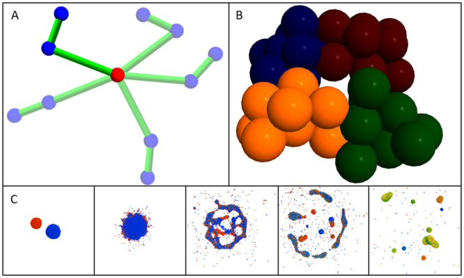
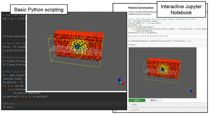
Tissue Forge is an open-source interactive environment for particle-based physics, chemistry and biology modeling and simulation. Tissue Forge allows users to create, simulate and explore models and virtual experiments based on soft condensed matter physics at multiple scales, from the molecular to the multicellular, using a simple, consistent interface. While Tissue Forge is designed to simplify solving problems in complex subcellular, cellular and tissue biophysics, it supports applications ranging from classic molecular dynamics to agent-based multicellular systems with dynamic populations. Tissue Forge users can build and interact with models and simulations in real-time and change simulation details during execution, or execute simulations off-screen and/or remotely in high-performance computing environments. Tissue Forge provides a growing library of built-in model components along with support for user-specified models during the development and application of custom, agent-based models. Tissue Forge includes an extensive Python API for model and simulation specification via Python scripts, an IPython console and a Jupyter Notebook, as well as C and C++ APIs for integrated applications with other software tools. Tissue Forge supports installations on 64-bit Windows, Linux and MacOS systems and is available for local installation via conda.


Conflict of interest statement

No competing interests.

Figures

Fig 1
Fig 1. Examples of Tissue Forge modeling features.
A: Five superimposed snapshots of a double pendulum implemented in Tissue Forge. Bonded interactions (represented as green cylinders) explicitly describe the interaction between a particular pair of particles, while a constant force acts on the blue particles in the downward direction. The red particle is fixed. B: Four Tissue Forge clusters representing biological cells, each consisting of ten particles whose color demonstrates cluster membership. Potentials describe particle interactions by whether they are in the same cluster (i.e., intracellular) or different clusters i.e., intercellular. C: Tissue Forge simulation of chemical flux during fluid droplet collision. Each particle represents a portion of fluid that carries an amount of a diffusive chemical, the amount of which varies from zero (blue) to one (red). When two droplets carrying different initial chemical amounts collide, resulting droplets tend towards homogeneous chemical distributions.
Fig 2
Fig 2. Tissue Forge simulation of a simple oscillator with two particles interacting via a harmonic potential.
Tissue Forge helps to orient the user by drawing a yellow box around the simulation domain, a white grid along the xy plane at the center of the domain, and an orientation glyph at the bottom right to demonstrate the axes of the simulation domain with reference to the camera view, where red points in the x direction, green in the y direction and blue in the z direction.
Fig 3
Fig 3. Tissue Forge Python deployment examples.
Sample use of the Python API to specify an interactive simulation of convection of a species near a species sink in a Python script (left) and in an interactive Jupyter Notebook (right).
Fig 4
Fig 4. Conceptual diagram of space discretization and task-based parallelism.
Space is discretized into subdomains called “cells” (shown here for N subdomains), and particles (listed as “PX”) are stored in memory by which cell contains them. For each simulation step, a set of threads, called “runners” (shown here for M runners) perform a pre-defined set of tasks. The “Sort” task builds ordered lists of particle indices according to proximity to neighboring cells for efficient pruning of inter-cell interactions between particle pairs outside of the cutoff distance. The “Force Self” task calculates interactions between particles of the same cell. The “Force Pair” task calculates interactions between particles of different cells using results from the Sort task. Task scheduling enforces task dependency. After all forces are calculated, particle positions are updated and particles that move into a neighboring cell are appropriately moved in memory, as demonstrated for particles P247 (from cell N to cell 1) and P233 (from cell 1 to cell 2).
Fig 5
Fig 5. Tissue Forge performance metrics for windowless and real-time rendering modes.
A: Computational cost per time step per particle for varying number of particles, varying cutoff distance and varying architecture when running windowless with fixed particle density and one implicit interaction. Computational cost generally increases with increasing cutoff distance. On a CPU, computational cost is lowest near 10k particles and then begins to increase. When offloading implicit interactions to a GPU, computational cost is generally less and tends towards a constant value. B. Computational cost for varying number of potentials defining implicit interactions with 10M particles and a cutoff of 5. Multiple potentials were implemented using potential arithmetic. Computational cost generally increases linearly with increasing number of potentials. C. Representative cost of solver stages when executing simulations from panel A with a cutoff of 5 in windowless (left) and real-time rendering (right) modes with implicit interactions calculated on a CPU (top) and GPU (bottom). Windowless mode simulated 10M particles. Real-time rendering mode simulated 10k particles. Size of bars for each mode represents relative cost of a simulation step. Bars are divided by solver stages that are ordered by execution order, and the area of each represents the portion of the total cost that the stage contributes. In both modes and architectures, force calculations make up the majority of the computational cost. As demonstrated in A, computing performance on a GPU is more efficient with increasing number of particles (Windowless), whereas computing performance on a CPU is more efficient for few particles (Real-time rendered).
Fig 6
Fig 6. Molecular modeling and simulation with Tissue Forge.
A: Classes of bonded interactions, where a measured property of the bond (length l for Bonds, angle θ for Angles, and planar angle ϕ for Dihedrals) is used as input to a potential function. B: Detailed view of thymine (left) and adenine (right) molecules constructed from Tissue Forge objects. Bonds shown as green cylinders, angles as blue arcs, and dihedrals as gold planes. C: Real-time simulation of a cloud of thymine and adenine molecules interacting via long-range potentials in a neutral medium.
Fig 7
Fig 7. Active pumping of a diffusive species across a deformable membrane separating two fluid-filled compartments.
A: Cut-plane views during simulation of two fluid-filled compartments separated by a deformable membrane, where each fluid is uniformly initialized with an initial concentration of a species. Particle color indicates species concentration with red as high, yellow and green as intermediate, and blue as low concentration. The membrane contains a particle that actively pumps the species from the lower to the upper compartment. B: Three-dimensional view of initial simulation state. C: Measurements of total species amounts in the lower (blue, circles), upper (red, triangles) and both (green, diamonds) compartments (left-hand vertical axis), and in the channel (magenta, squares, right-hand axis), during simulation.
Fig 8
Fig 8. Simulating fusion of multicellular, homotypic spheroids.
A: Spheroids of 12.5k cells each were individually pre-assembled, as in typical bioprinting practice. B: Two spheroids (green and blue) placed in close proximity fuse over time, as measured by the neck diameter along the y (blue circles) and z (red triangles) directions, which grows over time. The neck diameter along a direction is measured as the largest distance along the direction between any two particles at the mid-plane. Insets show the simulation at times 1, 50, 100, 150 and 200.
Fig 9
Fig 9. Two-dimensional agent-based model of cell proliferation and differential in the colonic crypt.
A: Cells represented as particles are arranged in a sheet as if fixed on an unfolded cylindrical surface, where the upper boundary is treated as the base of the crypt and periodic boundary conditions are applied along the horizontal direction, as in [16]. Each cell is assigned a state dynamics model of the cell cycle and unique clonal identification (visualized as a unique particle color). The cell cycle model of each cell progresses through G1, S, G2 and M phases with deterministic periods except for G1, the period of which is randomly selected from a normal distribution for each cell. When the cell cycle model of a cell transitions from the M phase to the G1 phase, the cell divides and copies its clonal identification to its progeny. Cells are removed when they reach the base of the crypt. B: Number of clones during simulation time, measured as the mean of ten simulation replicates. Over time, the crypt tends toward monoclonality.
Fig 10
Fig 10. Delta-Notch signaling in a cellular monolayer subject to environmental control via a soluble signal.
A: Lateral inhibition of Delta expression (cells with high Delta shown as red, cells with low Delta as blue) through contact-mediated Delta-Notch signaling without environmental regulation produces patterns in monolayers. Initially (left) Delta expression is approximately uniform but over time a pattern spontaneously emerges through contact-mediated signaling (right). B: Diffusion of a regulatory signal that induces Delta-Notch signaling along a uniform grid of particles representing point measurements of concentration. Initially (left) the cells are not exposed to the signal, but over time (right) the signal diffuses from the top boundary. C: When Delta-Notch signaling depends on induction via local concentration of the regulatory signal in B (not shown), pattern formation in the monolayer follows the propagation of the diffusive signal across the monolayer.

References

    1. Swat M., Thomas G., Belmonte J., Shirinifard A., Hmeljak D. & Glazier J. Multi-Scale Modeling of Tissues Using CompuCell3D. Methods In Cell Biology. 110 pp. 325–366 (2012), https://www.ncbi.nlm.nih.gov/pmc/articles/PMC3612985/ doi: 10.1016/B978-0-12-388403-9.00013-8 - DOI - PMC - PubMed
    1. Starruß J., Back W., Brusch L. & Deutsch A. Morpheus: a user-friendly modeling environment for multiscale and multicellular systems biology. Bioinformatics. 30, 1331–1332 (2014,5) - PMC - PubMed
    1. Graner F. & Glazier J. Simulation of biological cell sorting using a two-dimensional extended Potts model. Physical Review Letters. 69, 2013–2016 (1992,9), https://link.aps.org/doi/10.1103/PhysRevLett.69.2013, Publisher: American Physical Society; - DOI - PubMed
    1. Ghaffarizadeh A., Heiland R., Friedman S., Mumenthaler S. & Macklin P. PhysiCell: An open source physics-based cell simulator for 3-D multicellular systems. PLOS Computational Biology. 14, e1005991 (2018,2), https://journals.plos.org/ploscompbiol/article?id=10.1371/journal.pcbi.1..., Publisher: Public Library of Science: - PMC - PubMed
    1. Mirams G., Arthurs C., Bernabeu M., Bordas R., Cooper J., Corrias A., Davit Y., Dunn S., Fletcher A., Harvey D., Marsh M., Osborne J., Pathmanathan P., Pitt-Francis J., Southern J., Zemzemi N. & Gavaghan D. Chaste: An Open Source C++ Library for Computational Physiology and Biology. PLOS Computational Biology. 9, e1002970 (2013,3), https://journals.plos.org/ploscompbiol/article?id=10.1371/journal.pcbi.1..., Publisher: Public Library of Science; - PMC - PubMed