A degron-based approach to manipulate Eomes functions in the context of the developing mouse embryo
- PMID: 37871215
- PMCID: PMC10622880
- DOI: 10.1073/pnas.2311946120
A degron-based approach to manipulate Eomes functions in the context of the developing mouse embryo
Abstract
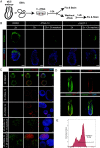
The T-box transcription factor Eomesodermin (Eomes), also known as Tbr2, plays essential roles in the early mouse embryo. Loss-of-function mutant embryos arrest at implantation due to Eomes requirements in the trophectoderm cell lineage. Slightly later, expression in the visceral endoderm promotes anterior visceral endoderm formation and anterior-posterior axis specification. Early induction in the epiblast beginning at day 6 is necessary for nascent mesoderm to undergo epithelial to mesenchymal transition (EMT). Eomes acts in a temporally and spatially restricted manner to sequentially specify the yolk sac haemogenic endothelium, cardiac mesoderm, definitive endoderm, and axial mesoderm progenitors during gastrulation. Little is known about the underlying molecular mechanisms governing Eomes actions during the formation of these distinct progenitor cell populations. Here, we introduced a degron-tag and mCherry reporter sequence into the Eomes locus. Our experiments analyzing homozygously tagged embryonic stem cells and embryos demonstrate that the degron-tagged Eomes protein is fully functional. dTAG (degradation fusion tag) treatment in vitro results in rapid protein degradation and recapitulates the Eomes-null phenotype. However in utero administration of dTAG resulted in variable and lineage-specific degradation, likely reflecting diverse cell type-specific Eomes expression dynamics. Finally, we demonstrate that Eomes protein rapidly recovers following dTAG wash-out in vitro. The ability to temporally manipulate Eomes protein expression in combination with cell marking by the mCherry-reporter offers a powerful tool for dissecting Eomes-dependent functional roles in these diverse cell types in the early embryo.
Keywords: Eomes; dTAG; degron; embryo; mouse.
Conflict of interest statement
The authors declare no competing interest.
Figures


Similar articles
-
Pivotal roles for eomesodermin during axis formation, epithelium-to-mesenchyme transition and endoderm specification in the mouse.Development. 2008 Feb;135(3):501-11. doi: 10.1242/dev.014357. Epub 2008 Jan 2. Development. 2008. PMID: 18171685 Free PMC article.
-
The T-box transcription factor Eomesodermin acts upstream of Mesp1 to specify cardiac mesoderm during mouse gastrulation.Nat Cell Biol. 2011 Aug 7;13(9):1084-91. doi: 10.1038/ncb2304. Nat Cell Biol. 2011. PMID: 21822279 Free PMC article.
-
Spatiotemporal sequence of mesoderm and endoderm lineage segregation during mouse gastrulation.Development. 2021 Jan 7;148(1):dev193789. doi: 10.1242/dev.193789. Development. 2021. PMID: 33199445
-
Eomesodermin-At Dawn of Cell Fate Decisions During Early Embryogenesis.Curr Top Dev Biol. 2017;122:93-115. doi: 10.1016/bs.ctdb.2016.09.001. Epub 2016 Oct 13. Curr Top Dev Biol. 2017. PMID: 28057273 Review.
-
Dose-dependent Nodal/Smad signals pattern the early mouse embryo.Semin Cell Dev Biol. 2014 Aug;32:73-9. doi: 10.1016/j.semcdb.2014.03.028. Epub 2014 Apr 1. Semin Cell Dev Biol. 2014. PMID: 24704361 Review.
Cited by
-
Degron tagging for rapid protein degradation in mice.Dis Model Mech. 2024 Apr 1;17(4):dmm050613. doi: 10.1242/dmm.050613. Epub 2024 Apr 26. Dis Model Mech. 2024. PMID: 38666498 Free PMC article.
-
Eomesodermin in conjunction with the BAF complex promotes expansion and invasion of the trophectoderm lineage.Nat Commun. 2025 May 31;16(1):5079. doi: 10.1038/s41467-025-60417-w. Nat Commun. 2025. PMID: 40450029 Free PMC article.
-
Cell-type specific, inducible and acute degradation of targeted protein in mice by two degron systems.Nat Commun. 2024 Nov 29;15(1):10129. doi: 10.1038/s41467-024-54308-9. Nat Commun. 2024. PMID: 39613744 Free PMC article.
-
Transcriptional remodeling by OTX2 directs specification and patterning of mammalian definitive endoderm.bioRxiv [Preprint]. 2024 May 30:2024.05.30.596630. doi: 10.1101/2024.05.30.596630. bioRxiv. 2024. Update in: Dev Cell. 2025 Aug 13:S1534-5807(25)00496-4. doi: 10.1016/j.devcel.2025.07.020. PMID: 38854146 Free PMC article. Updated. Preprint.
-
Fine-tuning of gene expression through the Mettl3-Mettl14-Dnmt1 axis controls ESC differentiation.Cell. 2025 Feb 20;188(4):998-1018.e26. doi: 10.1016/j.cell.2024.12.009. Epub 2025 Jan 17. Cell. 2025. PMID: 39826545
References
-
- Ciruna B. G., Rossant J., Expression of the T-box gene Eomesodermin during early mouse development. Mech. Dev. 81, 199–203 (1999). - PubMed
-
- Russ A. P., et al. , Eomesodermin is required for mouse trophoblast development and mesoderm formation. Nature 404, 95–99 (2000). - PubMed
-
- Strumpf D., et al. , Cdx2 is required for correct cell fate specification and differentiation of trophectoderm in the mouse blastocyst. Development 132, 2093–2102 (2005). - PubMed
MeSH terms
Substances
Grants and funding
LinkOut - more resources
Full Text Sources
Molecular Biology Databases

