. 2023 Oct 31;120(44):e2307593120.
doi: 10.1073/pnas.2307593120. Epub 2023 Oct 23.

Expression signature of human endogenous retroviruses in chronic lymphocytic leukemia

Affiliations

Expression signature of human endogenous retroviruses in chronic lymphocytic leukemia

Alessandro La Ferlita et al. Proc Natl Acad Sci U S A. .

Abstract

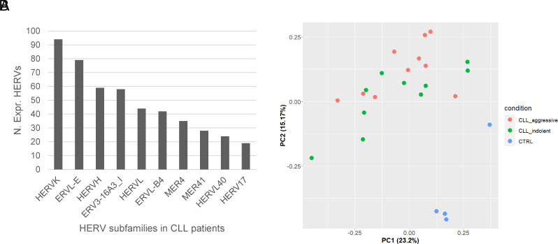
Chronic lymphocytic leukemia (CLL) is one of the most diagnosed forms of leukemia worldwide and it is usually classified into two forms: indolent and aggressive. These two forms are characterized by distinct molecular features that drive different responses to treatment and clinical outcomes. In this context, a better understanding of the molecular landscape of the CLL forms may potentially lead to the development of new drugs or the identification of novel biomarkers. Human endogenous retroviruses (HERVs) are a class of transposable elements that have been associated with the development of different human cancers, including different forms of leukemias. However, no studies about HERVs in CLL have ever been reported so far. Here, we present the first locus-specific profiling of HERV expression in both the aggressive and indolent forms of CLL. Our analyses revealed several dysregulations in HERV expression occurring in CLL and some of them were specific for either the aggressive or indolent form of CLL. Such results were also validated by analyzing an external cohort of CLL patients and by RT-qPCR. Moreover, in silico analyses have shown relevant signaling pathways associated with them suggesting a potential involvement of the dysregulated HERVs in these pathways and consequently in CLL development.

Keywords: CLL; HERVs; RNA-Seq.


Conflict of interest statement

The authors declare no competing interest.

Figures

Fig. 1.
Fig. 1.
Expressed HERVs in CLL. (A) Bar plot showing the most expressed HERV groups identified in CD5+ B cells taken from CLL patients; (B) PCA plot showing the distribution of CLL aggressive, CLL indolent, and normal B cell samples in a two-dimensional space using the expression of the top 200 most variable HERVs based on their MAD values.
Fig. 2.
Fig. 2.
Differentially expressed HERVs in CLL. (A) Volcano plots showing the differentially expressed HERVs found in either the aggressive or indolent form of CLL compared to normal B cells; (B) Venn diagrams showing the down-regulated and up-regulated HERVs found in common between the aggressive and indolent forms of CLL; (C) Bipartite graphs reporting the log2FC values of the differentially expressed HERVs found in common in the aggressive and indolent forms of CLL. P-values were calculated by applying one-tailed Wilcoxon signed rank test.
Fig. 3.
Fig. 3.
Distribution of differentially expressed HERVs in CLL. (A) Heatmaps with sample clustering of the differentially expressed HERVs identified in either the aggressive or indolent forms of CLL compared to normal B cells; (B) Circos plots showing the genomic distribution of the HERV loci found up (red dots) or down-regulated (blue dots) in the aggressive and indolent forms of CLL; (C) Bar plot showing the percentage of differentially expressed HERVs across the human chromosomes normalized by the number of HERV loci in the corresponding chromosome; (D) Bar plot reporting the top 10 most dysregulated HERV groups (% of dysregulated HERVs per group) identified in the aggressive (dark gray bars) and indolent (light gray bars) forms of CLL.
Fig. 4.
Fig. 4.
Validated differentially expressed HERVs in CLL. (A) Venn diagram reporting the number of HERVs identified as differentially expressed in the aggressive and indolent forms of CLL, in common between the two cohorts (internal and external); (B) Box plots showing the 2−ΔCt values of HERVL_17p11.2b and HUERSP3_11p15.1 in our internal cohort of CLL samples across the three conditions (CLL aggressive, CLL indolent, and CTRL). P-values were calculated by applying a one-tailed Wilcoxon rank sum test.
Fig. 5.
Fig. 5.
HERV-specific pathway analysis. Heatmaps that show the results of the HERV-specific pathway analysis for the aggressive (A) and indolent (B) forms of CLL. In detail, the columns are the enriched pathways while the rows are the differentially expressed HERVs. The values plotted in the heatmap are the “corrected accumulators” generated by MITHrIL as an indicator of the magnitude of pathway dysregulation. Corrected accumulator values with an associated not statistically significant P-value have been assigned the value 0 and plotted with the color white in the heatmaps.
Fig. 6.
Fig. 6.
Correlation networks between deregulated HERVs and hallmarks genes in CLL. Correlation networks for the aggressive (A) and indolent (B) forms of CLL where the blue nodes are the differentially expressed HERVs, the red nodes are the hallmark genes for CLL and the edge are either the positive (red) or negative (blue) correlation between them. Red and blue arrows by the HERV nodes indicate the up- or downregulation of the HERV, respectively, compared with normal B cell samples. HERVs with the highlighted borders are dysregulated in both CLL forms. Correlations between the differentially expressed HERVs and the hallmark genes that were not statistically significant were not plotted in the correlation networks.
Fig. 7.
Fig. 7.
Real-Time RT-PCR results in CLL samples treated with ibrutinib. Bar plot that shows the expression of HERVL_17p11.2b as 2−ΔCt in three CLL samples untreated or treated with 10 μm of ibrutinib at either 24 or 48 h (two biological replicates per condition).

References

    1. Pekarsky Y., Zanesi N., Croce C. M., Molecular basis of CLL. Semin. Cancer Biol. 20, 370–376 (2010). - PMC - PubMed
    1. Bullrich F., Croce C. M., Molecular biology of chronic lymphocytic leukemia. Basic and Clinical Oncology 2, 9–32 (2001).
    1. Cheson B. D., Chronic Lymphocytic Leukemia: Epidemiological, Familial, and Genetic Aspects. CLL 2, 1–30 (2001).
    1. Kipps T. J., et al. , Chronic lymphocytic leukaemia. Nat. Rev. Dis. Primers 3, 16096 (2017). - PMC - PubMed
    1. Pekarsky Y., Croce C. M., Noncoding RNA genes in cancer pathogenesis. Adv. Biol. Regul. 71, 219–223 (2019). - PMC - PubMed

MeSH terms