. 2023 Nov:67:102937.
doi: 10.1016/j.redox.2023.102937. Epub 2023 Oct 19.

Cardiomyocyte NOX4 regulates resident macrophage-mediated inflammation and diastolic dysfunction in stress cardiomyopathy

Affiliations

Cardiomyocyte NOX4 regulates resident macrophage-mediated inflammation and diastolic dysfunction in stress cardiomyopathy

Aleksandr E Vendrov et al. Redox Biol. 2023 Nov.

Abstract

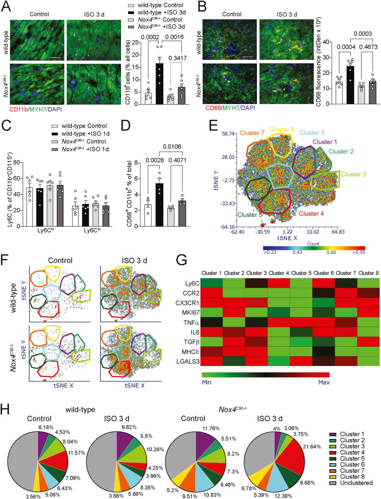
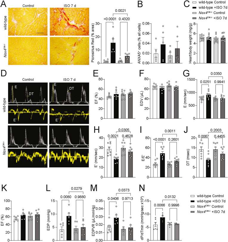
In acute sympathetic stress, catecholamine overload can lead to stress cardiomyopathy. We tested the hypothesis that cardiomyocyte NOX4 (NADPH oxidase 4)-dependent mitochondrial oxidative stress mediates inflammation and diastolic dysfunction in stress cardiomyopathy. Isoproterenol (ISO; 5 mg/kg) injection induced sympathetic stress in wild-type and cardiomyocyte (CM)-specific Nox4 knockout (Nox4CM-/-) mice. Wild-type mice treated with ISO showed higher CM NOX4 expression, H2O2 levels, inflammasome activation, and IL18, IL6, CCL2, and TNFα levels than Nox4CM-/- mice. Spectral flow cytometry and t-SNE analysis of cardiac cell suspensions showed significant increases in pro-inflammatory and pro-fibrotic embryonic-derived resident (CCR2-MHCIIhiCX3CR1hi) macrophages in wild-type mice 3 days after ISO treatment, whereas Nox4CM-/- mice had a higher proportion of embryonic-derived resident tissue-repair (CCR2-MHCIIloCX3CR1lo) macrophages. A significant increase in cardiac fibroblast activation and interstitial collagen deposition and a restrictive pattern of diastolic dysfunction with increased filling pressure was observed in wild-type hearts compared with Nox4CM-/- 7 days post-ISO. A selective NOX4 inhibitor, GKT137831, reduced myocardial mitochondrial ROS, macrophage infiltration, and fibrosis in ISO-injected wild-type mice, and preserved diastolic function. Our data suggest sympathetic overstimulation induces resident macrophage (CCR2-MHCII+) activation and myocardial inflammation, resulting in fibrosis and impaired diastolic function mediated by CM NOX4-dependent ROS.

Keywords: Cardiac fibroblasts; Cardiomyocyte mitochondria; Cardiomyopathy; Diastolic dysfunction; NADPH oxidase 4; Resident macrophages.


Conflict of interest statement

Declaration of competing interest Marschall S. Runge is a member of the Board of Directors at Eli Lilly and Company. Other authors have declared that no conflict of interest exists.

Figures

Image 1
Graphical abstract
Fig. 1
Fig. 1
ISO treatment induces NOX4-dependent mitochondrial oxidative stress in the heart. (A) Western blot analysis and quantification of NOX4 expression in the protein lysates from wild-type mice hearts 1 day after treatment with vehicle or 5 mg/kg ISO. Data are mean ± SEM (n = 4) of NOX4 expression fold change adjusted for GAPDH levels relative to wild-type control. (B–C) Representative fluorescence microscopy images and quantification of NOX4 colocalization with CM (MYH7 (B) and mitochondrial (ATP5G (C) markers in the transverse left ventricle sections from wild-type mice 1 day after treatment with vehicle or ISO stained for immunoreactive NOX4 (red), MYH7 or ATP5G (green), and DAPI (blue). Data are fluorescence integrated density (mean ± SEM, n = 7). (DF) Representative fluorescence microscopy images and quantification of DHE (D), MitoSOX (E), and MitoPY (F) fluorescence in the transverse left ventricle sections from wild-type and Nox4CM−/− mice 1 day after treatment with vehicle or ISO. Data are fluorescence integrated density (mean ± SEM, n = 8). (G) Hydrogen peroxide levels were measured using AmplexRed assay in the heart samples from wild-type and Nox4CM−/− mice 1 day after treatment with vehicle or ISO (mean ± SEM, n = 6). Scale is 100 μm.
Fig. 2
Fig. 2
Nox4 deletion reduces ISO-induced inflammasome activation in CM. (A) Western blot analysis of cleaved caspase 1 and IL18 levels in the protein lysates from wild-type and Nox4CM−/− mice 1 day after treatment with ISO. Data are fluorescence intensity fold change over wild-type control adjusted for GAPDH levels (mean ± SEM, n = 4). (B) Representative fluorescence microscopy images and quantification of IL18 expression in the transverse left ventricle sections from wild-type and Nox4CM−/− mice 1 day after treatment with vehicle or ISO stained for immunoreactive IL (red), MYH7 (green), and DAPI (blue). Data are fluorescence integrated density (mean ± SEM, n = 6). Scale is 100 μm. (C) Hydrogen peroxide levels were measured using AmplexRed assay in the cultured CM isolated from wild-type and Nox4CM−/− mice and treated with ISO (mean ± SEM, n = 6). (DG) Western blot analysis (D) and densitometric quantification of NLRP3 (E), cleaved caspase 1 (F), and cleaved IL18 (G) expression levels in isolated CM from wild-type and Nox4CM−/− mice treated with ISO for 3 h. Data are protein expression fold change relative to vehicle-treated wild-type CM and adjusted for GAPDH levels (mean ± SEM, n = 4). (H–K) ELISA analysis of IL18 (H), IL6 (I), CCL2 (J), and TNFα (K) levels in the heart protein lysates from wild-type and Nox4CM−/− mice 1 day after treatment with ISO (mean ± SEM, n = 6).
Fig. 3
Fig. 3
Multidimensional flow cytometry analysis of changes in macrophage subpopulations in the hearts after ISO treatment. (A) Representative fluorescence microscopy images and quantification of CD11b+ cells in the transverse left ventricle sections from wild-type and Nox4CM−/− mice 3 days after treatment with ISO stained for immunoreactive CD11b (red), MYH7 (green), and DAPI (blue). Data are fraction of CD11b+ cells as a percent of all cells in the FOV (mean ± SEM, n = 6). Scale is 100 μm. (B) Representative fluorescence microscopy images and quantification of CD68+ fluorescence in the left ventricle sections stained for immunoreactive CD68 (red), MYH7 (green), and DAPI (blue). Data are fluorescence integrated density adjusted for the number of cells in the FOV (mean ± SEM, n = 6). Scale is 100 μm. (C) Flow cytometry analysis of peripheral blood CD11b+CD115+Ly6C+ monocytes in wild-type and Nox4CM−/− mice 1 day after treatment with vehicle or ISO. Data are Ly6C+ cells as a percent of CD11b+CD115+ cells (mean ± SEM, n = 6). (D) Flow cytometry analysis of LV single cell suspension for infiltrating macrophages. Data are a fraction of CD68+CD11b+ cells as a percent of all LV cells (mean ± SEM, n = 4). (E) t-SNE analysis of heart flow cytometry data from wild-type and Nox4CM−/− mice 3 days after treatment with vehicle or ISO. CD68+CD11b+ cells (n = 100,000) were clustered based on the expression of CCR2, CX3CR1, IL6, LY6C, MHCII, MKI67, TNFα, TGFβ, and LGALS3. Distinct clusters were projected over t-SNE plot. (F) t-SNE clustering and identification of cardiac macrophage clusters in the control and ISO-treated wild-type and Nox4CM−/− mice (n = 4). (G) Heat map representation of macrophage markers relative mean fluorescence intensity for each distinct cluster. (H) Relative enrichment of cardiac macrophage clusters as a proportion of cells from the control and ISO-treated wild-type and Nox4CM−/− mice used in t-SNE analysis.
Fig. 4
Fig. 4
ISO-induced cardiac fibroblasts activation is reduced in Nox4CM−/− mice. (A) Flow cytometry analysis of LV single cell suspension for cardiac fibroblasts. Data are a fraction of FAP+ cells as a percent of all LV cells (mean ± SEM, n = 4). (B) t-SNE analysis of flow cytometry data from CD31CD3FAP+ heart cells suspension (n = 100,000) from wild-type and Nox4CM−/− mice 3 days after treatment with vehicle or ISO. Cells were clustered based on the expression of TNFα, TGFβ, IL10, MKI67, ACTA2, and COL1A1. Distinct clusters were projected over t-SNE plot (right panel). (C) t-SNE clustering and identification of cardiac fibroblast clusters in the control and ISO-treated wild-type and Nox4CM−/− mice (n = 4). (D) Heat map representation of fibroblast markers relative expression for each distinct cluster. (E) Relative enrichment of cardiac fibroblast clusters as a proportion of cells from the control and ISO-treated wild-type and Nox4CM−/− mice used in t-SNE analysis. (F) Representative fluorescence microscopy images and quantification of POSTN+ cells in the transverse left ventricle sections from wild-type and Nox4CM−/− mice 7 days after treatment with ISO stained for immunoreactive POSTN (red), MYH7 (green), and DAPI (blue). Data are a fraction of POSTN+ cells as a percent of all cells in the FOV (mean ± SEM, n = 6). (G) Representative fluorescence microscopy images and quantification of ACTA2+ cells in the transverse left ventricle sections from wild-type and Nox4CM−/− mice 3 days after treatment with ISO stained for immunoreactive ACTA2 (red), MYH7(green), and DAPI (blue). Data are a fraction of ACTA2+ cells as a percent of all cells in the FOV (mean ± SEM, n = 6). Scale is 100 μm.
Fig. 5
Fig. 5
CM Nox4 deletion attenuates interstitial fibrosis and preserves diastolic function in ISO-treated mice. (A) Representative microscopy images and quantification of picrosirius red staining in the transverse left ventricle sections from wild-type and Nox4CM−/− mice 7 days after treatment with vehicle or ISO. Data are Sirius red + area as a percent of total area (mean ± SEM, n = 6). Scale is 100 μm. (B) Flow cytometry analysis of LV single cell suspension for apoptotic cells from wild-type and Nox4CM−/− mice 1 day after treatment with vehicle or ISO. Data are a fraction of PIANXA5+ cells as a percent of all LV cells (mean ± SEM, n = 5). (C) Heart dry weight from wild-type and Nox4CM−/− mice 7 days after treatment with vehicle or ISO adjusted to the body weight (mean ± SEM, n = 12). (D) Representative pulsed-wave and tissue Doppler mode images from wild-type and Nox4CM−/− mice 7 days after treatment with vehicle or ISO. (EJ) Echocardiographic evaluation of ejection fraction (EF) (E), LV end-diastolic volume (EDV) (F), early diastolic filling wave peak velocity E (G), early diastolic lengthening velocity E′ (H), E/E′ ratio (I), and deceleration time (DT) (J) was performed in wild-type and Nox4CM−/− mice 7 days after treatment with vehicle or ISO. Data are mean ± SEM, n = 8. (K–N) Left ventricle pressure-volume relationship analysis of ejection fraction (EF) (K), end-diastolic pressure (EDP) (L), end-diastolic pressure-volume relationship (EDPVR) (M), and rate of pressure decrease (dP/dTmin) (N). Data are mean ± SEM, n = 6.
Fig. 6
Fig. 6
NOX4 inhibition attenuates cardiac inflammation and fibrosis and preserves diastolic function in ISO-treated mice. (AD) Western blot analysis (A) and densitometric quantification of NLRP3 (B), cleaved caspase 1 (C) and cleaved IL18 (D) expression levels in mouse CM treated with ISO and with vehicle or GKT137831 for indicated times. Data are protein expression fold change relative to untreated control and adjusted for GAPDH levels (mean ± SEM, n = 3). (EF) MitoSOX (E) and MitoPY1 (F) fluorescence were determined in microscopy images of the stained transverse left ventricle sections from wild-type mice 7 days after treatment with vehicle or ISO and GKT137831. Data are fluorescence integrated density (mean ± SEM, n = 5). (G) Representative fluorescence microscopy images and quantification of CD11b+ cells in the transverse left ventricle sections from wild-type mice 3 days after treatment with ISO and GKT137831 stained for immunoreactive CD11b (red), MYH7(green), and DAPI (blue). Data are fractions of CD11b+ cells as a percent of all cells in the FOV (mean ± SEM, n = 6). (H) Representative microscopy images and quantification of picrosirius red staining in the transverse left ventricle sections from wild-type mice 7 days after treatment with vehicle or ISO and GKT137831. Data are Sirius red + area as a percent of the FOV total area (mean ± SEM, n = 6). Scale is 100 μm. (I–N) Echocardiographic assessment of LV ejection fraction (I), LV end-diastolic volume (J), early diastolic filling E-wave velocity (K), early diastolic lengthening velocity E’ (L), E/E′ ratio (M), and deceleration time (N) was performed in wild-type mice 7 days after treatment with ISO and GKT137831. Data are mean ± SEM, n = 6.

References

    1. Templin C., Ghadri J.R., Diekmann J., Napp L.C., Bataiosu D.R., Jaguszewski M., Cammann V.L., Sarcon A., Geyer V., Neumann C.A., Seifert B., Hellermann J., Schwyzer M., Eisenhardt K., Jenewein J., Franke J., Katus H.A., Burgdorf C., Schunkert H., Moeller C., Thiele H., Bauersachs J., Tschope C., Schultheiss H.P., Laney C.A., Rajan L., Michels G., Pfister R., Ukena C., Bohm M., Erbel R., Cuneo A., Kuck K.H., Jacobshagen C., Hasenfuss G., Karakas M., Koenig W., Rottbauer W., Said S.M., Braun-Dullaeus R.C., Cuculi F., Banning A., Fischer T.A., Vasankari T., Airaksinen K.E., Fijalkowski M., Rynkiewicz A., Pawlak M., Opolski G., Dworakowski R., MacCarthy P., Kaiser C., Osswald S., Galiuto L., Crea F., Dichtl W., Franz W.M., Empen K., Felix S.B., Delmas C., Lairez O., Erne P., Bax J.J., Ford I., Ruschitzka F., Prasad A., Luscher T.F. Clinical features and outcomes of Takotsubo (stress) cardiomyopathy. N. Engl. J. Med. 2015;373:929–938. doi: 10.1056/NEJMoa1406761. - DOI - PubMed
    1. Sealove B.A., Tiyyagura S., Fuster V. Takotsubo cardiomyopathy. J. Gen. Intern. Med. 2008;23:1904–1908. doi: 10.1007/s11606-008-0744-4. - DOI - PMC - PubMed
    1. Medina de Chazal H., Del Buono M.G., Keyser-Marcus L., Ma L., Moeller F.G., Berrocal D., Abbate A. Stress cardiomyopathy diagnosis and treatment: JACC state-of-the-art review. J. Am. Coll. Cardiol. 2018;72:1955–1971. doi: 10.1016/j.jacc.2018.07.072. - DOI - PMC - PubMed
    1. Medeiros K., O'Connor M.J., Baicu C.F., Fitzgibbons T.P., Shaw P., Tighe D.A., Zile M.R., Aurigemma G.P. Systolic and diastolic mechanics in stress cardiomyopathy. Circulation. 2014;129:1659–1667. doi: 10.1161/CIRCULATIONAHA.113.002781. - DOI - PubMed
    1. Neil C., Nguyen T.H., Kucia A., Crouch B., Sverdlov A., Chirkov Y., Mahadavan G., Selvanayagam J., Dawson D., Beltrame J., Zeitz C., Unger S., Redpath T., Frenneaux M., Horowitz J. Slowly resolving global myocardial inflammation/oedema in Tako-Tsubo cardiomyopathy: evidence from T2-weighted cardiac MRI. Heart. 2012;98:1278–1284. doi: 10.1136/heartjnl-2011-301481. - DOI - PubMed

Publication types

LinkOut - more resources