. 2023 Dec;299(12):105379.
doi: 10.1016/j.jbc.2023.105379. Epub 2023 Oct 21.

AMPKα1 negatively regulates osteoclastogenesis and mitigates pathological bone loss

Affiliations

AMPKα1 negatively regulates osteoclastogenesis and mitigates pathological bone loss

Mariana S P Ribeiro et al. J Biol Chem. 2023 Dec.

Abstract

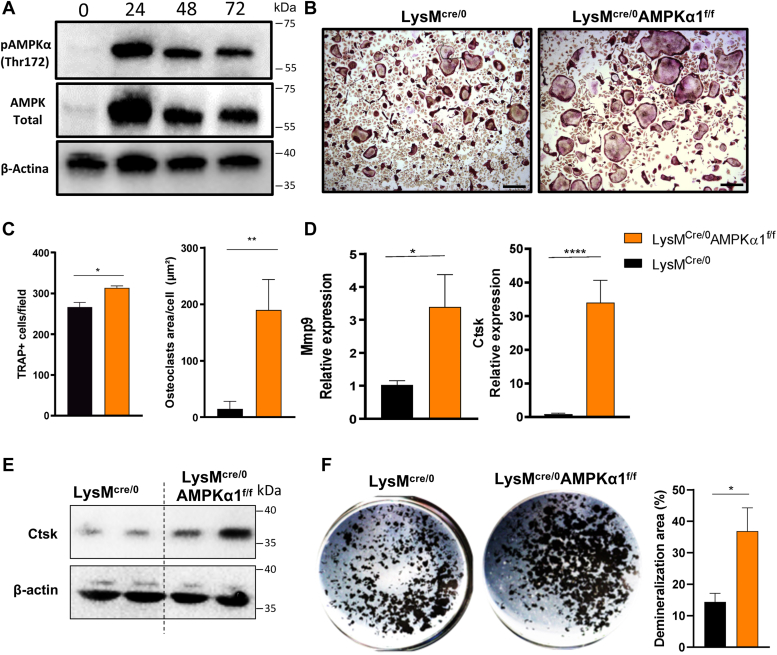
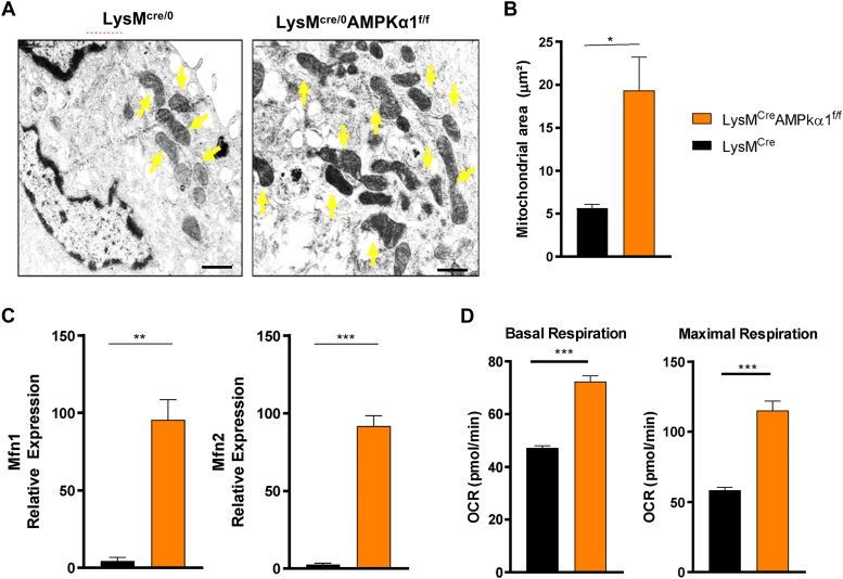
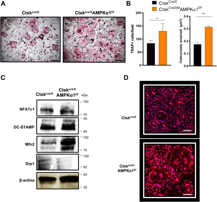
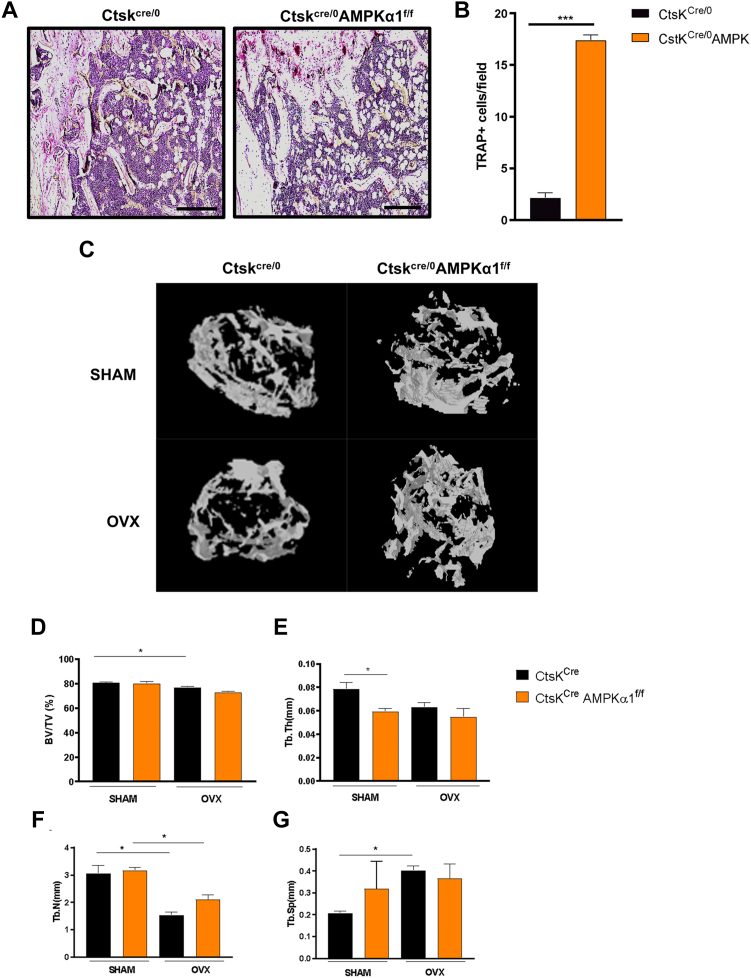
Osteoclasts are specialized cells responsible for bone resorption, a highly energy-demanding process. Focus on osteoclast metabolism could be a key for the treatment of osteolytic diseases including osteoporosis. In this context, AMP-activated protein kinase α1 (AMPKα1), an energy sensor highly expressed in osteoclasts, participates in the metabolic reconfiguration during osteoclast differentiation and activation. This study aimed to elucidate the role of AMPKα1 during osteoclastogenesis in vitro and its impact in bone loss in vivo. Using LysMcre/0AMPK⍺1f/f animals and LysMcre/0 as control, we evaluated how AMPKα1 interferes with osteoclastogenesis and bone resorption activity in vitro. We found that AMPKα1 is highly expressed in the early stages of osteoclastogenesis. Genetic deletion of AMPKα1 leads to increased gene expression of osteoclast differentiation and fusion markers. In addition, LysMcre/0AMPK⍺1f/f mice had an increased number and size of differentiated osteoclast. Accordingly, AMPKα1 negatively regulates bone resorption in vitro, as evidenced by the area of bone resorption in LysMcre/0AMPK⍺1f/f osteoclasts. Our data further demonstrated that AMPKα1 regulates mitochondrial fusion and fission markers upregulating Mfn2 and downregulating DRP1 (dynamics-related protein 1) and that Ctskcre/0AMPK⍺1f/f osteoclasts lead to an increase in the number of mitochondria in AMPK⍺1-deficient osteoclast. In our in vivo study, femurs from Ctskcre/0AMPK⍺1f/f animals exhibited bone loss associated with the increased number of osteoclasts, and there was no difference between Sham and ovariectomized group. Our data suggest that AMPKα1 acts as a negative regulator of osteoclastogenesis, and the depletion of AMPKα1 in osteoclast leads to a bone loss state similar to that observed after ovariectomy.

Keywords: AMPK; metabolism; osteoclast; osteoclastogenesis; osteoporosis.


Conflict of interest statement

Conflict of interest The authors declare that they have no conflicts of interest with the contents of this article.

Figures

Figure 1
Figure 1
AMPKα1 negatively regulates osteoclastogenesis.A, representative image of AMPKα1 and p-AMPKα1 protein expression during osteoclastogenesis, 0, 24, 48, and 72 h after RANKL stimulation. B, representative images of LysMcre/0 and LysMcre/0AMPKα1f/f osteoclasts differentiation by TRAP staining. The scale bar corresponds to 200 μm. C, quantification of osteoclasts number and area in vitro. D, gene expression of osteoclast function markers (MMP-9 and cathepsin K). E, protein expression of cathepsin K. F, demineralization area of LysMcre/0AMPKα1f/f and LysMcre/0 osteoclasts (n = 3, Student's t test ∗p < 0.05, ∗∗p < 0.01, ∗∗∗∗p < 0.0001). All experiments were performed three times. AMPKα1, AMP-activated protein kinase α1; MMP-9, matrix metallopeptidase 9; RANKL, receptor activator of nuclear factor-κB ligand; TRAP, tartrate-resistant acid phosphatase.
Figure 2
Figure 2
AMPKα1 deletion increases mitochondrial fusion and function in osteoclasts.A, representative image of mitochondrias from LysMcre/0 and LysMcre/0AMPKα1f/f differentiated osteoclasts. The scale bar corresponds to 1 μm. B, mitochondrial area in osteoclasts. C, mitofusin 1 and mitofusin 2 gene expressions. D, basal and maximal respiration from LysMcre/0AMPKα1f/f and LysMcre/0 osteoclasts in vitro (n = 5, Student's t test ∗p < 0.05, ∗∗p < 0.01, ∗∗∗p < 0.001). All experiments were performed three times. AMPKα1, AMP-activated protein kinase α1.
Figure 3
Figure 3
AMPKα1 deletion inhibits mitochondrial fission.A, representative images of Ctskcre/0 and Ctskcre/0AMPKα1f/f osteoclast differentiation by TRAP staining. The scale bar corresponds to 200 μm. B, quantification of osteoclast number and area in vitro. C, protein expression of osteoclasts and mitochondrial dynamics markers: NFATc1, DC-STAMP, mitofusin 2, Drp-1, and β-actin as positive control. D, representative image of Ctskcre/0 and Ctskcre/0AMPKα1f/f osteoclasts labeled with MitoTracker Red. The scale bar corresponds to 200 μm. (n = 5, Student's t test ∗p < 0.05 and ∗∗p < 0.01). All experiments were performed two times. AMPKα1, AMP-activated protein kinase α1; Drp1, dynamics-related protein 1; TRAP, tartrate-resistant acid phosphatase.
Figure 4
Figure 4
AMPKα1 deletion leads to increase in osteoclast’s differentiation and function in vivo.A, representative image of TRAP+ cells in a femur section of Ctskcre/0 and Ctskcre/0AMPKα1f/f mice. The scale bar corresponds to 200 μm. B, TRAP+ cells. C, representative image of a computed microtomography of Ctskcre/0 and Ctskcre/0AMPKα1f/f mice under or not under ovariectomy. Trabecular parameters: (D) bone volume/tissue volume, (E) trabecular thickness, (F) trabecular number, (G) trabecular separation (n = 3, Student's t test and ANOVA following by Tukey for multiple comparisons ∗p < 0.05, ∗∗∗p < 0.001). All experiments were performed three times. AMPKα1, AMP-activated protein kinase α1; TRAP, tartrate-resistant acid phosphatase.

References

    1. Armstrong A.P., Tometsko M.E., Glaccum M., Sutherland C.L., Cosman D., Dougall W.C. A RANK/TRAF6-dependent signal transduction pathway is essential for osteoclast cytoskeletal organization and resorptive function. J. Biol. Chem. 2002;277:44347–44356. - PubMed
    1. Kim J.H., Kim N. Regulation of NFATc1 in osteoclast differentiation. J. Bone Metab. 2014;21:233–241. - PMC - PubMed
    1. Lemma S., Sboarina M., Porporato P.E., Zini N., Sonveaux P., Di Pompo G., et al. Energy metabolism in osteoclast formation and activity. Int. J. Biochem. Cell Biol. 2016;79:168–180. - PubMed
    1. Da W., Tao L., Zhu Y. The role of osteoclast energy metabolism in the occurrence and development of osteoporosis. Front. Endocrinol. 2021;12 - PMC - PubMed
    1. Richter E.A., Ruderman N.B. AMPK and the biochemistry of exercise: implications for human health and disease. Biochem. J. 2009;418:261–275. - PMC - PubMed

Publication types