

Case Reports
doi: 10.1503/cmaj.221339.
Stroke in a 36-year-old man with neurosyphilis and HIV, diagnosed using high-resolution vessel wall imaging
Affiliations
- PMID: 37871950
- PMCID: PMC10593191
- DOI: 10.1503/cmaj.221339
Item in Clipboard
Case Reports
Stroke in a 36-year-old man with neurosyphilis and HIV, diagnosed using high-resolution vessel wall imaging
CMAJ.
.
No abstract available
Conflict of interest statement
Competing interests: Tarik Slaoui reports receiving honoraria for presentations for AbbVie and Lundbeck. No other competing interests were declared.
Figures
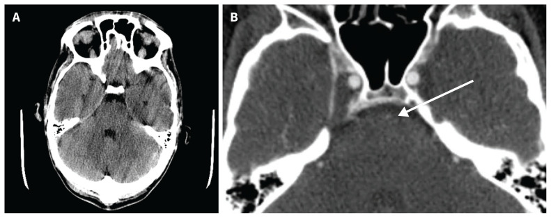
(A) Initial computed tomography (CT) scan of the head of a 36-year-old man, without contrast, showing no evidence of acute bleeding or apparent ischemic changes. (B) Initial head CT angiography showing severe stenosis of the basilar artery (arrow).
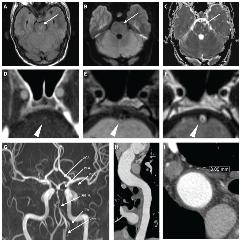
(A–C) Initial MRI scan of the patient’s head, showing (arrows) areas of hyperintensity within the left side of the pons on FLAIR (A) and corresponding areas of hypointensity on diffusion-weighted imaging (B) and hyperintensity on the apparent diffusion coefficient map (C) consistent with chronic infarction (> 3 wk old). (D–F) Vessel wall MRI of the basilar artery using 3D T1 SPACE before gadolinium (E) and after gadolinium injection (F) showing smooth, concentric enhancement of the basilar vessel wall (arrowheads). Sequence parameters: echo time 12 ms, repetition time 700 ms, voxel size 0.8 × 0.8 × 0.9 mm. Material: Siemens 3 T MAGNETOM Skyra MRI scanner, 20-channel head and neck coil. (D) Corresponding CT angiography showing severe stenosis of the basilar artery without evidence of vessel wall plaque or calcification. (G) Coronal MRI maximum intensity projection reconstruction of the intracranial arteries using time-of-flight technique showing multifocal stenosis of the basilar artery and proximal stenosis of the bilateral posterior cerebral arteries. (H–I) Computed tomography angiography of the thorax with reconstruction along the longitudinal axis of the aorta, showing increased diameter of the ascending aorta, measuring 43.1 mm × 38.4 mm (H), and increased thickness of the aortic wall (I) consistent with aortitis. Note: ACA = anterior cerebral artery, Bas. a. = basilar artery, CT = computed tomography, FLAIR = fluid-attenuated inversion recovery, ICA = internal carotid artery, MCA = middle cerebral artery, MRI = magnetic resonance imaging, PCA = posterior cerebral arteries, SPACE = Sampling Perfection with Application-optimized Contrast using different flip angle Evolution, Vert. a. = vertebral artery.
References
-
- Putaala J. Ischemic stroke in young adults. Continuum (Minneap Minn) 2020;26:386–414. - PubMed
-
- Yesilot Barlas N, Putaala J, Waje-Andreassen U, et al. . Etiology of first-ever ischaemic stroke in European young adults: the 15 cities young stroke study. Eur J Neurol 2013;20:1431–9. - PubMed
-
- Carod Artal FJ. Clinical management of infectious cerebral vasculitides. Expert Rev Neurother 2016;16:205–21. - PubMed
Publication types
MeSH terms
LinkOut - more resources
Full Text Sources
Medical