. 2023 Oct 23;14(1):6694.
doi: 10.1038/s41467-023-42478-x.

Acylation of glycerolipids in mycobacteria

Affiliations

Acylation of glycerolipids in mycobacteria

Shiva Kumar Angala et al. Nat Commun. .

Abstract

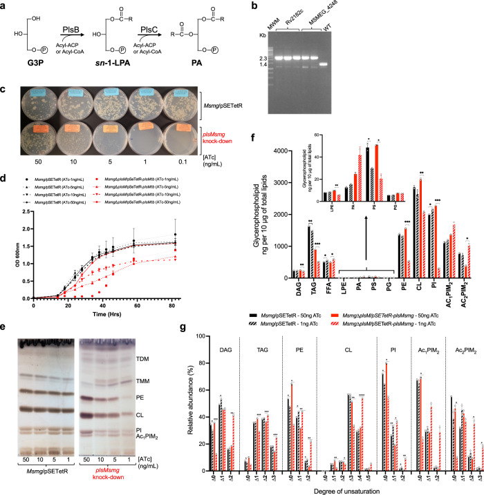
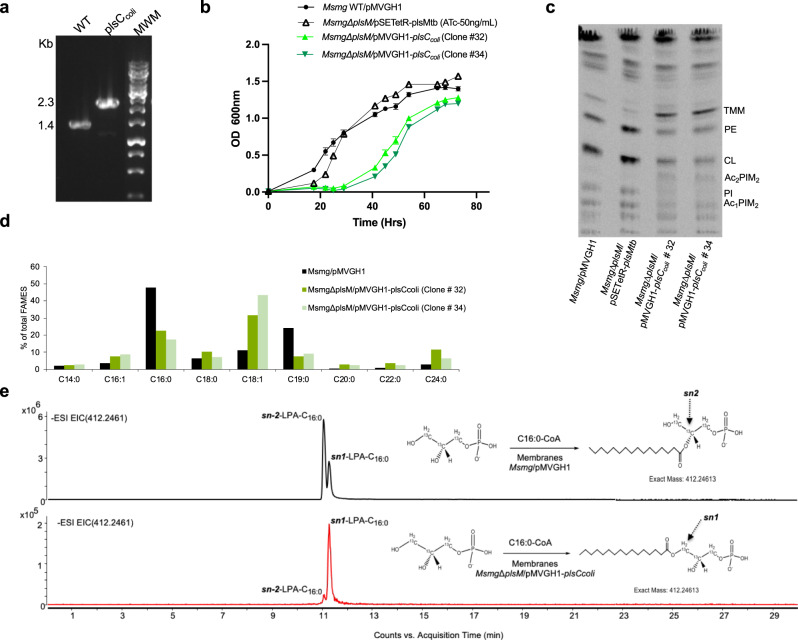
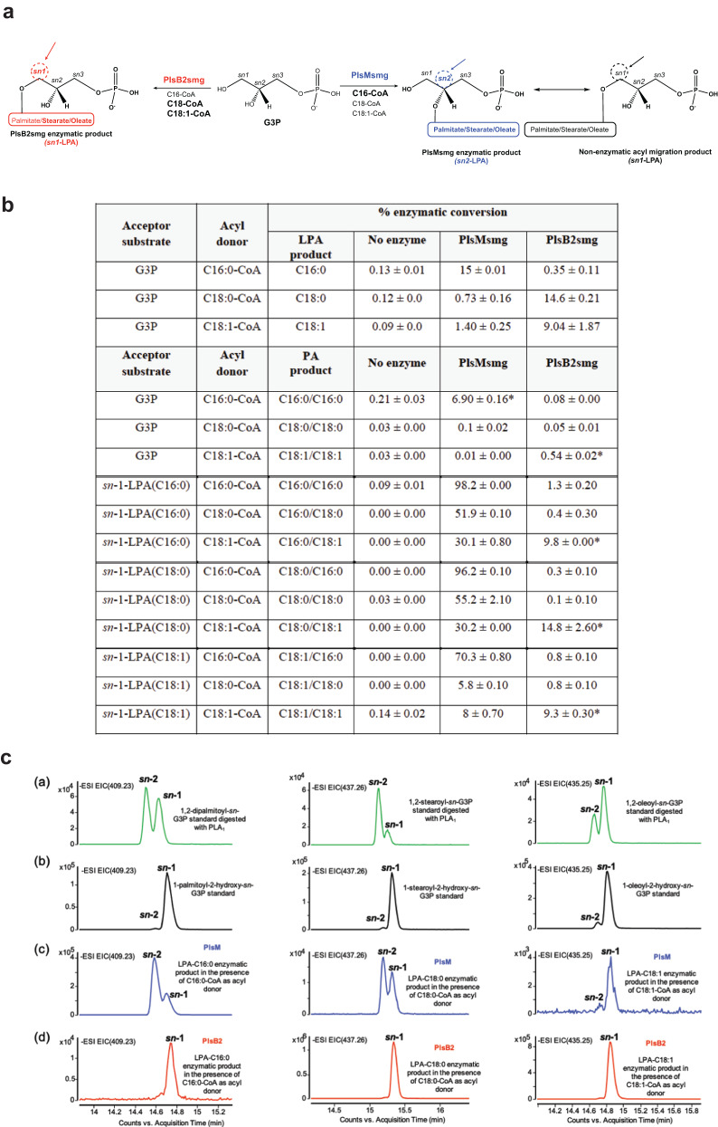
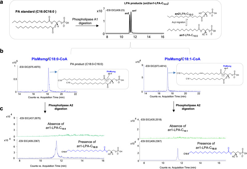
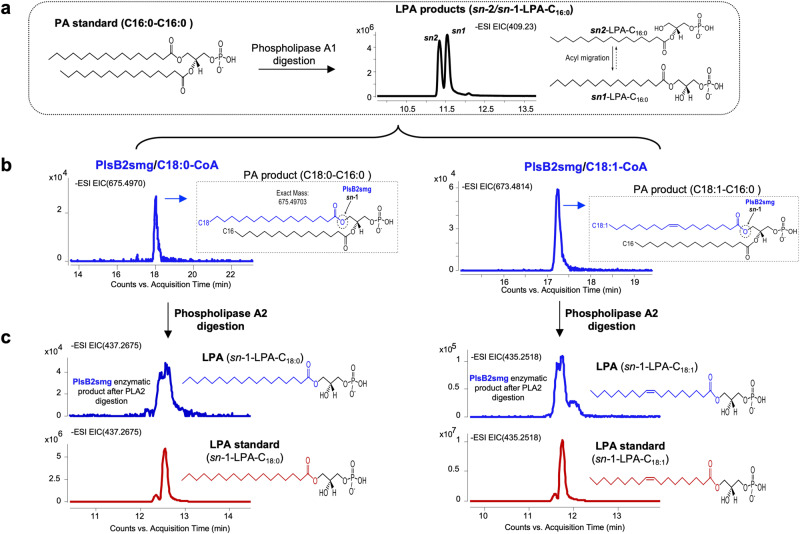
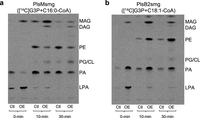
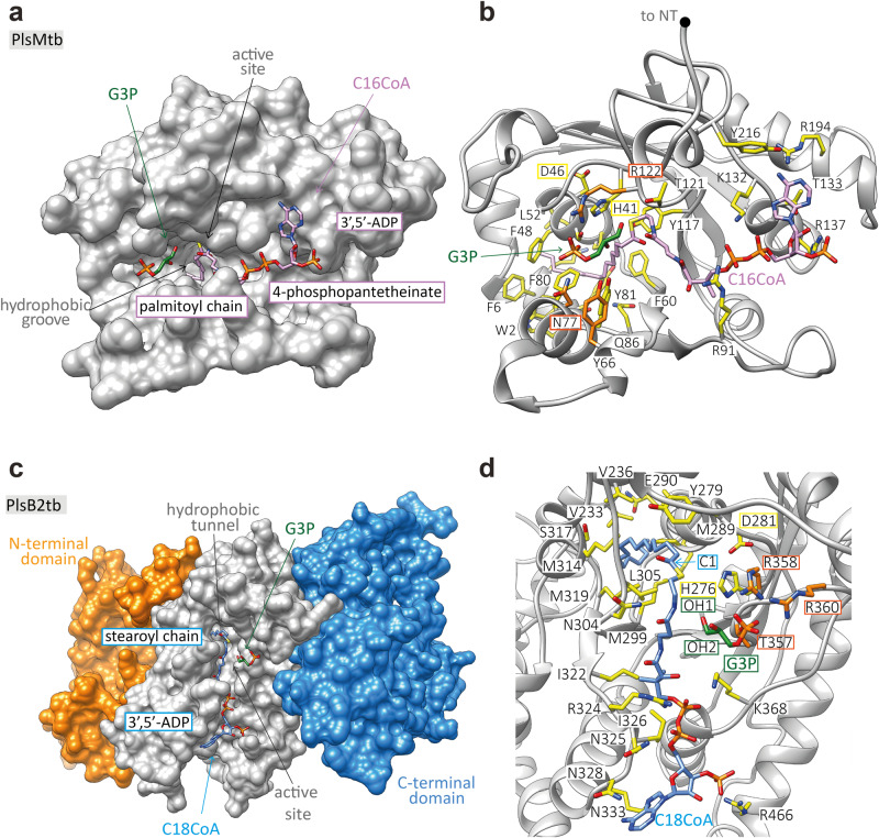
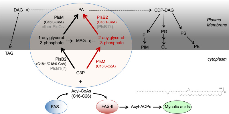
We report on the existence of two phosphatidic acid biosynthetic pathways in mycobacteria, a classical one wherein the acylation of the sn-1 position of glycerol-3-phosphate (G3P) precedes that of sn-2 and another wherein acylations proceed in the reverse order. Two unique acyltransferases, PlsM and PlsB2, participate in both pathways and hold the key to the unusual positional distribution of acyl chains typifying mycobacterial glycerolipids wherein unsaturated substituents principally esterify position sn-1 and palmitoyl principally occupies position sn-2. While PlsM selectively transfers a palmitoyl chain to the sn-2 position of G3P and sn-1-lysophosphatidic acid (LPA), PlsB2 preferentially transfers a stearoyl or oleoyl chain to the sn-1 position of G3P and an oleyl chain to sn-2-LPA. PlsM is the first example of an sn-2 G3P acyltransferase outside the plant kingdom and PlsB2 the first example of a 2-acyl-G3P acyltransferase. Both enzymes are unique in their ability to catalyze acyl transfer to both G3P and LPA.


Conflict of interest statement

The authors declare no competing interests.

Figures

Fig. 1
Fig. 1. plsM silencing in M. smegmatis abolishes phospholipid synthesis and leads to growth arrest.
a The PlsB/PlsC pathway to PA formation in E. coli. The PlsB-catalyzed transfer of a fatty acid to position sn-1 of G3P from acyl-ACP or acyl-CoA precedes the acylation of position sn-2 of sn-1-lysoPA by PlsC. b Allelic replacement at the plsM locus of Msmg mutants rescued with the plsM orthologs of Msmg (MSMEG_4248) and Mtb (Rv2182c) expressed from replicative pSETetR plasmids under control of an ATc-inducible (TET-ON) promoter was confirmed by PCR in two to three independent clones. The WT 1,394-bp amplification signal is replaced by a 2,312-bp fragment in the mutants due to the insertion of a 1.2 kb- kanamycin resistance cassette between the PstI and NruI restriction sites of MSMEG_4248. c Growth of the MsmgΔplsM/pSETetR-plsMtb conditional knock-down (red symbols) and Msmg/pSETetR control strain (black symbols) on 7H11-OADC plates and in, d, 7H9-ADC-tyloxapol at 37 °C in the presence of different concentrations of ATc. Shown in (d) are the means +/- SD of triplicate cultures (n = 3 biologically independent samples). Growth is totally inhibited in liquid culture in the conditional mutant at 1 ng/mL ATc until ATc regulation is lost (~40 h post-inoculation) and the strain starts replicating at a comparable rate to the control strain. e The phospholipid content of Msmg control and MsmgΔplsM/pSETetR-plsMtb cells grown on 7H11-OADC agar plates as shown in (c) were analyzed by TLC in the solvent system CHCl3:CH3OH:H2O (65:25:4 by vol.). The cells were collected on the same day and ~50 μg of total lipids were loaded per lane. f Lipids from Msmg control and MsmgΔplsM/pSETetR-plsMsmg duplicate cultures grown under permissive (50 ng/mL ATc) and non-permissive (1 ng/mL ATc) conditions in 7H9-OADC-tyloxapol at 37 °C to the same OD600 (~0.5–0.6) were quantitatively analyzed by LC/MS as described under Methods and the abundance of glycerolipids in the two strains under both culture conditions is shown as means ± SD of n = 2 biologically independent samples. g Relative abundance (in percentages) of saturated and unsaturated species within each glycerolipid category (DAG, TAG, PE, CL, PI, Ac1PIM2 and Ac2PIM2) in the same Msmg/pSETetR and MsmgΔplsM/pSETetR-plsMsmg strains grown under permissive (50 ng/mL ATc) and non-permissive (1 ng/mL ATc) conditions as in (f). Results are shown as means ± SD of n = 2 biologically independent samples. In panels (f, g) asterisks denote statistically significant differences between culture conditions pursuant to the two-sided unpaired Student’s t-test (f *p < 0.05, **p < 0.005, ***p < 0.0005; g *p < 0.01, **p < 0.001, ***p < 0.0001). The results presented in (cg) are representative of two to three independent experiments. CL cardiolipin, DAG diglycerides, FFA free fatty acids, G3P glycerol-3-phosphate, sn-1-LPA sn-1-lysophosphatidic acid, LPE lysophosphatidylethanolamine, PA phosphatidic acid, PE phosphatidylethanolamine, PG phosphatidylglycerol, PI phosphatidyl-myo-inositol, PS phosphatidylserine, Ac1PIM2 triacylated forms of phosphatidyl-myo-inositol dimannosides, Ac2PIM2 tetraacylated forms of phosphatidyl-myo-inositol dimannosides, TMM trehalose monomycolates, TDM trehalose dimycolates. Source data for panels (d, f, g) are provided as a Source Data file.
Fig. 2
Fig. 2. Effect of replacing PlsM by PlsC from E. coli in Msmg.
a Allelic replacement at the plsM locus of Msmg mutants rescued with plsCcoli was analyzed as in Fig. 1b. b Growth characteristics of WT Msmg mc2155 (black circles), MsmgΔplsM/pSETetR-plsMtb (black triangles), and MsmgΔplsM/pMVGH1-plsCcoli (clones # 32 and 34; light and dark green triangles, respectively) in 7H9-ADC-0.05% tyloxapol at 37 °C (in the presence of 50 ng/mL ATc for MsmgΔplsM/pSETetR-plsMtb). Shown are the means ± SD of three independent growth curves (n = 3 independent biological triplicate) for each strain. c De novo phospholipid synthesis in WT Msmg mc2155, MsmgΔplsM/pMVGH1-plsCcoli (clones # 32 and 34) and MsmgΔplsM/pSETetR-plsMtb grown in 7H9-ADC-0.05% tyloxapol at 37 °C (in the presence of 50 ng/mL ATc for MsmgΔplsM/pSETetR-plsMtb). [1,2-14C]acetic acid was added to bacterial cultures when they reached an OD600 nm of 0.8 after which cultures were incubated for another 8 h at 37 °C with shaking. [1,2-14C]acetic acid-derived lipids were analyzed by TLC in the solvent system CHCl3:CH3OH:H2O (70:20:2 by vol.). The same total counts (dpm) were loaded per lane. Ac2PIM2 are tetraacylated forms of phosphatidyl-myo-inositol dimannosides. Other lipid abbreviations are as in Fig. 1. The results shown are representative of two independent experiments. d Fatty acid composition of WT Msmg mc2155 and MsmgΔplsM/pMVGH1-plsCcoli (two different clones, # 32 and # 34; dark and light green bars, respectively) grown in 7H9-ADC-0.05% tyloxapol at 37 °C (same medium as used in panels b and c). C19:0: tuberculostearic acid. e Extracted ion chromatograms (EICs) showing the lysophosphatidic acid (LPA) products resulting from the incubation of membranes prepared from Msmg/pMVGH1 and MsmgΔplsM/pMVGH1-plsCcoli (clone # 32) with [13C]-G3P and C16:0-CoA. sn-2 palmitoyl transferase activity is dominant in Msmg/pMVGH1 whereas it is barely detectable in MsmgΔplsM/pMVGH1-plsCcoli. C16:0 in the latter strain is essentially transferred to position sn-1 of G3P. Source data for panels (b and d) are provided as a Source Data file.
Fig. 3
Fig. 3. G3P and LPA acyltransferase activity of PlsMsmg and PlsB2smg.
a Schematic representation of PlsMsmg and PlsB2smg acyltransferase activities in the presence of G3P and various acyl-CoA donors. The arrows point to the positions at which acyl groups are transferred by the two enzymes. Preferred acyl donors in each reaction are in bold letters. b Percentage enzymatic conversion of G3P and various 1-acyl-2-hydroxy-sn-G3P (sn-1-LPAs) to their corresponding LPA and PA products by PlsMsmg and PlsB2smg. Peak areas for substrates and enzymatic products were obtained from the integration of extracted ion chromatograms (EICs) and used to calculate percentage substrate conversion. The LPA and PA products reported for the assays that used G3P as the acceptor substrate are from the same reactions. Values for LPA products include both sn-1 and sn-2-LPAs to take into consideration the spontaneous transmigration of acyl chains. Assays were run as described under Methods for 1 h at 37 °C. The results shown are the means ± standard deviations of duplicate assays and are representative of at least two independent experiments using different enzyme preparations. Asterisks denote products resulting from the spontaneous transmigration of acyl chains between positions sn-1 and sn-2 as detailed in the text and Fig. S7. c G3P acyltransferase activity of PlsMsmg and PlsB2smg. (a) EICs showing LPAs generated from the enzymatic digestion of authentic phosphatidic acid (PA) standards (1,2-dipalmitoyl-sn-G3P, 1,2-stearoyl-sn-G3P and 1-palmitoyl-2-oleoyl-sn-G3P) with phospholipase A1 and analyzed by LC/MS. (b) EICs showing authentic standards of sn-1-palmitoyl-sn-2-hydroxy-G3P, sn-1-stearoyl-sn-2-hydroxy-G3P, and sn-1-oleoyl-sn-2-hydroxy-G3P analyzed by LC/MS. (c) EICs of PlsMsmg enzymatic products generated in the presence of G3P as acceptor substrate and C16:0-CoA, C18:0-CoA and C18:1-CoA as acyl donors. (d) EICs of PlsB2smg enzymatic products generated in the presence of G3P as acceptor substrate and C16:0-CoA, C18:0-CoA and C18:1-CoA as acyl donors. Non-radiolabeled PlsMsmg and PlsB2smg enzyme assays were run as described under Methods. The results shown for both enzymes are representative of at least two independent experiments using different enzyme preparations.
Fig. 4
Fig. 4. PlsMsmg does not display 2-acyl-G3P acyltransferase activity.
The products of PlsMsmg acyltransferase reactions using 1-hydroxy-2-palmitoyl-G3P as the acceptor substrate and C18:1-CoA or C18:0-CoA as acyl donors were analyzed by LC/MS. a shows the generation of LPA acceptor substrates from 1,2-dipalmitoyl-sn-G3P upon phospholipase A1 digestion. Both sn-1 and sn-2 LPAs are found as a result of the spontaneous (non-enzymatic) transmigration of C16:0 from position sn-2 to position sn-1. b Incubation of this LPA mixture with PlsMsmg in the presence of C18:1-CoA or C18:0-CoA yields PA products harboring C16:0/C18:0 or C16:0/C18:1 acyl chains. c Digestion of these PA products with phospholipase A2 solely yields sn-1 LPA products harboring a C16:0 chain indicating that PlsMsmg transferred C18:0 or C18:1 to the sn-2 position of 1-palmitoyl-2-hydroxy-G3P rather than to the sn-1 position of 1-hydroxy-2-palmitoyl-G3P.
Fig. 5
Fig. 5. PlsB2smg displays 2-acyl-G3P acyltransferase activity.
The products of PlsB2smg acyltransferase reactions using 1-hydroxy-2-palmitoyl-G3P as the acceptor substrate and C18:1-CoA or C18:0-CoA as acyl donors were analyzed by LC/MS. a The generation of sn-1 and sn-2 LPA acceptors from 1,2-dipalmitoyl-sn-G3P is as described in Fig. 4A. b Incubation of this LPA mixture with PlsB2smg in the presence of C18:1-CoA or C18:0-CoA yields PA products harboring C16:0/C18:0 or C16:0/C18:1 acyl chains. c Digestion of these PA products with phospholipase A2 yields sn-1 LPAs with C18:0 or C18:1 chains confirming that the PlsB2smg-mediated acyl transfer occurred at position sn-1 of 1-hydroxy-2-palmitoyl-G3P. sn-1 LPAs comigrate with authentic sn-1-LPA standards (red traces).
Fig. 6
Fig. 6. Effect of overexpressing plsMsmg and plsB2smg on glycerolipid synthesis by E. coli membranes.
a Phospholipid synthesis by E. coli membranes prepared from control cells (Ctl; harboring an empty pET28a plasmid) and plsMsmg overexpressing cells (OE). Reaction mixtures were as described under Methods with [14C(U)]G3P as the radiolabeled acceptor substrate and C16:0-CoA as the acyl donor. At the indicated time points, reactions were terminated and the products analyzed by TLC in the solvent system CHCl3:CH3OH:H2O (65:25:4 by vol.). b Phospholipid synthesis by E. coli membranes prepared from control cells (Ctl; harboring an empty pET14b plasmid) and plsB2smg overexpressing cells (OE). Reaction mixtures were as described under Methods with [14C(U)]G3P as the radiolabeled acceptor substrate and C18:1-CoA as the acyl donor. At the indicated time points, reactions were terminated and the products analyzed as described above. The results shown are representative of two (PlsB2) to three (PlsM) independent experiments. LPA, lysophosphatidic acid; PA, phosphatidic acid; CL, cardiolipin; PG, phosphatidylglycerol; PE, phosphatidylethanolamine; MAG, monoacylglycerol; DAG, diacylglycerol. Source data are provided as a Source Data file.
Fig. 7
Fig. 7. A model for PlsMtb and PlsB2tb substrates recognition.
a Surface representation of the predicted PlsMtb 3D structure, showing the palmitoyl moiety of C16:0-CoA deeply buried into a hydrophobic groove. This hydrophobic pocket runs perpendicular with respect to a main groove where the CoA moiety is located. The 4-phosphopantetheinate moiety of C16:0-CoA is placed in the hydrophobic groove entrance, nearby the catalytic site, with the adenosine 3´,5´-ADP moiety extended along the protein surface. The G3P acceptor substrate is positioned in the opposite site of the CoA group, in a binding pocket mainly decorated by polar residues. b The palmitoyl moiety of C16:0-CoA makes interactions with W2, H41, S47, F48, P51, L52, F60, F80, Y81, S84, Q86 along the groove’s walls and with F6, K7 and Y3 in the bottom, supporting PlsMtb’s specificity for C16-length donor substrates. The 4-phosphopantetheinate moiety of C16:0-CoA is placed nearby the catalytic residues H41 and D46 and stabilized by Y117 with the adenosine 3´,5´-ADP moiety making interactions with residues R91, T121, R137, T133, R194, Y216 and K132. The G3P is stabilized by the R122, Y66 and N77 residues. c Surface representation of the predicted PlsB2tb 3D structure, showing the stearoyl moiety of C18:0-CoA deeply buried into a hydrophobic tunnel of the acyltransferase domain. This hydrophobic tunnel runs perpendicular with respect to a main groove where the CoA moiety is located. The 4-phosphopantetheinate moiety of C18:0-CoA is placed in the hydrophobic groove entrance, nearby the catalytic site, with the adenosine 3´,5´-ADP moiety extended along the protein surface. The G3P acceptor substrate is positioned in the opposite site of the CoA group, in a binding pocket mainly decorated by polar residues. d The stearoyl moiety of C18:0-CoA makes interactions with E290, M289, V233, P286, S317, M314, M319, M299, V236, V283 and Y279 along the groove’s walls. The 4-phosphopantetheinate moiety of C18:0-CoA is placed nearby the catalytic residues H276 and D281 and further stabilized by L305, N304 and I322, with the adenosine 3´,5´-ADP moiety making interactions with residues N325, I326, K368, N328, K333, R466 and R324. G3P is stabilized by the R358, R360 and T357 residues.
Fig. 8
Fig. 8. Proposed pathway for glycerolipid synthesis in mycobacteria.
The proposed steps leading to the biosynthesis of phosphatidic acid (PA) and derived glycerolipids in Mtb based on earlier biochemical studies and the present work are shown. The acyl-CoA products of FAS-I serve as the acyl donors in the biosynthesis of PA from glycerol-3-phosphate (G3P). The initial substrates of FAS-II are medium length (C16–C26) keto-acyl-ACP resulting from the condensation of the acyl-CoA products of FAS-I with malonyl-ACP. The processive addition of multiple malonate units to these precursors leads to the elongation of the meromycolate chain (C48–C54) of mycolic acids. The prototypical structure of an Mtb alpha-mycolate is shown. Glycerophospholipids and di- and tri-acylglycerol (DAG and TAG) arise from the central intermediate, PA. The catalytic activity of the enzymes in gray font has not been confirmed. “Other PlsCs” refers, in particular, to Mtb PlsC homologs Rv2483c and Rv3816c which are conserved in other Mycobacterium species, including M. leprae [Table S1]. Other putative PlsC candidates otherwise include Rv3814c, Rv3815c and Rv3026c in Mtb. PS, phosphatidylserine; PI, phosphatidyl-myo-inositol; PIM, phosphatidylinositol mannosides; TAG, triglycerides. Other lipid abbreviations are as in Fig. 6.

References

    1. Bansal-Mutalik R, Nikaido H. Mycobacterial outer membrane is a lipid bilayer and the inner membrane is unusually rich in diacyl phosphatidylinositol dimannosides. Proc. Natl Acad. Sci. USA. 2014;111:4958–4963. doi: 10.1073/pnas.1403078111. - DOI - PMC - PubMed
    1. Daffé M, Crick DC, Jackson M. Genetics of capsular polysaccharides and cell envelope (glyco)lipids. Microbiol Spectr. 2014;2:MGM2-0021-2013. doi: 10.1128/microbiolspec.MGM2-0021-2013. - DOI - PubMed
    1. Okuyama H, Kankura T, Nojima S. Positional distribution of fatty acids in phospholipids from Mycobacteria. J. Biochem. 1967;61:732–737. doi: 10.1093/oxfordjournals.jbchem.a128607. - DOI - PubMed
    1. Goren, M. B. in The mycobacteria. A sourcebook, 1st edn, Vol. 1 (eds. Kubica, G. P. & Wayne, L. G.) 379–415 (Marcel Dekker, 1984).
    1. Zhang YM, Rock CO. Membrane lipid homeostasis in bacteria. Nat. Rev. Microbiol. 2008;6:222–233. doi: 10.1038/nrmicro1839. - DOI - PubMed

Publication types

MeSH terms

Substances

LinkOut - more resources