. 2023 Oct 23;11(1):53.
doi: 10.1038/s41413-023-00289-2.

HuR-mediated nucleocytoplasmic translocation of HOTAIR relieves its inhibition of osteogenic differentiation and promotes bone formation

Affiliations

HuR-mediated nucleocytoplasmic translocation of HOTAIR relieves its inhibition of osteogenic differentiation and promotes bone formation

Yuheng Li et al. Bone Res. .

Abstract

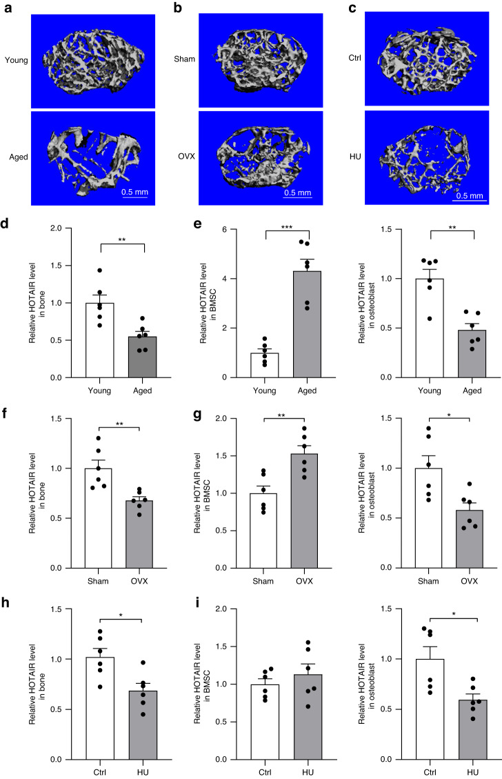
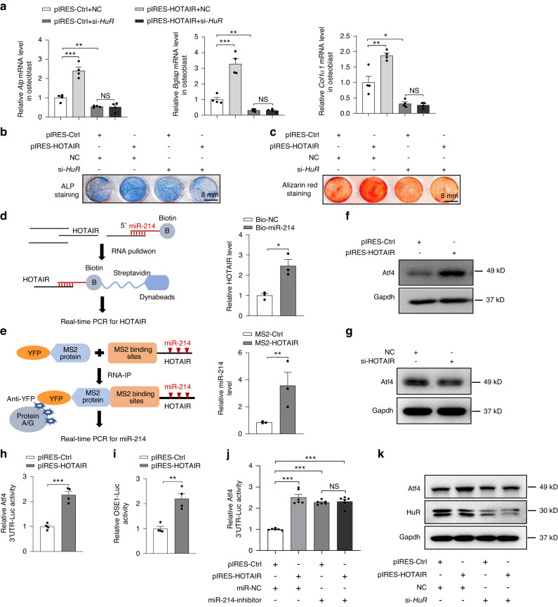
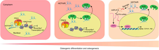
Bone marrow mesenchymal stem cell (BMSC) osteogenic differentiation and osteoblast function play critical roles in bone formation, which is a highly regulated process. Long noncoding RNAs (lncRNAs) perform diverse functions in a variety of biological processes, including BMSC osteogenic differentiation. Although several studies have reported that HOX transcript antisense RNA (HOTAIR) is involved in BMSC osteogenic differentiation, its effect on bone formation in vivo remains unclear. Here, by constructing transgenic mice with BMSC (Prx1-HOTAIR)- and osteoblast (Bglap-HOTAIR)-specific overexpression of HOTAIR, we found that Prx1-HOTAIR and Bglap-HOTAIR transgenic mice show different bone phenotypes in vivo. Specifically, Prx1-HOTAIR mice showed delayed bone formation, while Bglap-HOTAIR mice showed increased bone formation. HOTAIR inhibits BMSC osteogenic differentiation but promotes osteoblast function in vitro. Furthermore, we identified that HOTAIR is mainly located in the nucleus of BMSCs and in the cytoplasm of osteoblasts. HOTAIR displays a nucleocytoplasmic translocation pattern during BMSC osteogenic differentiation. We first identified that the RNA-binding protein human antigen R (HuR) is responsible for HOTAIR nucleocytoplasmic translocation. HOTAIR is essential for osteoblast function, and cytoplasmic HOTAIR binds to miR-214 and acts as a ceRNA to increase Atf4 protein levels and osteoblast function. Bglap-HOTAIR mice, but not Prx1-HOTAIR mice, showed alleviation of bone loss induced by unloading. This study reveals the importance of temporal and spatial regulation of HOTAIR in BMSC osteogenic differentiation and bone formation, which provides new insights into precise regulation as a target for bone loss.


Conflict of interest statement

The authors declare no competing interests.

Figures

Fig. 1
Fig. 1
Distinct expression of HOTAIR in BMSCs and osteoblasts. a Representative image showing three-dimensional trabecular architecture by micro-CT reconstruction at the distal femurs from young mice (2 months) and aged mice (24 months) (n = 6 for each group). Scale bar, 0.5 mm. b Representative images showing three-dimensional trabecular architecture by micro-CT reconstruction at the distal femurs from sham- or ovariectomy (OVX)-operated mice for 6 months (n = 6 for each group). Scale bar, 0.5 mm. c Representative images showing three-dimensional trabecular architecture by micro-CT reconstruction at the distal femurs from mice that were hindlimb unloaded (HU) or control (Ctrl) treated for 28 days (n = 6 for each group). Scale bar, 0.5 mm. d qRT‒PCR analysis of HOTAIR levels in bone tissue from young and aged mice (n = 6 for each group). e qRT‒PCR analysis of HOTAIR levels in Sca-1+CD29+CD45CD11b bone marrow mesenchymal stem cells (BMSCs) and ALP+ osteoblasts separated by cell sorting with fluorescence-activated cell sorting (FACS) from young and aged mice (n = 6 for each group). f qRT‒PCR analysis of HOTAIR levels in bone tissue from mice that underwent OVX or sham operation (n = 6 for each group). g qRT‒PCR analysis of HOTAIR levels in bone marrow mesenchymal stem cells (BMSCs) and osteoblasts separated by cell sorting with FACS from OVX and sham mice (n = 6 for each group). h qRT‒PCR analysis of HOTAIR levels in bone tissue from mice treated with hindlimb unloading (HU) or control (Ctrl) treatment (n = 6 for each group). i qRT‒PCR analysis of HOTAIR levels in bone marrow mesenchymal stem cells (BMSCs) and osteoblasts separated by cell sorting with FACS from Ctrl and HU mice (n = 6 for each group). All data are the mean ± s.e.m. *P < 0.05, **P < 0.01. *** P < 0.001. NS, no significance
Fig. 2
Fig. 2
HOTAIR overexpression in MSCs delays bone development. a, b Alcian blue and Alizarin red staining of the whole skeletons of WT or Prx1-HOTAIR TG mice at 5 days old. Scale bar, 1 cm. c Representative images showing three-dimensional trabecular architecture by micro-CT reconstruction from WT and Prx1-HOTAIR TG male mice at 1 month old. Scale bar, 0.5 mm. d Representative images showing the three-dimensional trabecular architecture and cortical architecture as shown by micro-CT reconstruction at the distal femurs from WT and Prx1-HOTAIR TG mice at 1 month old. Scale bar, 0.5 mm. e Micro-CT measurements of BV/TV, BMD, Tb.N, Tb.Th, Tb.Sp, and Cort.Th in the distal femurs of mice. n = 10 for each group. BV/TV, ratio of bone volume to tissue volume; BMD, bone mineral density; Tb.N, trabecular number; Tb.Th, trabecular thickness; Tb.Sp, trabecular separation; Cort.Th, cortical bone thickness. f Representative images showing new bone formation assessed by double calcein labeling in WT and Prx1-HOTAIR TG mice. Scale bar, 20 μm. g Quantification of mineral apposition rate (MAR) and osteoblast number to bone surface (N.ob/BS) (n = 6 for each group). h The maximal (max.) load at failure determined by three-point bending of femurs from WT (n = 10) and Prx1-HOTAIR TG (n = 10) mice. i Histological images for Ocn staining of the proximal tibia from WT and Prx1-HOTAIR TG mice. Scale bar, 100 μm. j ELISA analysis of the PINP protein level in the serum from WT (n = 10) and Prx1-HOTAIR TG (n = 10) mice. k qRT-PCR analysis of Alp, Runx2 and Sp7 mRNA levels in bone tissues collected from WT (n = 10) and Prx1-HOTAIR TG (n = 10) mice. Two-tailed unpaired Student’s t test was used for statistical evaluations of two group comparisons. All data are the mean ± s.e.m. *P < 0.05, **P < 0.01, ***P < 0.001
Fig. 3
Fig. 3
HOTAIR overexpression in osteoblasts increases bone formation. a Schematic representation of the transgenic construct used to generate osteoblast-specific HOTAIR overexpression transgenic mouse lines. b qRT‒PCR analysis of HOTAIR levels in bone and other tissues from 6-month-old WT and osteoblast-specific HOTAIR-overexpressing mice (Bglap-HOTAIR TG) (n = 6). c Representative images showing three-dimensional trabecular architecture by micro-CT reconstruction at 6 months of age. Scale bar, 0.5 mm. d Representative images showing the three-dimensional trabecular architecture and cortical architecture as shown by micro-CT reconstruction at the distal femurs at 6 months of age. Scale bar, 0.5 mm. e Micro-CT measurements of BV/TV, BMD, Tb.N, Tb.Th, Tb.Sp, and Cort.Th in the distal femurs of mice (n = 10 for each group). f Representative images showing new bone formation assessed by double calcein labeling. Scale bar, 20 μm. g Quantification of mineral apposition rate (MAR) and osteoblast number to bone surface (N.ob/BS) (n = 6 for each group). h The maximal (max.) load at failure determined by three-point bending of femurs from WT (n = 10) and Ocn-HOTAIR TG (n = 10) mice. i Histological images for Ocn staining of the proximal tibia. Scale bar, 100 μm. j ELISA analysis of the PINP protein level in the serum from WT (n = 10) and Bglap-HOTAIR TG (n = 10) mice. k qRT-PCR analysis of Alp, Bglap and Col1a1 mRNA levels in bone tissues collected from WT (n = 10) and Bglap-HOTAIR TG (n = 10) mice. Two-tailed unpaired Student’s t test was used for statistical evaluations of two group comparisons. All data are the mean ± s.e.m. *P < 0.05, **P < 0.01, ***P < 0.001
Fig. 4
Fig. 4
The expression and function of HOTAIR in BMSCs and osteoblasts. a qRT‒PCR analysis of HOTAIR levels in BMSCs isolated from WT and Prx-HOTAIR TG mice. b qRT‒PCR analysis of Sp7, Runx2, and Alp mRNA levels in BMSCs isolated from WT and Prx-HOTAIR TG mice. c Representative images of ALP staining (top) in BMSCs cultured with osteogenic medium for 3 days and Alizarin red staining (bottom) in BMSCs cultured with osteogenic medium for 21 days. Scale bar, 8 mm. d qRT‒PCR analysis of HOTAIR levels in BMSCs cultured with osteogenic medium for 3 days. e qRT‒PCR analysis of Sp7, Runx2, and Alp mRNA levels in BMSCs cultured with osteogenic medium for 3 days. f Representative images of ALP staining in BMSCs cultured with osteogenic medium for 3 days and Alizarin red staining (below) in BMSCs cultured with osteogenic medium for 21 days. Scale bar, 8 mm. g qRT‒PCR analysis of HOTAIR levels in primary osteoblasts isolated from WT and Bglap-HOTAIR TG mice and cultured with osteogenic medium for 3 days. h qRT‒PCR analysis of Alp, Bglap, and Col1a1 mRNA levels in primary osteoblasts cultured with osteogenic medium for 3 days. i Representative images of ALP staining (top) in primary osteoblasts cultured with osteogenic medium for 3 days. Alizarin red staining (below) in primary osteoblasts cultured with osteogenic medium for 14 days. j qRT‒PCR analysis of HOTAIR levels in primary osteoblasts cultured with osteogenic medium for 3 days. k qRT‒PCR analysis of Alp, Bglap, and Col1a1 mRNA levels in primary osteoblasts cultured with osteogenic medium for 3 days. l Representative images of ALP staining (top) in primary osteoblasts cultured with osteogenic medium for 3 days. Alizarin red staining (below) in primary osteoblasts cultured with osteogenic medium for 14 days. Scale bar, 8 mm. All data are the mean ± s.e.m. from three independent experiments. Two-tailed unpaired Student’s t test was used for statistical evaluations of two group comparisons. *P < 0.05, **P < 0.01, ***P < 0.001
Fig. 5
Fig. 5
HuR is essential for the nucleocytoplasmic translocation of HOTAIR. a RNA-fluorescent in situ hybridization (FISH) was conducted to detect HOTAIR localization using digoxigenin (DIG) labeling probes specific for HOTAIR sequences (red). Nuclei were stained with Hoechst (blue). Scale bar, 20 μm. b Detection of U6 and 18 S RNA using RNA probes labeled with Cy3 (red). Nuclei were stained with Hoechst (blue). Scale bar, 20 μm. c RNA FISH to analyze the HOTAIR localization in BMSCs cultured with osteogenic medium for 0 days and 3 days. Scale bar, 20 μm. d KEGG analysis of the HOTAIR binding proteins in BMSCs through biotin-labeled full-length HOTAIR or biotin-labeled control RNA-based RNA pulldown by mass spectrometry. e Western blot analysis of HOTAIR binding protein HuR in BMSCs by biotinylated RNA pulldown. f qRT‒PCR analysis of HOTAIR levels by RNA immunoprecipitation in BMSCs with a HuR antibody, and rabbit IgG was used as a control. g Western blot analysis of HuR protein levels in BMSCs cultured with osteogenic medium for 0 days and 3 days. h Structured illumination microscopy (SIM) images for the colocalization of HuR and HOTAIR in BMSCs cultured with osteogenic medium for 0 days and 3 days. FISH was conducted to detect HOTAIR using DIG labeling probes specific for HOTAIR sequences and TRITC secondary antibody (red). Immunofluorescence was performed to detect HuR using a HuR antibody and FITC secondary antibody (green). Scale bar, 10 μm. i The distribution of HOTAIR in BMSCs cultured with osteogenic medium for 0 days and 3 days. Scale bar, 10 μm. All data are the mean ± s.e.m. from three independent experiments. Two-tailed unpaired Student’s t test was used for statistical evaluations of two group comparisons. **P < 0.01
Fig. 6
Fig. 6
HuR mediated the effect of HOTAIR on osteoblast function by sponging miR-214 in the cytoplasm. a qRT‒PCR analysis of Alp, Bglap, and Col1a1 mRNA levels in primary osteoblasts transfected with pIRES-HOTAIR and pIRES-Ctrl with or without HuR knockdown and cultured with osteogenic medium for 3 days. b, c Representative images of ALP staining (b) in primary osteoblasts cultured with osteogenic medium for 3 days and Alizarin red staining (c) in primary osteoblasts cultured with osteogenic medium for 14 days. Scale bar, 8 mm. d Analysis of the interaction of HOTAIR and miR-214 by biotin-labeled miR-214 probe-based RNA pulldown assays. Left, schematic diagram of the miR-214-based RNA pulldown assay. Right, qRT‒PCR was performed with RIP samples from osteoblasts using biotin-labeled miR-214 probes or biotin-labeled negative control (NC). e Analysis of the interaction of HOTAIR and miR-214 by MS2-based pulldown assay. Left, schematic diagram of the MS2-based RNA immunoprecipitation (RIP) assay. Right, qRT‒PCR was performed with RIP samples from osteoblasts transfected with pcDNA3.1-12 x MS2 (control vector), pcDNA3.1-HOTAIR-12xMS2, or YFP-MS2. f Western blot analysis of Atf4 protein levels in osteoblasts transfected with pIRES-HOTAIR or pIRES-Ctrl. g Western blot analysis of Atf4 protein levels in osteoblasts transfected with si-HOTAIR and NC. h The luciferase activity of Atf4 3’UTR in osteoblasts transfected with pIRES-HOTAIR or pIRES-Ctrl. i The luciferase activity of OSE1 in osteoblasts transfected with pIRES-HOTAIR or pIRES-Ctrl. j The luciferase activity of Atf4 3’UTR in osteoblasts transfected with pIRES-HOTAIR or pIRES-Ctrl with or without miR-214 inhibition. k Western blot analysis of Atf4 and HuR protein levels in primary osteoblasts transfected with pIRES-HOTAIR and pIRES-Ctrl with or without HuR knockdown and cultured with osteogenic medium for 3 days. All data are the mean ± s.e.m. from three independent experiments. Statistical analysis with more than two groups was performed with one-way analysis of variance (ANOVA) with Tukey’s multiple comparison test to determine group differences. *P < 0.05, **P < 0.01, ***P < 0.001. NS, not significant
Fig. 7
Fig. 7
Osteoblast-specific overexpression of HOTAIR alleviates unloading-induced bone loss. a Representative image showing three-dimensional distal femur trabecular architecture by micro-CT reconstruction from the indicated groups of mice. Representative images of six independent tissues in each group. Ctrl, control group, HU, hindlimb unloading group. Scale bar, 0.5 mm. b Micro-CT measurements for BV/TV, BMD, Tb.N and Tb.Sp at the distal femurs. n = 10 for each group. c Representative images of Ocn staining of the proximal tibia. Scale bar, 100 μm. d ELISA analysis of PINP protein levels in the serum. n = 10 for each group. e qRT-PCR analysis of Alp, Bglap, and Col1a1 mRNA levels in bone tissues. n = 10 for each group. f, g qRT-PCR analysis of HOTAIR and miR-214 levels in bone tissues. n = 10 for each group. h Western blot analysis of Atf4 protein levels in bone tissue. All data are the mean ± s.e.m. Statistical analysis with more than two groups was performed with two-way analysis of variance (ANOVA) with the Šídák post hoc test to determine group differences. All data are the mean ± s.e.m. *P < 0.05, **P < 0.01, ***P < 0.001
Fig. 8
Fig. 8
Model of the distinct function of HOTAIR in BMSC osteogenic differentiation and osteogenesis. In BMSCs, nuclear HOTAIR interacts with EZH2 and enhances the modification of H3K27me3 in the promoter regions of Runx2 and Sp7 genes, which leads to the inhibition of osteogenic differentiation of BMSCs. In osteoblasts, HOTAIR is located in the cytoplasm to sponge miR-214 and promotes osteoblast function by alleviating Atf4 translation. Moreover, HuR mediates the nucleocytoplasmic translocation of HOTAIR in the progression of osteogenic differentiation of BMSCs

References

    1. Gao Q, et al. Bone marrow mesenchymal stromal cells: identification, classification, and differentiation. Front. Cell Dev. Biol. 2021;9:787118. - PMC - PubMed
    1. Salhotra A, Shah HN, Levi B, Longaker MT. Mechanisms of bone development and repair. Nat. Rev. Mol. Cell Biol. 2020;21:696–711. - PMC - PubMed
    1. Long F. Building strong bones: molecular regulation of the osteoblast lineage. Nat. Rev. Mol. Cell Biol. 2011;13:27–38. - PubMed
    1. Franceschi RT, Ge C, Xiao G, Roca H, Jiang D. Transcriptional regulation of osteoblasts. Ann. N. Y. Acad. Sci. 2007;1116:196–207. - PubMed
    1. Quinn JJ, Chang HY. Unique features of long non-coding RNA biogenesis and function. Nat. Rev. Genet. 2016;17:47–62. - PubMed

Publication types