. 2023 Oct 23;13(1):18119.
doi: 10.1038/s41598-023-43567-z.

Chronic dehydration induces injury pathways in rats, but does not mimic histopathology of chronic interstitial nephritis in agricultural communities

Affiliations

Chronic dehydration induces injury pathways in rats, but does not mimic histopathology of chronic interstitial nephritis in agricultural communities

Gerd Schreurs et al. Sci Rep. .

Abstract

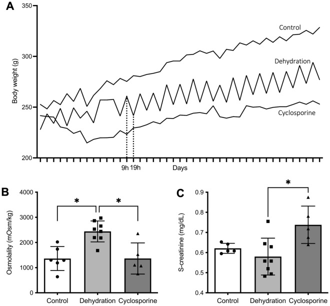
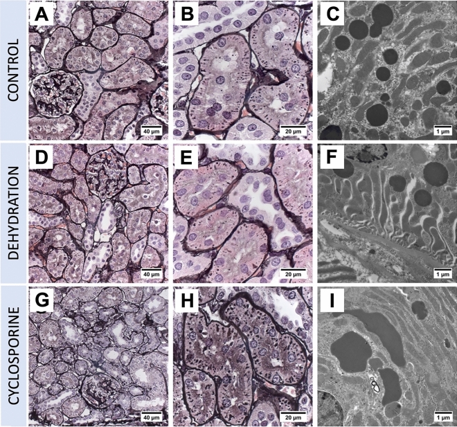
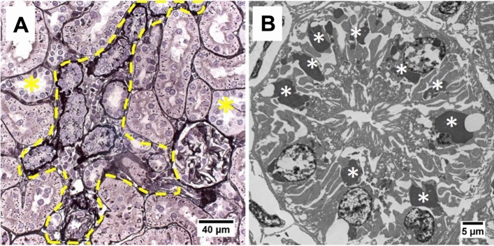
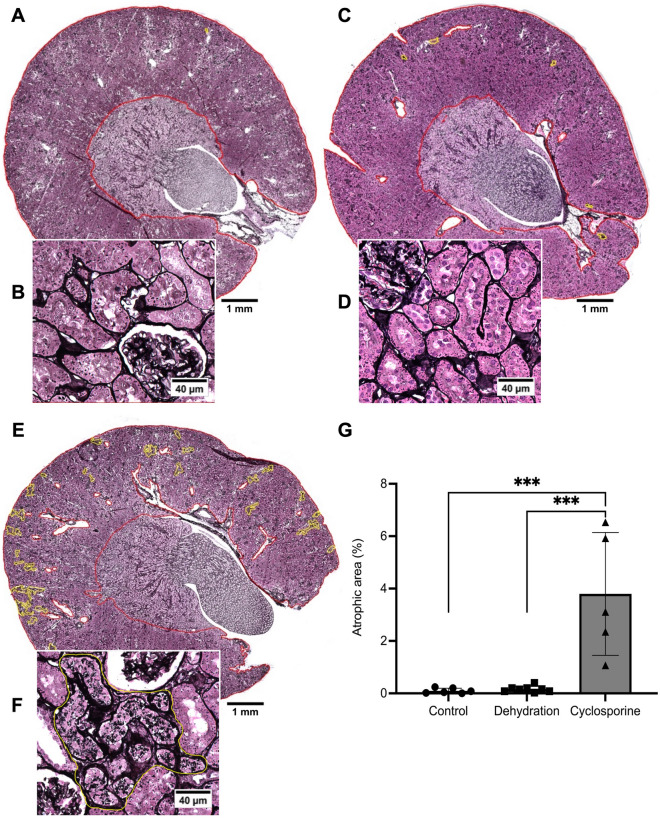
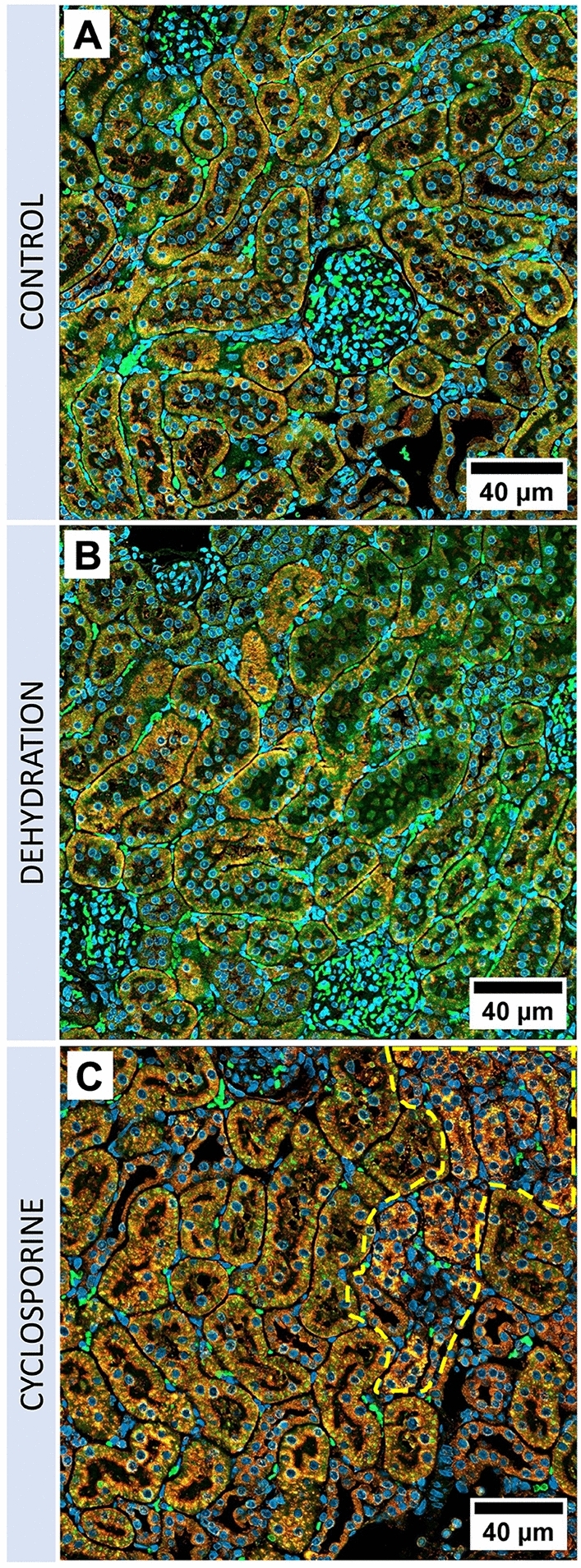
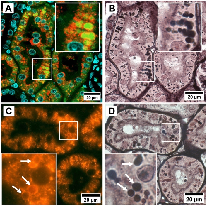
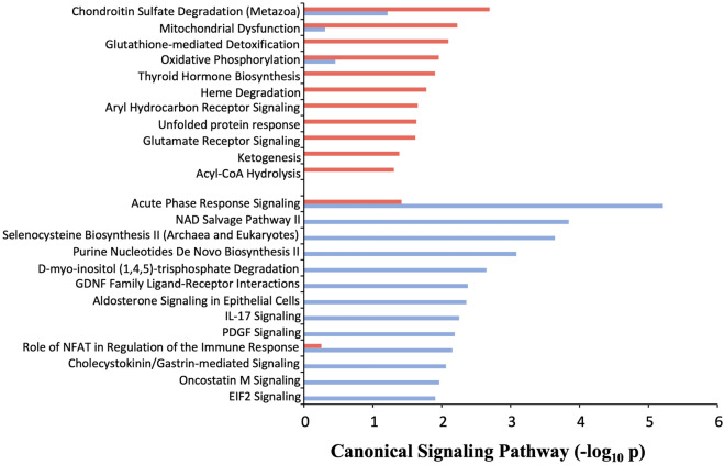
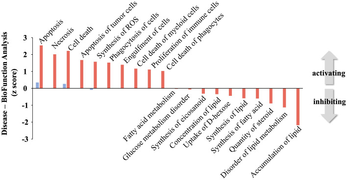
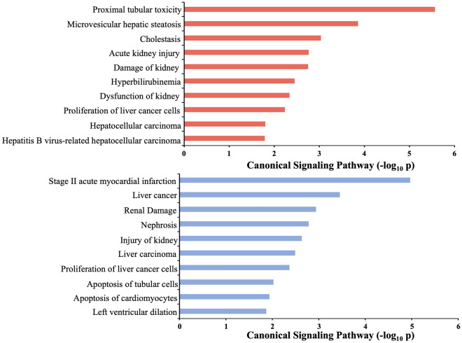
CINAC-patients present renal proximal tubular cell lysosomal lesions which are also observed in patients experiencing calcineurin inhibitor (CNI) nephrotoxicity, suggesting that CINAC is a toxin-induced nephropathy. An alternative hypothesis advocates chronic dehydration as a major etiological factor for CINAC. Here, we evaluated histological and molecular changes in dehydrated versus toxin exposed rats. Wistar rats were divided in 3 groups. Group 1 (n = 6) had free access to drinking water (control group). Group 2 (n = 8) was water deprived for 10 h per 24 h, 5 days/week and placed in an incubator (37 °C) for 30 min/h during water deprivation. Group 3 (n = 8) underwent daily oral gavage with cyclosporine (40 mg/kg body weight). After 28 days, renal function, histopathology and proteomic signatures were analysed. Cyclosporine-treated rats developed focal regions of atrophic proximal tubules with associated tubulo-interstitial fibrosis. PASM staining revealed enlarged argyrophilic granules in affected proximal tubules, identified as lysosomes by immunofluorescent staining. Electron microscopy confirmed the enlarged and dysmorphic phenotype of the lysosomes. Overall, these kidney lesions resemble those that have been previously documented in farmers with CINAC. Dehydration resulted in none of the above histopathological features. Proteomic analysis revealed that dehydration and cyclosporine both induce injury pathways, yet of a clear distinct nature with a signature of toxicity only for the cyclosporine group. In conclusion, both cyclosporine and dehydration are injurious to the kidney. However, dehydration alone does not result in kidney histopathology as observed in CINAC patients, whereas cyclosporine administration does. The histopathological analogy between CINAC and calcineurin inhibitor nephrotoxicity in rats and humans supports the involvement of an as-yet-unidentified environmental toxin in CINAC etiology.


Conflict of interest statement

The authors declare no competing interests.

Figures

Figure 1
Figure 1
Clinical parameters. (A) Body weights were measured daily at the start (09h00) and end (19h00) of the water retention period. The daily recurrent drop reflects the dehydration vs. rehydration periods (marked with dotted line). (B) Osmolality measurements of urine of dehydrated animals, collected after 10 h of dehydration, compared to control and cyclosporine-exposed animals, collected after 24 h, at the end of the study period on week 4. (C) Serum creatinine concentrations in control, dehydrated, or cyclosporine-administrated rats measured at study endpoint at 4 weeks of treatment. Data represents individual serum creatinine values and means ± SEM. *P < 0.05.
Figure 2
Figure 2
Renal histopathology in rats on PASM-silver stain and TEM of controls (AC), dehydration (DF) or cyclosporine exposure (GI). PASM stain of cyclosporine administrated rat renal tissue showed focally dispersed atrophic regions of affected proximal tubular cells holding an increased number of enlarged and dysmorphic lysosomes (G,H). On TEM, these lysosomes are enlarged and dysmorphic (I). In control and dehydrated rats no PTC damage was observed (A,B,D,E) and lysosomes appear normal and spherical in shape on TEM (C,F).
Figure 3
Figure 3
Renal histopathology of cyclosporine-exposed rats on PASM stain (A) and TEM of popoff paraffin-embedded tissue (B). PASM stain revealed affected atrophic PTC with an increased lysosomal number and thickened basement membrane with surrounded mild cellular infiltration (marked within yellow dotted line). Distal tubules (yellow asterisks) are unaffected. On TEM, prominent PTC dysmorphic lysosomes (white asterisks) appear as large as nuclei after CNI administration.
Figure 4
Figure 4
Evaluation of renal cortical atrophy. Whereas insignificant signs of atrophy were noted in control (A,B) and dehydrated (C,D) animals, atrophy clearly presented itself in the cyclosporin exposed group (E,F) in patches scattered throughout the cortex. The patches (marked by yellow lines) contain atrophic tubules with proximal epithelial cells filled with an increased number of enlarged argyrophilic granules in a context of mild interstitial fibrosis. (G) Area% of the combined atrophic patches over the delineated tissue (in red) consisting of cortex and the OSOM, but excluding medulla. Data presented as mean per animal ± SEM. ***P < 0.001.
Figure 5
Figure 5
Immunofluorescence microscopy of control, dehydrated, and cyclosporine-administrated renal tissue. Fluorescence microscopy of red/yellow-stained lysosomal-associated membrane protein 1 (LAMP 1) demonstrating the increase of perinuclear lysosomal staining in proximal tubules of cyclosporine-administrated animals (C) as compared to control (A) and dehydrated (B) animals. Blue represents Hoechst-stained nuclei, green is emitted renal tissue autofluorescence. Affected atrophic PTCs with an increased lysosomal load are marked within the yellow dotted line.
Figure 6
Figure 6
Enlarged intracellular granules are lysosomes. Immunofluorescent staining for red stained lysosomal-associated membrane protein 1 demonstrating enlarged green autofluorescent lysosomes in several proximal tubules in detail (A); Immunofluorescent red stained lysosomal-associated membrane protein 1 stain reveals large cytoplasmic granules are delineated by this lysosomal membrane marker in detail (white arrows) (C). PASM staining of same section (B,D) revealed that the same lysosomes are argyrophilic. Images are from a cyclosporine-administrated animal.
Figure 7
Figure 7
IPA canonical signalling pathway enrichment analysis of cyclosporine or dehydration DEP lists. IPA qualitative signalling pathway nature of both cohorts demonstrating cyclosporine exposure (red) associated with metabolic dysfunction pathways while dehydration (blue) is predominantly linked with inflammatory processes and vasculature-related receptor tyrosine kinase pathways.
Figure 8
Figure 8
IPA-based disease & bio-function pathway enrichment analysis of cyclosporine or dehydration DEP lists. Cyclosporine administration in rats upregulates proteins (red) involved in processes linked to cellular pathology and oxidative stress, while downregulating proteins involved linked with fatty acid metabolism and glucometabolic dysfunction. Dehydration cohort proteins (blue) depict a minimal degree of classical disease/bio-function annotation, suggesting a less coherent molecular signalling response.
Figure 9
Figure 9
IPA-based toxicity pathway enrichment analysis of dehydration and cyclosporine. Both dehydration (blue) and cyclosporine (red) DEP cohorts demonstrate renal injury pathways through distinct proteomic cell signalling responses. Cyclosporine exposure is significantly associated to renal toxicity-induced injury pathways while dehydration is linked to renal damage/injury without a toxicity link.

References

    1. Gooneratne IK, Ranaweera AKP, Liyanarachchi NP, Gunawardane N, Lanerolle RD. Epidemiology of chronic kidney disease in a Sri Lankan population. Int. J. Diabetes Dev. Ctries. 2008;28:60–64. doi: 10.4103/0973-3930.43101. - DOI - PMC - PubMed
    1. Trabanino RG, Aguilar R, Silva CR, Mercado MO, Merino RL. Nefropatía terminal en pacientes de un hospital de referencia en El Salvador. Rev. Panam. Salud Publica Pan Am. J. Public Health. 2002;12:202–206. doi: 10.1590/S1020-49892002000900009. - DOI - PubMed
    1. Jayatilake N, Mendis S, Maheepala P, Mehta FR. Chronic kidney disease of uncertain aetiology: Prevalence and causative factors in a developing country. BMC Nephrol. 2013 doi: 10.1186/1471-2369-14-180. - DOI - PMC - PubMed
    1. Coca SG, Singanamala S, Parikh CR. Chronic kidney disease after acute kidney injury: A systematic review and meta-analysis. Kidney Int. 2012;81:442–448. doi: 10.1038/ki.2011.379. - DOI - PMC - PubMed
    1. Weaver VM, Fadrowski JJ, Jaar BG. Global dimensions of chronic kidney disease of unknown etiology (CKDu): A modern era environmental and/or occupational nephropathy? BMC Nephrol. 2015;16:145. doi: 10.1186/s12882-015-0105-6. - DOI - PMC - PubMed

Publication types

LinkOut - more resources