[Preprint]. 2023 Oct 10:2023.10.10.561731.
doi: 10.1101/2023.10.10.561731.

Ventral hippocampus neurons encode meal-related memory

Affiliations

Ventral hippocampus neurons encode meal-related memory

Léa Décarie-Spain et al. bioRxiv. .

Update in

  • Ventral hippocampus neurons encode meal-related memory.
    Décarie-Spain L, Gu C, Tierno Lauer L, Subramanian KS, Chehimi SN, Kao AE, Gao SX, Deng I, Bashaw AG, Klug ME, Rea JJ, Waldow AI, Galbokke AH, Moody O, Donohue KN, Yang M, de Lartigue G, Myers KP, Crist RC, Reiner BC, Hayes MR, Kanoski SE. Décarie-Spain L, et al. Nat Commun. 2025 Jun 11;16(1):4898. doi: 10.1038/s41467-025-59687-1. Nat Commun. 2025. PMID: 40500290 Free PMC article.

Abstract

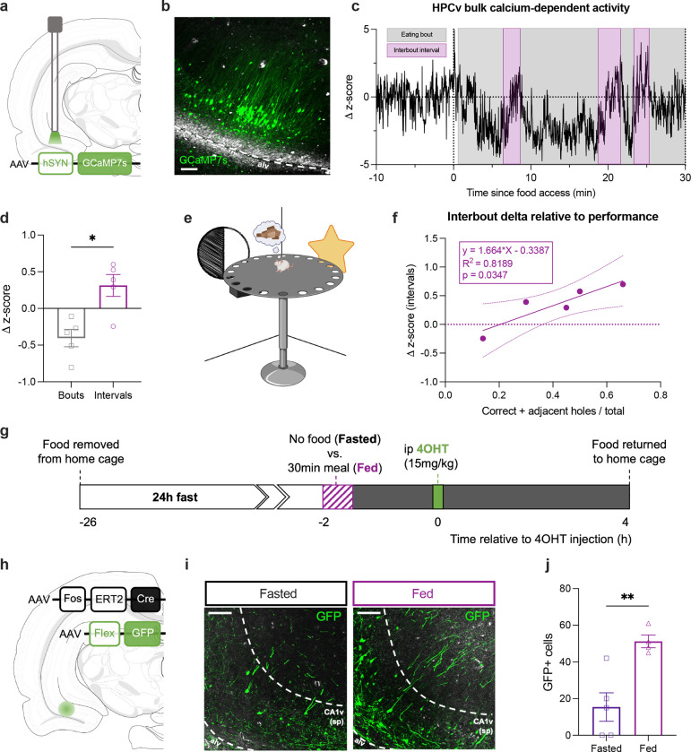
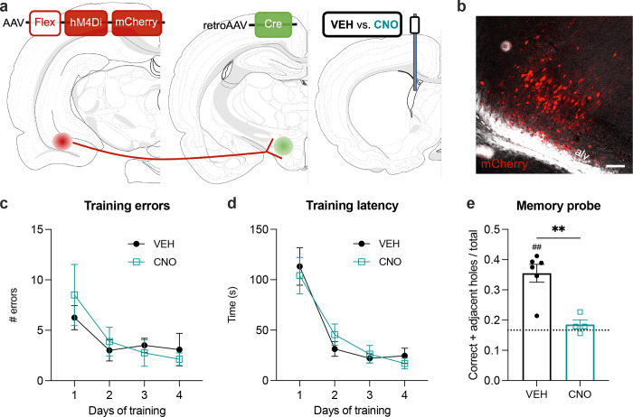
The ability to encode and retrieve meal-related information is critical to efficiently guide energy acquisition and consumption, yet the underlying neural processes remain elusive. Here we reveal that ventral hippocampus (HPCv) neuronal activity dynamically elevates during meal consumption and this response is highly predictive of subsequent performance in a foraging-related spatial memory task. Targeted recombination-mediated ablation of HPCv meal-responsive neurons impairs foraging-related spatial memory without influencing food motivation, anxiety-like behavior, or escape-mediated spatial memory. These HPCv meal-responsive neurons project to the lateral hypothalamic area (LHA) and single-nucleus RNA sequencing and in situ hybridization analyses indicate they are enriched in serotonin 2a receptors (5HT2aR). Either chemogenetic silencing of HPCv-to-LHA projections or intra-HPCv 5HT2aR antagonist yielded foraging-related spatial memory deficits, as well as alterations in caloric intake and the temporal sequence of spontaneous meal consumption. Collective results identify a population of HPCv neurons that dynamically respond to eating to encode meal-related memories.


Conflict of interest statement

CONFLICT OF INTEREST BCR and MRH both receive research funding from Novo Nordisk and Boehringer Ingelheim that was not used in support of these studies. MRH receives research funding from Eli Lilly & Co., Gila Therapeutics, and Pfizer that was not used in support of these studies. MRH is CEO of Cantius Therapeutics, LLC which pursues biological work unrelated to the current study. All other authors declare no competing interests.

Figures

Figure 1.
Figure 1.. Dynamic changes in ventral hippocampus calcium-dependent activity during a meal are predictive of performance during a foraging-related spatial memory task.
(a) Diagram of viral approach for fiber photometry. (b) Representative photomicrograph of viral expression and optic fiber placement in the ventral CA1 relative to the alveus (alv); scale bar 100 um. (c) Representative trace of a single animal of the increase in ventral CA1 calcium-dependent activity during the interbout intervals (purple). (d) Average change in z-score for fluorescence over the course of an eating bout versus an interval. (e) Foraging-related spatial memory task apparatus. (f) Simple linear regression of the increase during interbout intervals predictive of subsequent performance in a separate foraging-related spatial memory task. (g) Timeline for intraperitoneal (ip) injection of 4-hydroxitamoxifen (4OHT) under the Fasted or Fed state. (h) Diagram of the viral approach for 4OHT-inducible expression of green fluorescent protein (GFP) in ventral CA1 neurons active in a Fasted or Fed state. (i) Representative photomicrographs of the pyramidal layer of the ventral CA1 (CA1v(sp)) with GFP+ cell bodies from neurons that were active in the Fasted or Fed state, relative to the alveus (alv); scare bar 100 um. (i) Quantification of GFP+ cell bodies under the Fasted or Fed state in the ventral CA1. Data are presented as mean ± SEM. For Fig. 1d, paired t-test (n=5). For Fig. 1j, unpaired t-test (n=3–5/group). *p<0.05, **p<0.01.
Figure 2.
Figure 2.. Ablation of ventral hippocampus meal-responsive neurons selectively impairs foraging-related spatial memory.
(a) Diagram of viral approach for 4-hydroxytamoxifen (4OHT)-inducible Cre-dependent expression of diphteria toxin (dTA) (Fasted and Fed) or green fluorescent protein (Control) in ventral CA1 neurons active in the Fasted or Fed state. (b) Average daily food intake following 4OHT intraperitoneal administration to induce dTA-mediated lesion (Fasted and Fed) versus Control. (c) Body weight following intraperitoneal administration of 4OHT. (d) Diagram of Barnes maze apparatus for the foraging-related spatial memory task. (e) Average number of errors and (f) latency to find the food during training for the foraging-related spatial memory task. (g) Performance index during the probe for the foraging-related spatial memory task. (h) Diagram of Barnes maze apparatus for the escape-based spatial memory task. (i) Average number of errors and (j) latency to find the escape box during training for the escape-based spatial memory task. (k) Performance index during the probe for the escape-based spatial memory task. Data are presented as mean ± SEM. For Fig. 2b, one-way ANOVA (n=9–10/group). For Fig. 2c, two-way ANOVA, Tukey post hoc (n=9–11/group). For Fig. 2e–f and Fig. 2i–j, two-way ANOVA (n=9–10/group and n=6–7/group). For Fig. 2g, Kruskal-Wallis, multiple comparisons, (n=9–11/group). For Fig. 2k, two-tailed unpaired t-test, (n=6–7/group); *p<0.05, **p<0.01, ***p<0.005. For Fig. 2g and Fig. 2k, one-sample t-test, different from chance set at 0.1667; ##p<0.01, ###p<0.005, ####p<0.001.
Figure 3.
Figure 3.. Ventral hippocampus meal- but not fast-responsive neurons project to the lateral hypothalamic area.
(a) Left: Diagram of a coronal section of the nucleus accumbens (ACB). Middle: Representative photomicrographs of axonal green fluorescent protein (GFP)+ expression in the ACB from ventral CA1 neurons active in the Fasted or Fed state, relative to the anterior commissure (ac); scale bar 100 um. Right: Average number of GFP+ pixels in the ACB. (b) Left: Diagram of a coronal section of the lateral septum (LS). Middle: Representative photomicrographs of axonal GFP+ expression in the LS from ventral CA1 neurons active in the Fasted or Fed state, relative to the lateral ventricle (lv); scale bar 100 um. Right: Average number of GFP+ pixels in the LS. (c) Left: Diagram of a coronal section of the lateral hypothalamic area (LHA). Middle: Representative photomicrographs of axonal GFP+ expression in the LHA from ventral CA1 neurons active in the Fasted or Fed state, relative to the fornix (fx) and 3rd ventricle (3v); scale bar 100 um. Right: Average number of GFP+ pixels in the LHA. Data are presented as mean ± SEM. For Fig. 3a–c, two-tailed unpaired t-test (n=4–5/group); *p<0.05.
Figure 4.
Figure 4.. Chemogenetic inhibition of a ventral hippocampus to lateral hypothalamus pathway impairs foraging-related spatial memory.
(a) Diagram of viral approach for expression of hM4Di receptors in ventral CA1 neurons projecting to the lateral hypothalamic area and administration of vehicle (VEH) or clozapine-N-oxyde (CNO) through a lateral ventricle (LV) cannula. (b) Representative photomicrograph of mCherry expression for placement validation of viral injections, relative to the alveus (alv); scale bar 100 um. (c) Average number of errors and (d) latency to find the food during training for the foraging-related spatial memory task. (e) Performance index during the probe for the foraging-related spatial memory task, 1h following LV administration of VEH or CNO. Data are presented as mean ± SEM. For Fig. 5c–d, two-way ANOVA (n=4–6/group). For Fig. 5e, two-tailed unpaired t-test (n=4–6/group); **p<0.01. For Fig. 5e, one-sample t-test, different from chance set at 0.1667; ##p<0.01.
Figure 5.
Figure 5.. Meal consumption primarily alters the transcriptional profile of ventral hippocampus endothelial cells and CA1v excitatory neurons and engages 5HT2aR expressing cells.
(a) Uniform manifold approximation and projection (UMAP) of the ventral hippocampus (HPCv) samples identifying 17 clusters. (b) Annotation of cellular subtypes with known makers of HPCv cellular subtypes. The size and color of dots are proportional to the percentage of cells expressing the gene (Pct. Exp) and the average expression levels of the gene (Avg. Exp.), respectively. The cluster numbers and colors are matched to that of the UMAP. (c) Volcano plot depicting the number of significant differential expression events induced by meal consumption. (d) Number of genes with meal consumption-altered increased (right) or decreased (left) expression per cluster. (e) Heatmaps of select genes enriched in Fos+ cells under a Fasted or Fed state.
Figure 6.
Figure 6.. Ventral hippocampus 5HT2aR expressing neurons are engaged by a meal, project to the lateral hypothalamic rea, and are functionally required for foraging-related spatial memory.
(a) Representative photomicrograph of fluorescent in situ hybridization for Fos (red) and Htr2a (green) in the ventral CA1, relative to the alveus (alv); scale bar 100um. (b) Percentage of ventral CA1 Fos+ cells that co-express Htr2a in rats perfused under a Fasted or Fed state. (c) Diagram of approach for ventral CA1 administration of vehicle (VEH) or the 5HT2aR antagonist M100907 (1g/hemisphere). (d) Average number of errors and (e) latency to find food during the training for the foraging-related spatial memory task. (f) Performance during the probe for the foraging-related spatial memory task, 5min following ventral CA1 infusion of VEH or M100907. Data are presented as mean ± SEM. For Fig. 6b and Fig. 6f, two-tailed unpaired t-test (n=7–9/group and n=5–6/group). For Fig. 6d–e, two-way ANOVA (n=7–9/group); *p<0.05. For Fig. 6f, one-sample t-test, different from chance set at 0.1667; ##p<0.01.
Figure 7.
Figure 7.. Blockade of ventral hippocampus to lateral hypothalamic area signaling or 5HT2aR signaling increases food intake by reducing temporal intervals between meals during spontaneous feeding.
(a) Average 2h chow intake, (b) meal size, (c) meal frequency and (d) inter-meal intervals following ventral CA1 administration of vehicle (VEH) or M100907. (e) Average 2h chow intake, (f) meal size, (g) meal frequency and (h) inter-meal intervals following lateral ventricle administration of vehicle (VEH) or clozapine-N-oxide (CNO) in rats expressing hM4Di receptors in ventral CA1 neurons projecting to the lateral hypothalamic area. Data are presented as mean ± SEM. Two-tailed paired t-test (n=7/group and n=5–6/group); *p<0.05, **p<0.01, ***p<0.001.

Similar articles

References

    1. Higgs S. (2005). Memory and its role in appetite regulation. Physiol Behav 85, 67–72. 10.1016/J.PHYSBEH.2005.04.003. - DOI - PubMed
    1. Brunstrom J.M., Burn J.F., Sell N.R., Collingwood J.M., Rogers P.J., Wilkinson L.L., Hinton E.C., Maynard O.M., and Ferriday D. (2012). Episodic Memory and Appetite Regulation in Humans. PLoS One 7. 10.1371/journal.pone.0050707. - DOI - PMC - PubMed
    1. Higgs S., and Donohoe J.E. (2011). Focusing on food during lunch enhances lunch memory and decreases later snack intake. Appetite 57, 202–206. 10.1016/j.appet.2011.04.016. - DOI - PubMed
    1. Collins R., and Stafford L.D. (2015). Feeling happy and thinking about food. Counteractive effects of mood and memory on food consumption. Appetite 84, 107–112. 10.1016/j.appet.2014.09.021. - DOI - PubMed
    1. Parent M.B., Higgs S., Cheke L.G., and Kanoski S.E. (2022). Memory and eating: A bidirectional relationship implicated in obesity. Neurosci Biobehav Rev 132, 110–129. 10.1016/j.neubiorev.2021.10.051. - DOI - PMC - PubMed

Publication types