

This is a preprint.
A red-emitting carborhodamine for monitoring and measuring membrane potential
- PMID: 37873283
- PMCID: PMC10592620
- DOI: 10.1101/2023.10.06.561080
A red-emitting carborhodamine for monitoring and measuring membrane potential
Update in
-
A red-emitting carborhodamine for monitoring and measuring membrane potential.Proc Natl Acad Sci U S A. 2024 Apr 2;121(14):e2315264121. doi: 10.1073/pnas.2315264121. Epub 2024 Mar 29. Proc Natl Acad Sci U S A. 2024. PMID: 38551837 Free PMC article.
Abstract
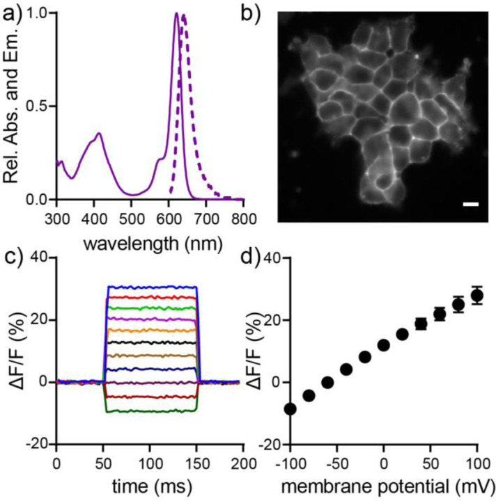
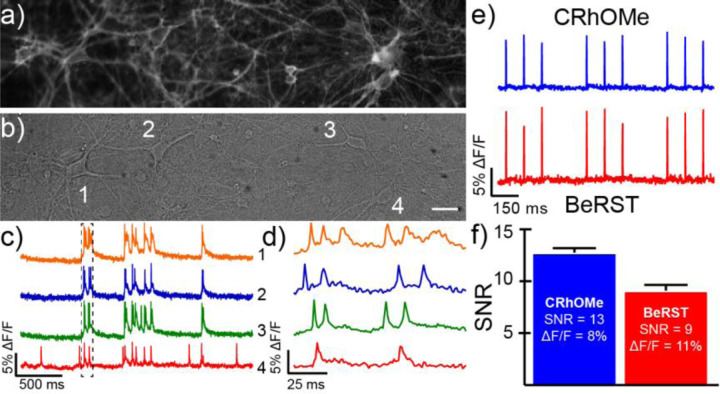
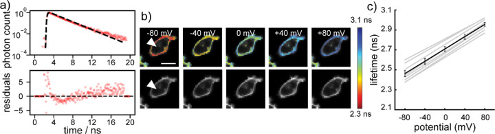
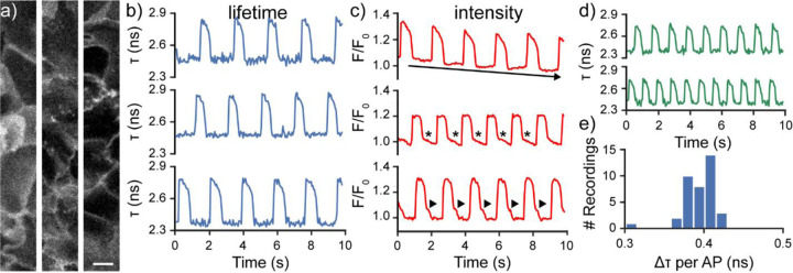
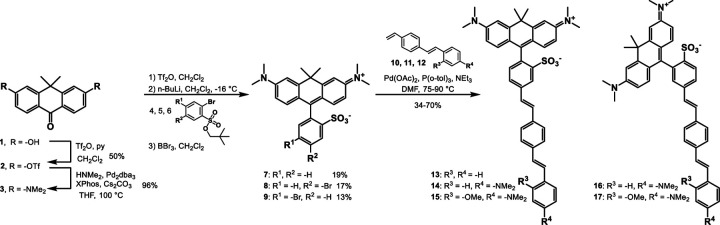
Biological membrane potentials, or voltages, are a central facet of cellular life. Optical methods to visualize cellular membrane voltages with fluorescent indicators are an attractive complement to traditional electrode-based approaches, since imaging methods can be high throughput, less invasive, and provide more spatial resolution than electrodes. Recently developed fluorescent indicators for voltage largely report changes in membrane voltage by monitoring voltage-dependent fluctuations in fluorescence intensity. However, it would be useful to be able to not only monitor changes, but also measure values of membrane potentials. This study discloses a new fluorescent indicator which can address both. We describe the synthesis of a new sulfonated tetramethyl carborhodamine fluorophore. When this carborhodamine is conjugated with an electron-rich, methoxy (-OMe) containing phenylenevinylene molecular wire, the resulting molecule, CRhOMe, is a voltage-sensitive fluorophore with red/far-red fluorescence. Using CRhOMe, changes in cellular membrane potential can be read out using fluorescence intensity or lifetime. In fluorescence intensity mode, CRhOMe tracks fast-spiking neuronal action potentials with greater signal-to-noise than state-of-the-art BeRST (another voltage-sensitive fluorophore). CRhOMe can also measure values of membrane potential. The fluorescence lifetime of CRhOMe follows a single exponential decay, substantially improving the quantification of membrane potential values using fluorescence lifetime imaging microscopy (FLIM). The combination of red-shifted excitation and emission, mono-exponential decay, and high voltage sensitivity enable fast FLIM recording of action potentials in cardiomyocytes. The ability to both monitor and measure membrane potentials with red light using CRhOMe makes it an important approach for studying biological voltages.
Figures

References
-
- Bean B. P., The action potential in mammalian central neurons. Nature reviews. Neuroscience 2007, 8 (6), 451–65. - PubMed
-
- Nattel S.; Carlsson L., Innovative approaches to anti-arrhythmic drug therapy. Nature Reviews Drug Discovery 2006, 5 (12), 1034–1049. - PubMed
-
- Sanguinetti M. C.; Tristani-Firouzi M., hERG potassium channels and cardiac arrhythmia. Nature 2006, 440 (7083), 463–9. - PubMed
-
- Nichols C. G., KATP channels as molecular sensors of cellular metabolism. Nature 2006, 440 (7083), 470–6. - PubMed
-
- Tsuchiya W.; Okada Y., Membrane potential changes associated with differentiation of enterocytes in the rat intestinal villi in culture. Developmental Biology 1982, 94 (2), 284–290. - PubMed
Publication types
Grants and funding
LinkOut - more resources
Full Text Sources