[Preprint]. 2024 Jun 17:2023.10.05.560950.
doi: 10.1101/2023.10.05.560950.

A murine model of Trypanosoma brucei-induced myocarditis and cardiac dysfunction

Affiliations

A murine model of Trypanosoma brucei-induced myocarditis and cardiac dysfunction

Nathan P Crilly et al. bioRxiv. .

Update in

Abstract

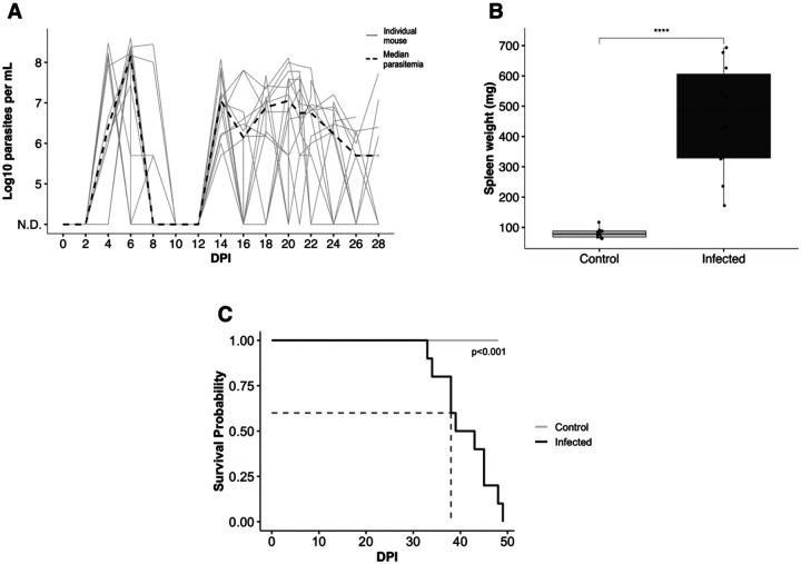
Trypanosoma brucei is a protozoan parasite that causes human and animal African trypanosomiases (HAT and AAT). Cardiac symptoms are commonly reported in HAT patients, and intracardiac parasites with accompanying myocarditis have been observed in both natural hosts and animal models of T. brucei infection. Despite the importance of T. brucei as a cause of cardiac dysfunction and the dramatic socioeconomic impact of African trypanosomiases in sub-Saharan Africa, there are currently no reproducible murine models of T. brucei-associated cardiomyopathy. We present the first clinically relevant, reproducible murine model of cardiac dysfunction in chronic T. brucei infection. Similar to humans, mice showed histological evidence of myocarditis and elevation of serum NT-proBNP with electrocardiographic abnormalities. Serum NT-proBNP levels were elevated prior to the development of severe ventricular dysfunction. On flow cytometry, myocarditis was associated with an increase of most myocardial immune cell populations, including multiple T cell and macrophage subsets, corroborating the notion that T. brucei-associated cardiac damage is an immune-mediated event. This novel mouse model represents a powerful and practical tool to investigate the pathogenesis of T. brucei-mediated heart damage and supports the development of therapeutic options for T. brucei-associated cardiac disease.


Conflict of interest statement

Conflict-of-interest statement The authors have declared that no conflicts of interest exist

Figures

Figure 1.
Figure 1.
T. brucei infection causes waxing and waning waves of parasitemia and massive splenomegaly, with terminal endpoint reached within 50 dpi A. Quantification of median and individual parasitemia in a representative experiment. Parasitemia first becomes detectable at 4–6 dpi. B. Weight of spleens of infected mice at 28 dpi(n=6) compared to uninfected, age-matched controls. Infected mice exhibit significant splenomegaly (Student’s t-test, p=3.7E-06). C. Kaplan-Meier survival curve of mice infected with T. brucei vs. uninfected control animals. Infected mice have a median survival time=39 dpi (p=4.32E-05).
Figure 2.
Figure 2.
T. brucei infection causes elevated NT-proBNP, electrocardiographic abnormalities and heart failure with preserved ejection fraction A. Measurement of plasma NT-proBNP at 28 dpi. Plasma NT-proBNP is significantly elevated at 28 dpi (n=12) compared to age-matched uninfected controls (Student’s t-test, p=0.00037). B. Echocardiographic measurements at 28 dpi. At 28 dpi, infected mice (n=12) exhibit significantly increased ejection fraction compared to uninfected age-matched controls, as measured by sedated echocardiography (Student’s t-test, p=0.0029). C. Electrocardiographic measurements at 28 dpi. At 28 dpi, infected mice (n=10) exhibit significantly decreased heart rate (HR, Student’s t-test, p= 0.00011), increased QRS interval, (Student’s t-test, p=0.00281), increased QT interval (Student’s t-test, p=0.00493), and increased QTc (Student’s t-test, p=0.01294) compared to uninfected age-matched controls.
Figure 3.
Figure 3.
T. brucei infection causes elevated NT-proBNP and decreased heart function at terminal endpoint (33 dpi). A. Measurement of plasma NT-proBNP at 33 dpi. Plasma NT-proBNP is significantly elevated at 33 dpi in infected mice (n=6) compared to uninfected age-matched controls (Student’s t-test, p=2.8e-05). B. Echocardiographic measurements at 33 dpi. At 33 dpi, infected mice (n=6) exhibit significantly decreased ejection fraction compared to uninfected age-matched controls, as measured by awake echocardiography (Student’s t-test, p=0.011)
Figure 4.
Figure 4.
T. brucei parasites occupy extravascular spaces in the heart Representative immunofluorescence microphotographs of the cardiac ventricle at 14 dpi at 20x magnification. T. brucei parasites (tdTomato—red) localize separately from CD31-lined vascular structures (Alexa Fluor 488—green), confirming the extravascular localization of parasites. Nuclei are stained with Hoechst (blue).
Figure 5.
Figure 5.
T. brucei infection causes myocarditis at 28 dpi A. Representative brightfield microphotographs of the heart of infected mice at 28 dpi, compared to uninfected age-matched controls at 10x magnification. Inflammation is present in all layers and regions of the heart and is composed largely of mononuclear cells, primarily lymphocytes, plasma cells, and histiocytes. Inflammation is most severe at the atrioventricular junction. B. Histological grading of myocarditis (n=6) at 28 dpi, compared to equal numbers of age-matched controls. The average grade at 28 dpi is ‘2’, indicating multifocal moderate myocarditis without extensive associated necrosis. Histological grade is significantly higher in infected than uninfected mice (Student’s t test, p=1.5e-8). C. Measurement of the percentage of collagen in longitudinal sections of heart (n=6) at 28 dpi, compared to equal numbers of age-matched controls. There is no significant difference between infected and control mice (Student’s t test, p=0.31).
Figure 6.
Figure 6.
The intracardiac immune cell population during T. brucei infection is characteristic of immune-mediated myocarditis A. Flow cytometric quantification of CD45+ cells per mg. At 28 dpi, CD45+ cells are markedly increased in the hearts of infected mice (n=12) compared to uninfected age matched controls, supportive of myocarditis (Student’s t-test, p= 4.6e-7). B. Flow cytometric quantification of myeloid and lymphoid cell populations. Most immune cell populations are significantly increased in the hearts of infected mice at 28 dpi. Statistical comparisons were made using One-way Anova. *p<0.05, **p<0.01, ***p<0.001. C. Visualization of magnitude of changes in immune cell populations using the log2-fold change in cells/mg compared to uninfected age-matched control mice calculated based on the average cells/mg in the hearts of uninfected control mice.

References

    1. Holmes P. Tsetse-transmitted trypanosomes - Their biology, disease impact and control [preprint]. J Invertebr Pathol. 2013;112(SUPPL.1). 10.1016/j.jip.2012.07.014. - DOI - PubMed
    1. Kennedy PGE, Rodgers J. Clinical and neuropathogenetic aspects of human African trypanosomiasis [preprint]. Front Immunol. 2019;10(JAN). 10.3389/fimmu.2019.00039. - DOI - PMC - PubMed
    1. Spickler AR. African Animal Trypanosomiasis [Internet]. IO State University Center for Food Security and Public Health. 2018.
    1. Balmer O, et al. Phylogeography and taxonomy of Trypanosoma brucei. PLoS Negl Trop Dis. 2011;5(2):e961. - PMC - PubMed
    1. Franco JR, et al. The elimination of human African trypanosomiasis: Achievements in relation to WHO road map targets for 2020. PLoS Negl Trop Dis. 2022;16(1). 10.1371/JOURNAL.PNTD.0010047. - DOI - PMC - PubMed

Publication types