[Preprint]. 2023 Oct 2:2023.09.29.560022.
doi: 10.1101/2023.09.29.560022.

Stem cell-specific ecdysone signaling regulates the development and function of a Drosophila sleep homeostat

Affiliations

Stem cell-specific ecdysone signaling regulates the development and function of a Drosophila sleep homeostat

Adil R Wani et al. bioRxiv. .

Update in

Abstract

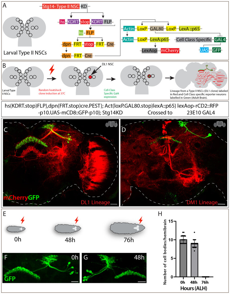
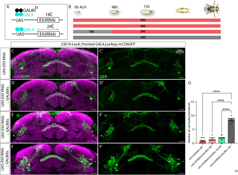
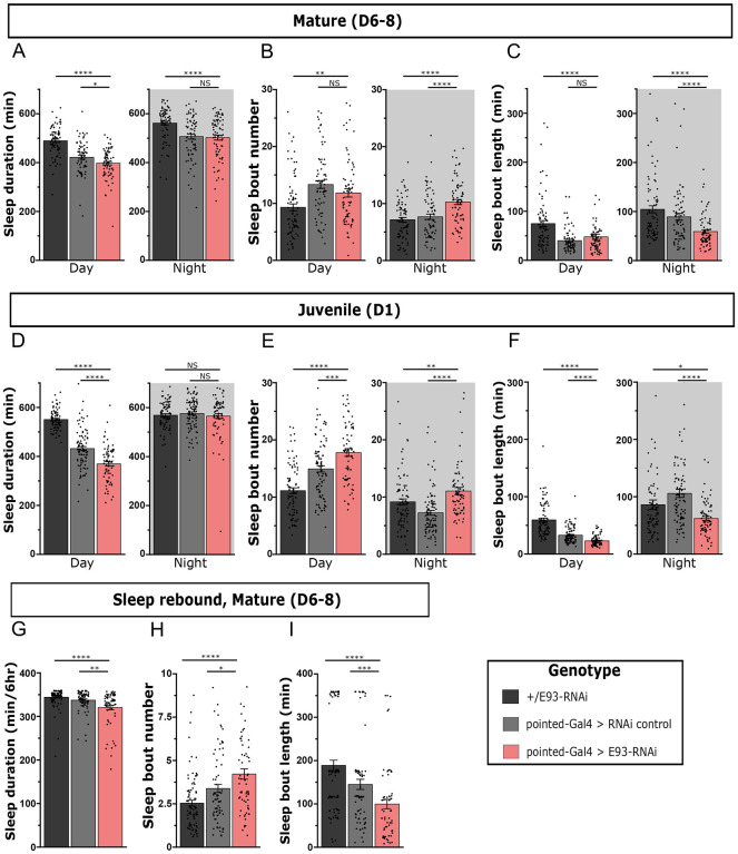
Complex behaviors arise from neural circuits that are assembled from diverse cell types. Sleep is a conserved and essential behavior, yet little is known regarding how the nervous system generates neuron types of the sleep-wake circuit. Here, we focus on the specification of Drosophila sleep-promoting neurons-long-field tangential input neurons that project to the dorsal layers of the fan-shaped body neuropil in the central complex (CX). We use lineage analysis and genetic birth dating to identify two bilateral Type II neural stem cells that generate these dorsal fan-shaped body (dFB) neurons. We show that adult dFB neurons express Ecdysone-induced protein E93, and loss of Ecdysone signaling or E93 in Type II NSCs results in the misspecification of the adult dFB neurons. Finally, we show that E93 knockdown in Type II NSCs affects adult sleep behavior. Our results provide insight into how extrinsic hormonal signaling acts on NSCs to generate neuronal diversity required for adult sleep behavior. These findings suggest that some adult sleep disorders might derive from defects in stem cell-specific temporal neurodevelopmental programs.


Conflict of interest statement

Declaration of interests The authors declare no competing interests.

Figures

Figure 1
Figure 1. Sleep-promoting dFB neurons are generated by Type II NSC lineages
A) Schematics of larval Type II NSCs (8 per lobe: DM1–6, DL1–2), which divide asymmetrically over 120 hours ALH to generate INPs and express early and late TTFs. The temporally expressed EcR mediates the switch from early to late transition. The Type II NSC TTFs and INP temporal factors are thought to contribute to the formation and diversification of neural lineages of the Drosophila central complex. We are investigating the role of ecdysone signaling in the specification and function of dFB sleep-promoting neurons, which are part of the Drosophila sleep-wake circuit. B) Schematics showing intersectional genetic strategy for Type II NSC lineage analysis. The Worniu-GAL4, Ase-GAL80 combination drives the expression of FLP in all Type II NSCs, which excises a stop and makes LexAopmCD8GFP functional in all Type II NSCs and their progeny. This allows dFB neurons to be labeled in green if produced from Type II NSCs. C) The FLP expression in Type II NSCs labels all adult dFB neurons (green) (max projection), and nc82 labels neuropil (magenta) (projections showing only FB). The expression of GFP reporter in dFB neurons confirms that they are part of Type II NSC lineages. Scale bars, 20μm, n = 8 adult brains.
Figure 2
Figure 2. Sleep-promoting dFB neurons are generated by late DL1 and DM1 Type II NSCs
A) Schematics of CLIn intersectional genetics explaining how different genetic elements work in a sequence to label different Type II NSC lineages. The CLIn flies use Type II NSC-specific promotor, stg, to express KD recombinase in Type II-specific manner. The KD recombinase removes the stop sequence, bringing the FLP in frame with the heat shock promotor only in Type II NSCs. The FLP expression removes the stop sequence upon heat shock, making Cre recombinase active in Type II NSCs. The Cre recombinase makes LexA::p65 functional, making the lineage-specific expression of reporter mCherry possible. The removal of GAL80 by Cre recombinase also removes the inhibition of GAL4, making the expression of mCD8GFP in a class-specific manner. B) Schematics of how CLIn allows lineage analysis of Type II NSCs. The stochastic heat-sensitive FLP event in a single Type II NSC labels all neurons and glia born from that particular NSC. C) Single DL1 NSC clone induced at 0h ALH labels most dFB neurons (green). All the lineages from DL1 NSC are labeled in red (mCherry) and dFB neurons in green. D) Single DM1 NSC clone induced at 0h ALH labels 1–2 dFB neurons. The DM1 lineages are labeled in red (mCherry), and dFB neurons in green. E) Schematics representing heat shock given at different time points during larval development. The red lightning bolt symbol indicates heat shock given at three different time points 0h, 48h, and 76h after larval hatching (ALH). F-H) Clones induced at 0h and 48h ALH label all the dFB neurons (F, G), while clones induced at 76h ALH didn’t label any dFB neurons (not shown) H) Quantification of dFB neuron cell bodies labeled per hemibrain when clones are induced at 0h, 48h, and 76h ALH. Scale bars, 20μm, n = 16 adult hemibrains.
Figure 3
Figure 3. Ecdysone signaling regulates dFB neuron specification
A) Genetic elements of EcR-FlpStop2.0 scheme. The expression of FLP recombinase in Type II NSCs flips the tdTomato sequence in frame with a UAS promoter, allowing it to label mutant cells specifically under the control of the Type II specific GAL4 in red color. The FLP event also inverts the STOP sequence - transcription-based disruption (Tubα1 terminator and 10x Ribozyme sequence) and translation disruption (MHC splice acceptor paired with STOP codons) - to generate a premature stop, disrupting EcR expression and function. B) Schematics showing typical Type II NSC expressing EcR at 55h ALH that leads to expression of other EcR-induced downstream genes; upon removal of ecdysone receptor, the expression of target genes is disrupted. C, C’) Shows a control brain with GFP-labeled dFB neurons and their projection pattern in the FB. D, D’) Upon EcR loss of function in Type II NSCs, dFB neurons are not specified (dashed line annotates the FB). E, E’) Blocking ecdysone signaling in Type II NSCs using EcR-DN results in significant loss of dFB neurons. F-H’) In control brains, the dFB neurons project to layer 6 of the FB (F, F’); in EcR loss of function (G, G’) and EcR-DN (H, H’) animals, the surviving dFB neurons miss target to the ectopic FB layers indicated by arrows. I) One-way ANOVA test quantification of dFB neuron cell bodies. Error bars represent SEM; * p<0.05, **p<0.01, ***p<0.001, **** p<0.0001, NS, non-significant. Scale bars, 20μm, n = 10 adult hemibrains.
Figure 4
Figure 4. Ecdysone signaling regulates dFB neural fate specification via E93
A-A”) The dFB neurons labeled in green (A) express E93 in cell bodies (A’, A”). B, B’) Control dFB neurons project normally to the FB. C, C’) Complete loss of dFB neurons upon E93 knock-down in Type-II NSCs. D, D’) No change in cell body number and morphology of dFB neurons upon E93 overexpression in Type-II NSCs. E) One-way ANOVA test quantification of dFB neuron cell bodies per hemibrain. Error bars represent SEM; * p<0.05, **p<0.01, ***p<0.001, **** p<0.0001, NS, non-significant. Scale bars, 20μm, n = 14 adult hemibrains.
Figure 5
Figure 5. E93 expression in a restricted time window regulates dFB neuronal fate.
A) Schematics of TARGET system showing GAL80ts mediated restricted knockdown ofE93. At 18°C, GAL80ts will be active, preventing the expression of E93RNAi by inhibiting Pointed-GAL4. At higher temperatures, 29°C, GAL80ts will be inactive, allowing the expression of E93RNAi temporally. B) Schematics of the experimental setup showing E93 RNAi flies growing at different temperatures and under different conditions across the larval life cycle from 0h ALH to 120h ALH. The E93RNAi experimental flies containing GAL80ts were initially grown at 18°C and later shifted to 29°C around 40h ALH to make E93 RNAi expression possible in late Type II NSCs. C, C’) Shows loss of dFB neurons labeled with GFP at 29°C upon E93 knockdown (E93 RNAi without dicer) D, D’) Loss of dFB neurons can be seen upon E93 RNAi combined with Gal80ts grown continuously at 29°C (GAL80ts will be inactive at 29°C) E, E’) Significant loss of dFB neurons can be seen when the UAS-E93 RNAi was restricted to the late Type II NSCs using GAL80ts; the flies were grown at 18°C till 40h ALH and then shifted to 29°C to make GAL80ts inactive. F, F’) The dFB neuron number is normal when flies expressing UAS-E93RNAi combined with Gal80ts are grown continuously under 18°C. G) One-way ANOVA test quantification of dFB neuron cell bodies per hemibrain. Error bars represent SEM; * p<0.05, **p<0.01, ***p<0.001, **** p<0.0001, NS, non-significant. Scale bars, 20μm, n = 12 adult hemibrains.
Figure 6
Figure 6. Knockdown of E93 in larval Type II NSCs impairs adult sleep.
Quantification of day and night sleep duration (A), sleep bout number (B), and sleep bout length (C) in mature adult flies expressing E93-RNAi under control of pointed-GAL4 (red) compared to genetic controls (black, gray). n=79,74,74 from left to right. Quantification of sleep duration (D), sleep bout number (E), and sleep bout length (F) in juvenile adult E93-RNAi flies and controls. n=85,94,74 from left to right. Quantification of sleep duration (G), sleep bout number (H), and sleep bout length (I) in mature adult E93-RNAi flies and controls over 6 hours following a night (12 hours) of sleep deprivation. n=93,82,67 from left to right. Error bars represent SEM; *p<0.05, **p<0.01, ***p<0.001, ****p<0.0001, NS, non-significant by One-way ANOVA with Mann-Whitney multiple comparisons test corrections.

Similar articles

References

    1. Kohwi M., and Doe C.Q. (2013). Temporal Fate Specification and Neural Progenitor Competence During Development. Nat. Rev. Neurosci. 14, 823–838. - PMC - PubMed
    1. Molnár Z., Clowry G.J., Šestan N., Alzu’bi A., Bakken T., Hevner R.F., Hüppi P.S., Kostović I., Rakic P., Anton E.S., et al. (2019). New insights into the development of the human cerebral cortex. J. Anat. 235, 432–451. 10.1111/joa.13055. - DOI - PMC - PubMed
    1. Oberst P., Agirman G., and Jabaudon D. (2019). Principles of progenitor temporal patterning in the developing invertebrate and vertebrate nervous system. Curr. Opin. Neurobiol. 56, 185–193. 10.1016/j.conb.2019.03.004. - DOI - PubMed
    1. El-Danaf R.N., Rajesh R., and Desplan C. (2023). Temporal regulation of neural diversity in Drosophila and vertebrates. Semin. Cell Dev. Biol. 142, 13–22. 10.1016/j.semcdb.2022.05.011. - DOI - PMC - PubMed
    1. Malin J., and Desplan C. (2021). Neural specification, targeting, and circuit formation during visual system assembly. Proc. Natl. Acad. Sci. 118. 10.1073/pnas.2101823118. - DOI - PMC - PubMed

Publication types