[Preprint]. 2024 Nov 13:2023.10.03.560703.
doi: 10.1101/2023.10.03.560703.

Palatal segment contributions to midfacial anterior-posterior growth

Affiliations

Palatal segment contributions to midfacial anterior-posterior growth

Ian C Welsh et al. bioRxiv. .

Update in

Abstract

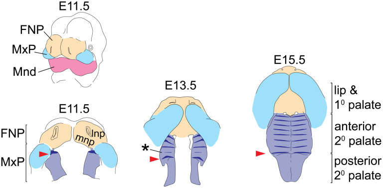
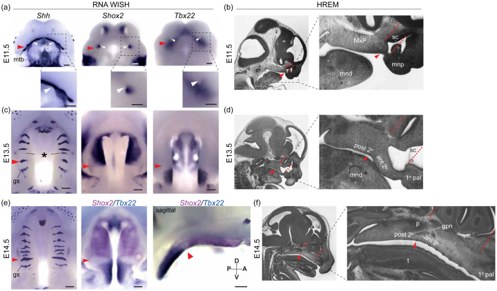
Anterior-posterior (A-P) elongation of the palate is a critical aspect of integrated midfacial morphogenesis. Reciprocal epithelial-mesenchymal interactions drive secondary palate elongation that is coupled to the periodic formation of signaling centers within the rugae growth zone (RGZ). However, the relationship between RGZ-driven morphogenetic processes, the differentiative dynamics of underlying palatal bone mesenchymal precursors, and the segmental organization of the upper jaw has remained enigmatic. A detailed ontogenetic study of these relationships is important because palatal segment growth is a critical aspect of normal midfacial growth, can produce dysmorphology when altered, and is a likely basis for evolutionary differences in upper jaw morphology. We completed a combined whole mount gene expression and morphometric analysis of normal murine palatal segment growth dynamics and resulting upper jaw morphology. Our results demonstrated that the first formed palatal ruga (ruga 1), found just posterior to the RGZ, maintained an association with important nasal, neurovascular and palatal structures throughout early midfacial development. This suggested that these features are positioned at a proximal source of embryonic midfacial directional growth. Our detailed characterization of midfacial morphogenesis revealed a one-to-one relationship between palatal segments and upper jaw bones during the earliest stages of palatal elongation. Growth of the maxillary anlage within the anterior secondary palate is uniquely coupled to RGZ-driven morphogenesis. This may help drive the unequaled proportional elongation of the anterior secondary palate segment prior to palatal shelf fusion. Our results also demonstrated that the future maxillary-palatine suture, approximated by the position of ruga 1 and consistently associated with the palatine anlage, formed predominantly via the posterior differentiation of the maxilla within the expanding anterior secondary palate. Our ontogenetic analysis provides a novel and detailed picture of the earliest spatiotemporal dynamics of intramembranous midfacial skeletal specification and differentiation within the context of the surrounding palatal segment AP elongation and associated rugae formation.

Keywords: Mus musculus; RRID:IMSR_JAX:000664; craniofacial; face elongation; gene expression; palatogenesis; rugae; secondary palate.


Figures

Figure 1 –
Figure 1 –
Tissue origins of the midfacial complex and rugae position during secondary palate morphogenesis – The upper and lower jaws are formed from the frontonasal process (FNP - tan), and branchial arch 1 derived maxillary and mandibular processes (MxP - pale blue and Mnd – magenta, respectively). From E11.5 to E15.5, outgrowth and fusion of the medial nasal process (mnp) with the superficial portion of the MxP frame out the lip and primary (1°) palate, while the A-P elongation and medially directed growth of the palatal shelves from the internal portion of the MxP gives rise to the secondary palate (2° pal – light purple). Shh expression (dark blue) highlights the dynamics of rugae formation and illustrates regional expansion of the anterior secondary palate. At E11.5, ruga 1 (red arrowheads) forms at the anterior extent of the nascent palatal shelf and subsequently defines the caudal end of the rugae growth zone (asterisk) where new rugae form prior to being displaced anteriorly. Additional abbreviation: lateral nasal process (lnp).
Figure 2 -
Figure 2 -
Flowchart illustrating this study’s research design
Figure 3 –
Figure 3 –
Anatomically homologous epithelial landmarks - Landmarks collected for the quantification of midfacial and palatal shape across limb based embryonic (E) developmental stages, from (a-c) palatal and (d-f) oblique facial views. Scale bars: 0.5mm. See also Supporting Table 1.
Figure 4 –
Figure 4 –
Palate segment length measurements – Identification of landmarks used to estimate midline lengths of the primary palate (yellow), anterior secondary palate (dark purple), and posterior secondary palate (light purple) on (a) E11.0, (b) E14.0, (c) E15.0 epithelial, (d) P1 epithelial, (e) P1 skeletal (after removing some neurocranial and facial bones to help with visualization), and (f) adult skeletal surfaces. Scale bars: 1mm. See also Supporting Tables 1 and 2.
Figure 5 –
Figure 5 –
A-P molecular heterogeneity and anatomical relationships during secondary palate morphogenesis - (a, c, e) RNA WISH for Shh, Shox2, and Tbx22 expression, and (b, d, f) sagittal sections of wildtype embryos imaged with high resolution episcopic microscopy (HREM) to provide histological resolution of the primary (1° pal), anterior secondary (ant 2°), and posterior secondary palate (post 2°) relative to surrounding facial structure during midfacial outgrowth at (a, b) E11.5 (c, d) E13.5, and (e, f) E14.5. Throughout midfacial outgrowth, ruga 1 (red arrowheads) marks the shared A-P expression boundary of Shox2 and Tbx22 in the anterior and posterior secondary palate, respectively. Double label WISH for Shox2 (magenta) and Tbx22 (blue) at E14.5 highlights mutually exclusive anterior and posterior expression domains organized relative to ruga 1 (oral view left, sagittal view right). White arrowheads mark the choanae, black asterisk marks the RGZ. Red dashed lines mark position of coronal planes passing through the primary-secondary palate junction and posterior wall of the nasal capsule to highlight coordinated elongation of the anterior secondary palate and overlying sinus cavity (sc). Regions in dashed boxes of (a) and in HREM images shown enlarged below or to the right, respectively. Additional abbreviations: mandible (mnd), molar tooth bud (mtb), maxillary process (MxP), medial nasal process (mnp), tongue (t), greater palatine nerve (gpn), geschmacksstreifen (gs), palatine (p), posterior domain of Shh expression (pd). Scale bars: 250um
Figure 6 –
Figure 6 –
Major axes of embryonic midfacial and palatal shape variation - C57BL/6J embryo specimens plotted along the (a) first and second principal component axes of shape (i.e., PC1 and PC2), (b) along PC1 and PC3, and (c) along PC1 and PC4. Circle color indicates the limb based developmental age categories of each specimen, with the yellow bordered circles indicating the mean PC scores for each age category with more than one specimen. The proportions of midfacial and palatal shape variance associated with each principal component are provided.
Figure 7 –
Figure 7 –
Palatal landmark growth trajectories – The positions of palatal landmarks are identified on representative C57BL/6J specimens at limb-derived developmental ages (a) E11, (b) E12.5, (c) E14, and (d) E15. (e) The average palatal landmark positions are plotted for each developmental age category that had more than one specimen. These landmark positions represent palatal shape after the removal of overall midfacial and palatal scale (as measured by centroid size) during Procrustes superimposition. The arrows indicate the trajectory of shape change for each landmark between ages. Black=E11; Light Blue = E12; Orange = E12.5; Green = E13; Dark Blue = E14; Red = E15. Scale bars: 0.5mm
Figure 8 –
Figure 8 –
Palate segment length across ontogeny – The midline projected mean lengths (in mm) of the primary palate, anterior secondary palate, and posterior secondary palate for all (a) E11-E15 specimens and (b) postnatal specimens. (c) Palate segment length relative to overall midline palate length for the all specimens. Whole-day limb bud derived age categories and postnatal age categories used for pairwise statistical comparisons are indicated by brackets above the bars. Comparable segment lengths were collected from epithelial and skeletal surfaces for a single set of P1 specimens.
Figure 9 -
Figure 9 -
Palate segment length comparisons - Boxplots of palate segment specific lengths (in mm) for (a) whole-day limb dub derived embryonic age categories and (b) postnatal age categories. (c) Palate segment length relative to overall midline palate length for all specimens. Outliers are specimen values that are 1.5 times the interquartile range outside of the 25th or 75th quartile of that segment’s measures. Significant differences are indicated for each age that a given segment’s median value is different than the next younger age category (* when p-value is < 0.05; ** when p-value < 0.001). Significant differences for a given segment’s P1 skeletal measurement median when compared to the same segment’s P1 epithelial measurement (^ when p-value < 0.05; ^^ when p-value < 0.001). Dashed lines illustrate ontogenetic trends of palatal segment proportional contributions to overall palatal length.
Figure 10 –
Figure 10 –
Segmental organization of skeletal specification and growth during A-P morphogenesis of the midface – WISH time course for (a) Runx2, (b) Sp7, and (c) Phospho1 between E11.5 and E15.5. Expression of Runx2 in osteoprogenitors highlights facial domains with osteogenic potential during midfacial morphogenesis. Expression of Sp7 and Phospho1 in committed and differentiating osteoblasts delineates the growth dynamics of individual skeletal anlagen during midfacial morphogenesis. Curved white dashed arrows in (b) and (c) indicate medial expansion of maxillary anlage. Abbreviations: maxilla (mx), palatine (p), premaxilla (pmx), pterygoid (ptg), palatal process of the maxilla (ppmx), palatal process of the palatine (ppp). Scale bars: 250um
Figure 11 –
Figure 11 –
Double label RNA WISH time course for Shh (cyan) and Sp7 (dark purple) between E12.5 and E15.5 - (a) Expression of Sp7 in committed and differentiating osteoblasts delineates the growth dynamics of individual skeletal anlagen and Shh expression in rugae provides a temporally ordered set of A-P landmarks (regions in dashed boxes enlarged below). (b) Summary model of skeletal growth dynamics during midfacial outgrowth. Growth of the premaxilla (yellow) and palatine (pale blue) anlagen towards their characteristic shape occurs largely at the site of initial specification. Following initial specification external to the oral cavity, the maxilla (purple) grows into the anterior of the anterior secondary palate (curved white and black dashed arrows in a and b, respectively) towards the position of ruga 1 (red arrowhead) and palatine as expansion of the anterior secondary palate (double headed red arrow) separates the primary and posterior secondary palate. (c) Summary of the position of epithelial landmarks (see also Fig. 3) selected to capture segmental growth dynamics of the primary palate (yellow) and anterior secondary palate (purple) and posterior secondary palate (pale blue) during midfacial outgrowth. Abbreviations: geschmacksstreifen (gs), maxilla (mx), palatine (p), premaxilla (pmx), pterygoid (ptg), palatal process of the maxilla (ppmx), palatal process of the palatine (ppp). Scale bars: 250um

References

    1. Adams D.C., Collyer M.L., Kaliontzopoulou A., 2020. geomorph: Software for geometric morphometric analyses. R package version 3.3.1.https://cran.r-project.org/package=geomorph
    1. Bush J.O., Jiang R., 2012. Palatogenesis: morphogenetic and molecular mechanisms of secondary palate development. Development 139, 231–243. - PMC - PubMed
    1. Cignoni P., Callieri M., Corsini M., Dellepiane M., Ganovelli F., Ranzuglia G., 2008. Meshlab: an open-source mesh processing tool., in: Eurographics Italian Chapter Conference. pp. 129–136. 10.2312/LocalChapterEvents/ItalChap/ItalianChapConf2008/129-136 - DOI
    1. Collyer M.L., Adams D.C., 2018. RRPP: An R package for fitting linear models to high-dimensional data using residual randomization. Methods in Ecology and Evolution.
    1. Dash S., Trainor P.A., 2020. The Development, Patterning, and Evolution of Neural Crest Cell Differentiation into Cartilage and Bone. Bone 137, 115409. 10.1016/j.bone.2020.115409 - DOI - PubMed

Publication types