[Preprint]. 2023 Oct 10:2023.10.10.561783.
doi: 10.1101/2023.10.10.561783.

A label-free approach for relative spatial quantitation of c-di-GMP in microbial biofilms

Affiliations

A label-free approach for relative spatial quantitation of c-di-GMP in microbial biofilms

Catherine S McCaughey et al. bioRxiv. .

Update in

Abstract

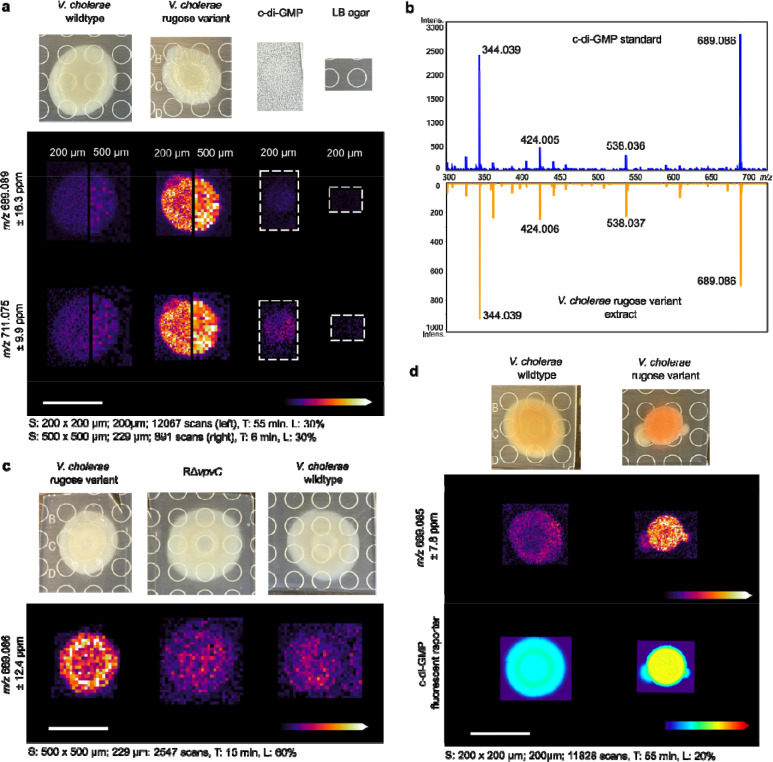
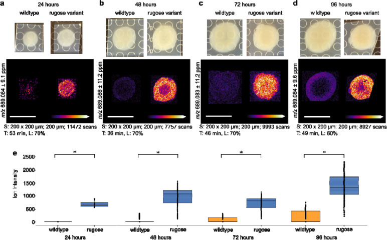
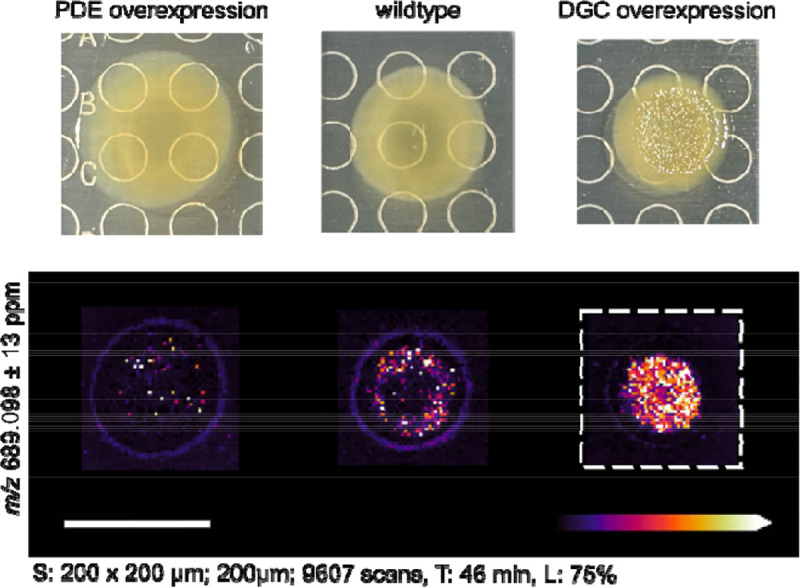
Microbial biofilms represent an important lifestyle for bacteria and are dynamic three dimensional structures. Cyclic dimeric guanosine monophosphate (c-di-GMP) is a ubiquitous signaling molecule that is known to be tightly regulated with biofilm processes. While measurements of global levels of c-di-GMP have proven valuable towards understanding the genetic control of c-di-GMP production, there is a need for tools to observe the local changes of c-di-GMP production in biofilm processes. We have developed a label-free method for the direct detection of c-di-GMP in microbial colony biofilms using matrix-assisted laser desorption ionization mass spectrometry imaging (MALDI-MSI). We applied this method to the enteric pathogen Vibrio cholerae, the marine symbiont V. fischeri, and the opportunistic pathogen Pseudomonas aeruginosa PA14 and detected spatial and temporal changes in c-di-GMP signal that accompanied genetic alterations in factors that synthesize and degrade the compound. We further demonstrated how this method can be simultaneously applied to detect additional metabolites of interest in a single experiment.


Figures

Figure 1
Figure 1
Spatial detection and validation of c-di-GMP in V cholerae wildtype and rugose variant strains. a) Ion images of the [M-H] (m/z 689.089) and [M-2H+Na]-(m/z 711.075) adducts of c-di-G in V. cholerae wildtype and rugose variant colonies. Data were acquired at both 200 μm and 500 μm raster to compare image resolution and sensitivity. An authentic standard of c-di-GMP (5 μL, 100 n was spotted on a sample of LB agar for comparison. b) MALDI-MS/MS spectrum of c-di-GMP compared to an extract of a V. cholerae rugose variant colony grown on LB agar. c) Ion images of cGMP in V. cholerae wildtype, rugose variant, and a rugose variant ΔvpvC (RΔvpvC). d) Ion images c-di-GMP in V. cholerae wildtype and the rugose variant compared to the c-di-GMP specific report Abundance of c-di-GMP is represented by heat maps showing the relative TurboRFP fluorescent si in the same bacterial colonies. Spot raster; size; scan number (S), acquisition time (T), and laser po (L) shown for each MSI experiment. All scales bars represent 1 cm.
Figure 2
Figure 2
Comparison of c-di-GMP spatial distribution in V. cholerae colonies over time. Ion images and boxplots comparing ion intensity for V. cholerae wildtype and rugose variant strains after a) 24 hours, b) 48 hours, c) 72 hours, and d) 96 hours of growth. Spot raster; size; scan number (S), acquisition time (T), and laser power (L) shown for each MSI experiment. All scale bars represent 1 cm. e) Box plots comparing ion intensity between V. cholerae wildtype and rugo variants grown on each day. The box plots are shown on a single graph, however the data from each time point were acquired as separate experiments. The line within each box plot represents t median, the edges of the box plots represent the upper and lower ends of the interquartile range a the whiskers represent the minimum and maximum quartiles. Outliers are shown beyond the whiskers of the box plots. Asterisks indicate statistical significance (p < 0.001) in a Wilcox Rank Sum test which was calculated using the SciLs MSI analysis software.
Figure 3
Figure 3
Comparison of c-di-GMP spatial distribution in V. fischeri colonies. The low c-di-GMP (PDE overexpression) and high c-di-GMP (DGC overexpression) strains contain a plasmid with an inducible promoter for the overexpression of the PDE VF_0087 and DGC MifA, respectively. The wildtype strain contains the vector control only. Spot raster; size; scan number (S), acquisition time (T), and laser power (L) shown for each MSI experiment. All scale bars represent 1 cm.
Figure 4
Figure 4
Ion images of c-di-GMP and other putatively identified metabolites in P. aeruginosa PA14. Ion images of metabolites detected in a) negative mode ionization and b) positive mode ionization. The following compound abbreviations are used: pyocyanin (PYO), phenazine-1-carboxamid (PCN), phenazine-1-carboxylic acid (PCA), Pseudomonas quinolone signal (PQS), 4-hydroxy-2-heptyquinoline-N-oxide (HQNO) 2-heptyl-4-quinolone (HHQ), and 4-hydroxy-2-nonylquinoline (HNQ) Table 2 shows the ppm error for all putatively identified compounds. Spot raster; size; scan number (S), acquisition time (T), and laser power (L) shown for each MSI experiment. All scale bars represent 1 cm.

References

    1. Römling U., Galperin M. Y. & Gomelsky M. Cyclic di-GMP: the first 25 years of a universal bacterial second messenger. Microbiol. Mol. Biol. Rev. 77, 1–52 (2013). - PMC - PubMed
    1. Valentini M. & Filloux A. Biofilms and Cyclic di-GMP (c-di-GMP) Signaling: Lessons from Pseudomonas aeruginosa and Other Bacteria. J. Biol. Chem. 291, 12547–12555 (2016). - PMC - PubMed
    1. Jenal U., Reinders A. & Lori C. Cyclic di-GMP: second messenger extraordinaire. Nat. Rev. Microbiol. 15, 271–284 (2017). - PubMed
    1. Conner J. G., Zamorano-Sánchez D., Park J. H., Sondermann H. & Yildiz F. H. The ins and outs of cyclic di-GMP signaling in Vibrio cholerae. Curr. Opin. Microbiol. 36, 20–29 (2017). - PMC - PubMed
    1. Petchiappan A., Naik S. Y. & Chatterji D. Tracking the homeostasis of second messenger cyclic-di-GMP in bacteria. Biophys. Rev. 12, 719–730 (2020). - PMC - PubMed

Publication types

LinkOut - more resources