[Preprint]. 2023 Dec 3:2023.10.14.559618.
doi: 10.1101/2023.10.14.559618.

Cholinergic modulation of rearing in rats performing a spatial memory task

Affiliations

Cholinergic modulation of rearing in rats performing a spatial memory task

Skylar Cassity et al. bioRxiv. .

Update in

Abstract

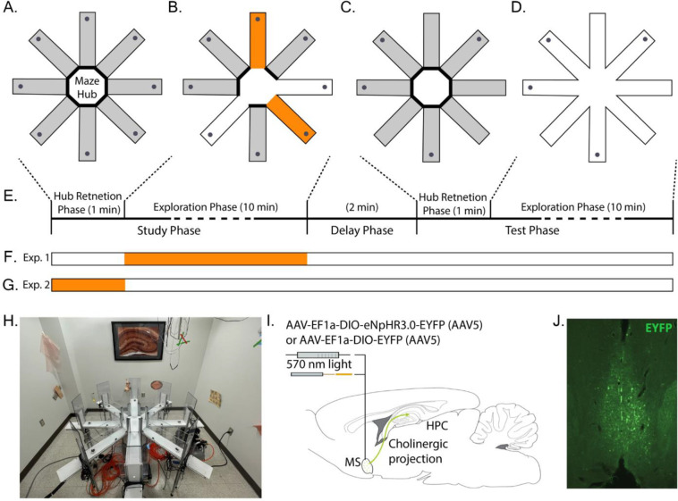
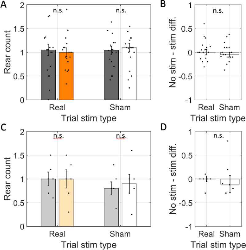
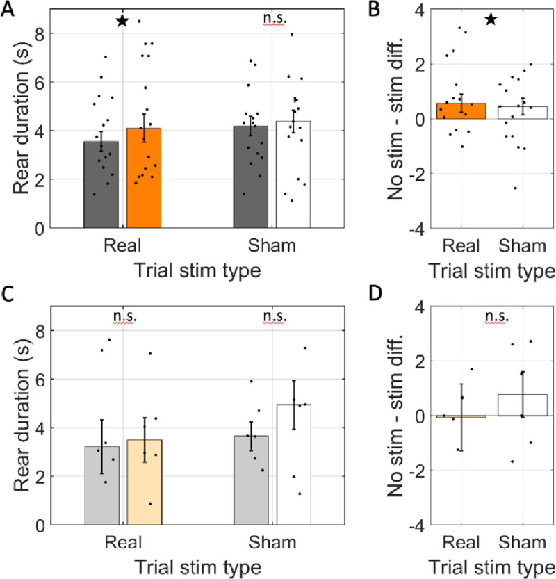
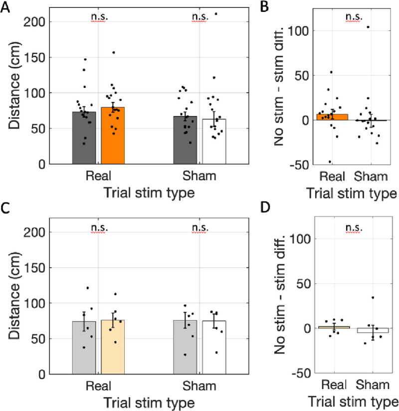
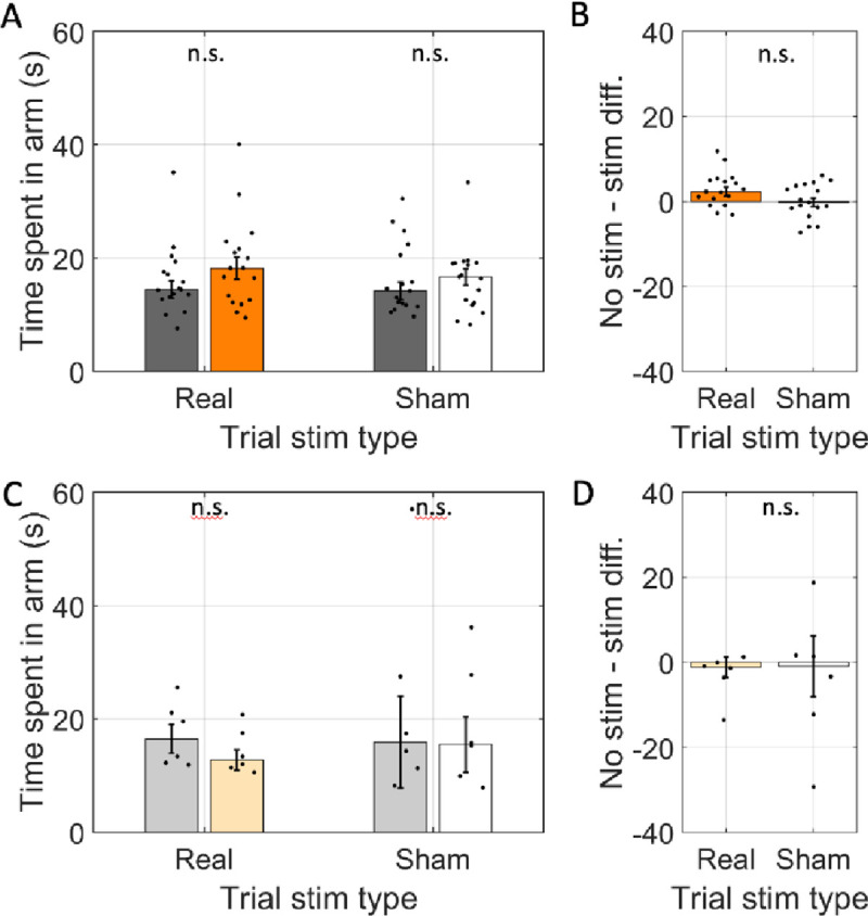
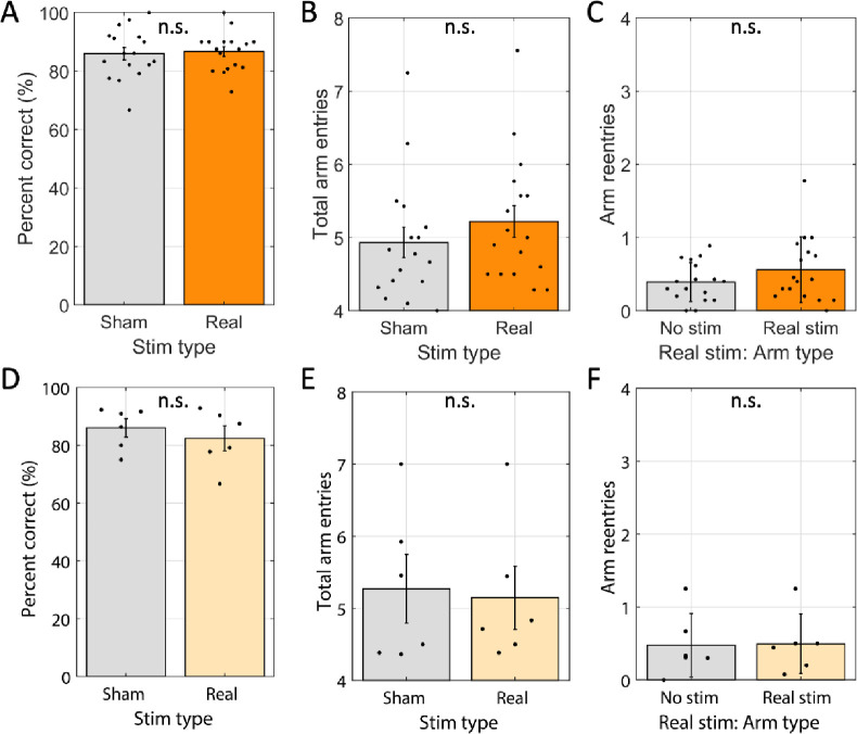
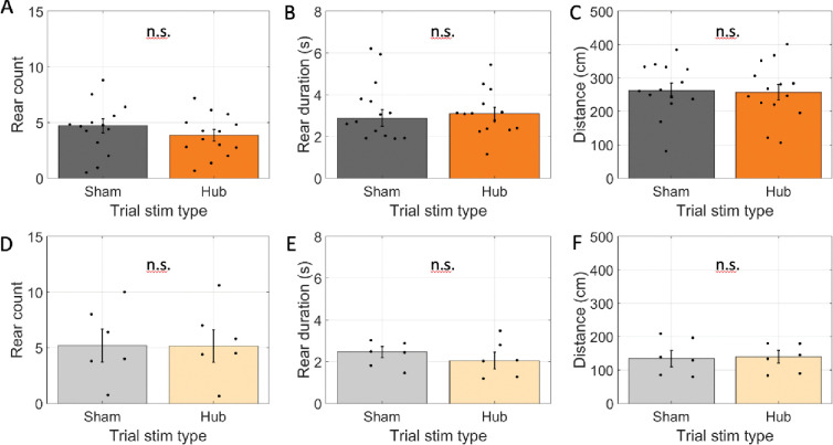
Spatial memory encoding depends in part on cholinergic modulation. How acetylcholine supports spatial memory encoding is not well understood. Prior studies indicate that acetylcholine release is correlated with exploration, including epochs of rearing onto hind legs. Here, to test whether elevated cholinergic tone increases the probability of rearing, we tracked rearing frequency and duration while optogenetically modulating the activity of choline acetyltransferase containing (i.e., acetylcholine producing) neurons of the medial septum in rats performing a spatial working memory task (n = 17 rats). The cholinergic neurons were optogenetically inhibited using halorhodopsin for the duration that rats occupied two of the four open arms during the study phase of an 8-arm radial arm maze win-shift task. Comparing rats' behavior in the two arm types showed that rearing frequency was not changed but the average duration of rearing epochs became significantly longer. This effect on rearing was observed during optogenetic inhibition but not during sham inhibition or in rats that received infusions of a fluorescent reporter virus (i.e., without halorhodopsin; n = 6 rats). Optogenetic inhibition of cholinergic neurons during the pre-trial waiting phase had no significant effect on rearing, indicating a context-specificity of the observed effects. These results are significant in that they indicate that cholinergic neuron activity in the medial septum is correlated with rearing not because it motivates an exploratory state but because it contributes to the processing of information acquired while rearing.

Keywords: Acetylcholine; Exploration; Hippocampus; Medial septum; Optogenetics; Rat; Rearing; Reversible inactivation; Spatial memory.


Conflict of interest statement

Competing Interests The authors have no competing interests to declare.

Figures

Figure 1:
Figure 1:
Overview of experimental approach. A-E. Rats performed a win-shift task on the 8-arm radial arm maze. A. Trials began with a pre-trial hub retention phase wherein the rat was placed in the hub with doors to all arms closed for 1 min. B. Study phase. A different set of four random doors opened on each trial. Rats could collect sucrose pellets from each (gray dots at arm ends). Two arms were randomly selected to be ‘stim-arms’ (orange) and the remaining two were ‘non-stim-arms’ (white). The laser was triggered throughout the time rats spent in in stim-arms. Rats were removed from the maze after collecting all pellets. C. After a 2 min. delay, rats were replaced in the hub with all doors closed for another 1 min. retention phase. D. A test phase began when all eight doors opened. Rats could collect pellets from arms not visited during the study phase. E. Timeline of a single trial. The times indicated for each exploration phase indicate the maximum allowed. Each phase was terminated once all available pellets were collected. F. Indication of timing of possible light delivery during real-stim trials. G. Indication of timing of light delivery during hub-stim trials. H. Photograph of experimental apparatus showing availability of distal cues around maze. I. Illustration of optogenetic modulation approach. ChAT::CRE+ rats were infused with either a virus packaged with CRE-dependent halorhodopsin and fluorescent reporter or a virus packaged with CRE-dependent reporter. The infusion targeted the medial septum, the dominant source of cholinergic input to the hippocampal formation (green arrow). An optical fiber was implanted just dorsal to the medial septum. J. Representative example of the distribution of EYFP expression in a rat from this study infused with CRE-dependent halorhodopsin and reporter.
Figure 2:
Figure 2:
Rear frequency was not changed by optogenetic inhibition. A. The average number of rears per arm (rear count) across rats transfected with halorhodopsin (n=17). Rear count is shown separately for arms with simulation (orange / white) and arms without stimulation (gray) on trials with the laser powered on (Trial stim type: real) and trials with the laser powered off (Trial stim type: sham). B. The difference in average rear count between arms with and without stimulation in real and sham stimulation trials. Rear count was not different across arm types in either trial type and trial types were statistically indistinguishable. C & D. Same as A & B but for rats transfected with reporter only (n=6). In all panels, dots indicate the mean over trials for individual rats, bar height indicates the median over rats, and error bars indicate standard error over rats. N.s. indicates p > 0.05.
Figure 3:
Figure 3:
Rear duration was increased by optogenetic inhibition. A. The average duration of individual rears across rats transfected with halorhodopsin (n=17). Rear duration is shown separately for rears in arms with simulation (real stimulation: orange, sham stimulation: white) and arms without stimulation (gray) on trials with the laser powered on (Trial stim type: real) and trials with the laser powered off (Trial stim type: sham). B. The difference in average rear duration between arms with and without stimulation in real and sham stimulation trials. Average rear duration was significantly greater in stim arms only in real stimulation trials. C & D. Same as A & B but for rats transfected with reporter only (n=6). In all panels, dots indicate the mean over trials for individual rats, bar height indicates the median over rats, and error bars indicate standard error over rats. ★ indicates p < 0.05. n.s. indicates p > 0.05.
Figure 4:
Figure 4:
Total distance traveled did not change with optogenetic inhibition. A. The cumulative distance traveled in real-stim-arms (orange) was not significantly different from in no-stim-arms (gray) across rats transfected with halorhodopsin (n=17) in trials with the laser powered on (Trial stim type: real) or trials with the laser powered off (Trial stim type: sham). B. The differences in distance traveled between arms with and without stimulation in real and sham stimulation trials were not significantly different. C & D. Same as A & B but for rats transfected with reporter only (n=6). In all panels, dots indicate the mean over trials for individual rats, bar height indicates the median over rats, and error bars indicate standard error over rats. n.s. indicates p > 0.05.
Figure 5:
Figure 5:
Average time spent in individual arms (i.e., dwell time) was not changed by optogenetic inhibition. A. The average time spent in individual arms across rats transfected with halorhodopsin (n=17). The time spent in individual arms is shown separately for arms with simulation (real stimulation: orange, sham stimulation: white) and arms without stimulation (gray) on trials with the laser powered on (Trial stim type: real) and trials with the laser powered off (Trial stim type: sham). B. The difference in time spent in arms with and without stimulation in real and sham stimulation trials. Average time spent was not significantly between arm types and was statistically indistinguishable between trial types. C & D. Same as A & B but for rats transfected with reporter only (n=6). In all panels, dots indicate the mean over trials for individual rats, bar height indicates the median over rats, and error bars indicate standard error over rats. n.s. indicates p > 0.05.
Figure 6:
Figure 6:
Win-shift performance was not changed by optogenetic inhibition. A. Average percentage of the first four arms entered containing reward (Percent correct) for sham stimulation trials (‘sham’) and real stimulation trials (‘real’) were statistically indistinguishable across rats transfected with halorhodopsin (n=17). B. Average number of arms entered to find the four rewards in sham stimulation and real stimulation trials were statistically indistinguishable across same rats as shown in A. C. For real stimulation trials, the average number of times each rat entered a no-stim arm relative to real stim arms were statistically indistinguishable across same rats shown in A. D–F. Same as A-C, respectively, but for rats that were transfected with reporter only (n = 6). Dots indicate the average over trials for individual rats, bar height indicates the mean over rats, and error bars indicate standard error over rats. n.s. indicates p > 0.05.
Figure 7:
Figure 7:
Optogenetic inhibition did not affect exploration when done in the pre-trial hub retention phase. A-C. Results for rats transfected with halorhodopsin. A. The number of rears during the pre-trial hub retention phase in trials with no laser stimulation (sham) and laser stimulation throughout the phase (Hub). B. The average duration of individual rearing events during the pre-trial hub retention phase. C. The total distance traveled during the pre-trial hub retention phase. D-F. Same as A-C but for rats transfected with reporter only. The average time spent in the maze hub was not significantly different between stim and sham trials. In all panels, dots indicate the average over trials for individual rats, bar height indicates the mean over rats, and error bars indicate standard error over rats. n.s. indicates p > 0.05.

References

    1. Abeelen J. H. F. van. (1989). Genetic control of hippocampal cholinergic and dynorphinergic mechanisms regulating novelty-induced exploratory behavior in house mice. Experientia 45, 839–845. - PubMed
    1. Acquas E., Wilson C. & Fibiger H. C. (1996). Conditioned and Unconditioned Stimuli Increase Frontal Cortical and Hippocampal Acetylcholine Release: Effects of Novelty, Habituation, and Fear. J Neurosci 16, 3089–3096. - PMC - PubMed
    1. Blokland A., Honig W. & Raaijmakers W.G.M. (1992). Effects of intra-hippocampal scopolamine injections in a repeated spatial acquisition task in the rat. Psychopharmacology 109, 373–376. - PubMed
    1. Dannenberg H, Pabst M., Braganza O, Schoch S, Niediek J. (2015). Synergy of direct and indirect cholinergic septo-hippocampal pathways coordinates firing in hippocampal networks. The Journal of neuroscience: the official journal of the Society for Neuroscience 35, 8394–8410. - PMC - PubMed
    1. Douchamps V., Jeewajee A., Blundell P., Burgess N. & Lever C. (2013). Evidence for Encoding versus Retrieval Scheduling in the Hippocampus by Theta Phase and Acetylcholine. The Journal of neuroscience : the official journal of the Society for Neuroscience 33, 8689–8704. - PMC - PubMed

Publication types