[Preprint]. 2023 Oct 2:2023.09.30.560293.
doi: 10.1101/2023.09.30.560293.

A Specialized Epithelial Cell Type Regulating Mucosal Immunity and Driving Human Crohn's Disease

Affiliations

A Specialized Epithelial Cell Type Regulating Mucosal Immunity and Driving Human Crohn's Disease

Jia Li et al. bioRxiv. .

Update in

  • Identification and multimodal characterization of a specialized epithelial cell type associated with Crohn's disease.
    Li J, Simmons AJ, Hawkins CV, Chiron S, Ramirez-Solano MA, Tasneem N, Kaur H, Xu Y, Revetta F, Vega PN, Bao S, Cui C, Tyree RN, Raber LW, Conner AN, Pilat JM, Jacobse J, McNamara KM, Allaman MM, Raffa GA, Gobert AP, Asim M, Goettel JA, Choksi YA, Beaulieu DB, Dalal RL, Horst SN, Pabla BS, Huo Y, Landman BA, Roland JT, Scoville EA, Schwartz DA, Washington MK, Shyr Y, Wilson KT, Coburn LA, Lau KS, Liu Q. Li J, et al. Nat Commun. 2024 Aug 22;15(1):7204. doi: 10.1038/s41467-024-51580-7. Nat Commun. 2024. PMID: 39169060 Free PMC article.

Abstract

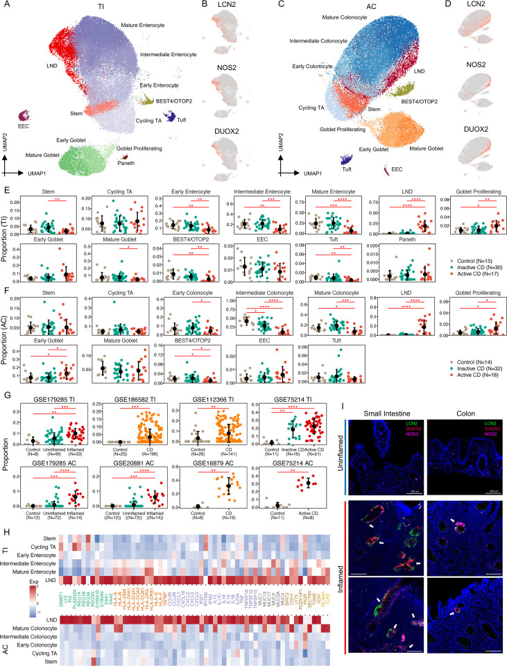
Crohn's disease (CD) is a complex chronic inflammatory disorder that may affect any part of gastrointestinal tract with extra-intestinal manifestations and associated immune dysregulation. To characterize heterogeneity in CD, we profiled single-cell transcriptomics of 170 samples from 65 CD patients and 18 non-inflammatory bowel disease (IBD) controls in both the terminal ileum (TI) and ascending colon (AC). Analysis of 202,359 cells identified a novel epithelial cell type in both TI and AC, featuring high expression of LCN2, NOS2, and DUOX2, and thus is named LND. LND cells, confirmed by high-resolution in-situ RNA imaging, were rarely found in non-IBD controls, but expanded significantly in active CD. Compared to other epithelial cells, genes defining LND cells were enriched in antimicrobial response and immunoregulation. Moreover, multiplexed protein imaging demonstrated that LND cell abundance was associated with immune infiltration. Cross-talk between LND and immune cells was explored by ligand-receptor interactions and further evidenced by their spatial colocalization. LND cells showed significant enrichment of expression specificity of IBD/CD susceptibility genes, revealing its role in immunopathogenesis of CD. Investigating lineage relationships of epithelial cells detected two LND cell subpopulations with different origins and developmental potential, early and late LND. The ratio of the late to early LND cells was related to anti-TNF response. These findings emphasize the pathogenic role of the specialized LND cell type in both Crohn's ileitis and Crohn's colitis and identify novel biomarkers associated with disease activity and treatment response.


Figures

Figure 1.
Figure 1.
A) Schematic for processing endoscopic and surgical specimens from TI and AC for non-IBD controls, inactive and active CD patients. B) Summary of the number of samples in each group. C) UMAP of 155,093 cells from endoscopy samples colored by cell types. D) Dotplot showing markers for each cell type. E) UMAP of 155,093 cells colored by tissue origin, TI (brown) or AC (blue). F) Proportion of each cell type in TI (brown) and AC samples (blue). G) UMAP of 155,093 cells colored by disease status, control (tan), inactive (green) or active CD (red). H) MDS plot of cellular compositional differences across all endoscopic specimens.
Figure 2.
Figure 2.
A) UMAP of immune and stromal cells, colored by cell type. B) Dotplot of markers in each cell type. C) Proportional changes of each immune and stromal cell type from non-IBD controls to inactive and active CD patients in TI. D) Proportional changes of each immune and stromal cell type from non-IBD controls to inactive and active CD patients in AC (* FDR<0.05, ** FDR<0.01, *** FDR<0.001, **** FDR<0.0001).
Figure 3.
Figure 3.
A) UMAP of 13 epithelial cell types in TI. B) UMAP labeled with expression of LCN2, NOS2, and DUOX2 in TI. C) UMAP of 12 epithelial cell types in AC. D) UMAP labeled with expression of LCN2, NOS2, and DUOX2 in AC. E) Proportional changes of each epithelial cell type from controls to inactive and active CD patients in TI. F) Proportional changes of each epithelial cell from controls to inactive and active CD patients in AC (* FDR<0.05, **FDR<0.01, ***FDR<0.001, **** FDR<0.0001). G) Proportional changes of LND cells with disease status in six independent cohorts (** FDR<0.01, ***FDR<0.001, **** FDR<0.0001). H) Heatmap of high expression of immune-related genes in the LND in both TI (top) and AC (bottom). I) HCR-FISH Co-staining of LCN2 (green), NOS2 (pink), and DUOX2 (red) on inflamed and uninflamed TI and AC tissues. The scale bar represents 100 μm.
Figure 4.
Figure 4.
A) Scatterplot of incoming and outgoing interaction strength of each cell type in TI. B) Scatterplot of incoming and outgoing interaction strength of each cell type in AC. C) Circle plots show the intercellular communication for LND-recruiting neutrophils, LND-plasma interaction via CCL28-CCR10, LND recruiting macrophage and neutrophils through SAA1-FPR2, and LND-mast/plasma interaction by TNFSF13-TNFRSF13B. D) Circle plots show the intercellular signaling of LND cells targeted by a variety of immune cells including IL10, LTA-TNFRSF1A, and TNFSF14-LTBR. E) Multiplex images of CD8+ , CD4+, and CD45+ cells in low and high LND in the TI (top) and AC (bottom). The scale bar represents 100 μm. F) DotPlot of marker genes in infiltrating lymocytes (ILs). G) The proportion differences of ILs between high and low LND patients.
Figure 5.
Figure 5.
A) Significance of cell-type specific expression of IBD/CD-risk genes in TI. B) Significance of cell-type specific expression of IBD/CD-risk genes in AC. C) Significance of cell-type specific expression of IBD/CD-risk genes in each CD TI specimen. D) Significance of cell-type specific expression of IBD/CD-risk genes in each CD AC specimen.
Figure 6.
Figure 6.
A) RNA velocity results mapped on the UMAP plot showing the predicted future transcriptional state of each cell. B) UMAP of early and late LND clusters in the TI. C) PAGA results mapped on the UMAP plot showing connectivity between cell types. D) Vilion plot comparing the developmental potential of each epithelial cell type predicted by CytoTRACE (** FDR<0.01, *** FDR<0.001, ****FDR<0.0001). E) Comparison of the expression of CXCL5, TNFRSF1A, and TNFRSF1B between early and late LND cells. F) Comparison of the ratio of late to early LND cells between anti-TNF responders and non-responders after the first dose of medication.
Figure 7.
Figure 7.
Spatial organization between LND and immune cells. A) H&E images for the four patient samples overlaid and colored by the expression of DUOX2. B) Dotplot of colocalization of LND markes (LCN2, NOS2, and DUOX2) and the general epithelial genes (KRT8 and KRT18) with immune signatures. Only significant colocalization (FDR<0.01) was included. Dot size denotes the z-value and color denotes the colocalization significance compared to random distribution. C) Dotplot of colocalization between epiethial and immune cells. Only significant colocalization (FDR<0.01) was included. Dot size denotes the z-value and color denotes the colocalization significance compared to random distribution.

References

    1. Arijs I., De Hertogh G., Lemaire K., Quintens R., Van Lommel L., Van Steen K., Leemans P., Cleynen I., Van Assche G., Vermeire S., et al. (2009). Mucosal gene expression of antimicrobial peptides in inflammatory bowel disease before and after first infliximab treatment. PLoS One 4, e7984. - PMC - PubMed
    1. Ayyaz A., Kumar S., Sangiorgi B., Ghoshal B., Gosio J., Ouladan S., Fink M., Barutcu S., Trcka D., Shen J., et al. (2019). Single-cell transcriptomes of the regenerating intestine reveal a revival stem cell. Nature 569, 121–125. - PubMed
    1. Badolato R., Wang J.M., Murphy W.J., Lloyd A.R., Michiel D.F., Bausserman L.L., Kelvin D.J., and Oppenheim J.J. (1994). Serum amyloid A is a chemoattractant: induction of migration, adhesion, and tissue infiltration of monocytes and polymorphonuclear leukocytes. J Exp Med 180, 203–209. - PMC - PubMed
    1. Banerjee A., Herring C.A., Chen B., Kim H., Simmons A.J., Southard-Smith A.N., Allaman M.M., White J.R., Macedonia M.C., McKinley E.T., et al. (2020). Succinate Produced by Intestinal Microbes Promotes Specification of Tuft Cells to Suppress Ileal Inflammation. Gastroenterology 159, 2101–2115 e2105. - PMC - PubMed
    1. Boland B.S., He Z., Tsai M.S., Olvera J.G., Omilusik K.D., Duong H.G., Kim E.S., Limary A.E., Jin W., Milner J.J., et al. (2020). Heterogeneity and clonal relationships of adaptive immune cells in ulcerative colitis revealed by single-cell analyses. Sci Immunol 5. - PMC - PubMed

Publication types