

Connective tissue growth factor: Role in trabecular meshwork remodeling and intraocular pressure lowering
- PMID: 37873757
- PMCID: PMC10657592
- DOI: 10.1177/15353702231199466
Connective tissue growth factor: Role in trabecular meshwork remodeling and intraocular pressure lowering
Abstract
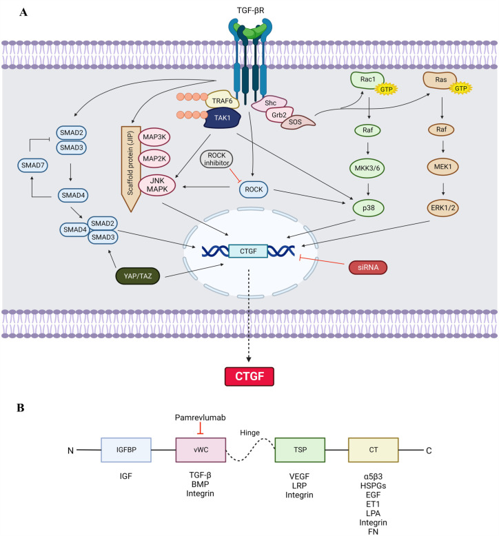
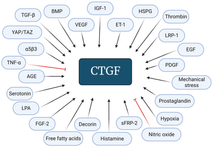
Connective tissue growth factor (CTGF) is a distinct signaling molecule modulating many physiological and pathophysiological processes. This protein is upregulated in numerous fibrotic diseases that involve extracellular matrix (ECM) remodeling. It mediates the downstream effects of transforming growth factor beta (TGF-β) and is regulated via TGF-β SMAD-dependent and SMAD-independent signaling routes. Targeting CTGF instead of its upstream regulator TGF-β avoids the consequences of interfering with the pleotropic effects of TGF-β. Both CTGF and its upstream mediator, TGF-β, have been linked with the pathophysiology of glaucomatous optic neuropathy due to their involvement in the regulation of ECM homeostasis. The excessive expression of these growth factors is associated with glaucoma pathogenesis via elevation of the intraocular pressure (IOP), the most important risk factor for glaucoma. The raised in the IOP is due to dysregulation of ECM turnover resulting in excessive ECM deposition at the site of aqueous humor outflow. It is therefore believed that CTGF could be a potential therapeutic target in glaucoma therapy. This review highlights the CTGF biology and structure, its regulation and signaling, its association with the pathophysiology of glaucoma, and its potential role as a therapeutic target in glaucoma management.
Keywords: Antiglaucoma; CTGF; TGF-β; extracellular matrix; fibrosis; remodeling.
Conflict of interest statement
Declaration of Conflicting InterestsThe author(s) declared no potential conflicts of interest with respect to the research, authorship, and/or publication of this article.
Figures
Similar articles
-
Transforming growth factor-beta2 utilizes the canonical Smad-signaling pathway to regulate tissue transglutaminase expression in human trabecular meshwork cells.Exp Eye Res. 2011 Oct;93(4):442-51. doi: 10.1016/j.exer.2011.06.011. Epub 2011 Jun 24. Exp Eye Res. 2011. PMID: 21722634 Free PMC article.
-
Src mediates TGF-β-induced intraocular pressure elevation in glaucoma.J Cell Physiol. 2019 Feb;234(2):1730-1744. doi: 10.1002/jcp.27044. Epub 2018 Aug 24. J Cell Physiol. 2019. PMID: 30144071
-
The aqueous humor outflow pathways in glaucoma: A unifying concept of disease mechanisms and causative treatment.Eur J Pharm Biopharm. 2015 Sep;95(Pt B):173-81. doi: 10.1016/j.ejpb.2015.04.029. Epub 2015 May 7. Eur J Pharm Biopharm. 2015. PMID: 25957840 Review.
-
A novel ocular function for decorin in the aqueous humor outflow.Matrix Biol. 2021 Mar;97:1-19. doi: 10.1016/j.matbio.2021.02.002. Epub 2021 Feb 11. Matrix Biol. 2021. PMID: 33582236
-
Trabecular meshwork ECM remodeling in glaucoma: could RAS be a target?Expert Opin Ther Targets. 2018 Jul;22(7):629-638. doi: 10.1080/14728222.2018.1486822. Epub 2018 Jun 14. Expert Opin Ther Targets. 2018. PMID: 29883239 Review.
Cited by
-
Challenging glaucoma with emerging therapies: an overview of advancements against the silent thief of sight.Front Med (Lausanne). 2025 Mar 26;12:1527319. doi: 10.3389/fmed.2025.1527319. eCollection 2025. Front Med (Lausanne). 2025. PMID: 40206485 Free PMC article. Review.
-
Regulation of TGF-β2-induced epithelial-mesenchymal transition and autophagy in lens epithelial cells by the miR-492/NPM1 axis.Biomol Biomed. 2024 Sep 6;24(5):1273-1289. doi: 10.17305/bb.2024.10249. Biomol Biomed. 2024. PMID: 38662949 Free PMC article.
-
Current state of signaling pathways associated with the pathogenesis of idiopathic pulmonary fibrosis.Respir Res. 2024 Jun 17;25(1):245. doi: 10.1186/s12931-024-02878-z. Respir Res. 2024. PMID: 38886743 Free PMC article. Review.
References
-
- Guha M, Xu ZG, Tung D, Lanting L, Natarajan R. Specific down-regulation of connective tissue growth factor attenuates progression of nephropathy in mouse models of type 1 and type 2 diabetes. FASEB J 2007;21:3355–68 - PubMed
-
- Ozcan AA, Ozdemir N, Canataroglu A. The aqueous levels of TGF-β2 in patients with glaucoma. Int Ophthalmol 2004;25:19–22 - PubMed
Publication types
MeSH terms
Substances
LinkOut - more resources
Full Text Sources
Medical
Miscellaneous