Characteristics of distal femoral articular cartilage in 6 weeks posttraumatic osteoarthritis by a subcritical impact
- PMID: 37874329
- PMCID: PMC10978303
- DOI: 10.1002/jor.25721
Characteristics of distal femoral articular cartilage in 6 weeks posttraumatic osteoarthritis by a subcritical impact
Abstract
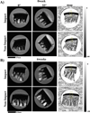
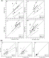
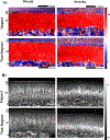
Traumatized knee greatly contributes to osteoarthritis (OA) of the knee in young adults. To intervene effectively before the onset of severe structural disruption, detection of the disease at the early onset is crucial. In this study, we put together the findings for the detection of OA from the femoral knee joint cartilage of the rabbit at 6 weeks posttrauma. Articular cartilage samples are taken from the impacted and nonimpacted joints at 0 week (serving as the control group) and at 6 weeks posttrauma by minimal force. The samples were imaged using microscopic magnetic resonance imaging (µMRI) at 11.7 µm/pixel and polarized light microscopy (PLM) at 1 µm/pixel. In addition, an inductively coupled plasma - optical emission spectrometry analysis was performed using the adjacent cartilage samples. The outcomes of this study demonstrate an increase in T2 values in 6 weeks samples compared to the 0 week samples by µMRI technique, indicating a general increase of tissue hydration within cartilage. PLM detects a decrease in the average thickness of the superficial zones in the posttraumatic osteoarthritis samples, significant in the impacted femurs. There was an average increasing trend of maximum retardation in the tide mark in comparison to the reported calcium concentration (mg/L) in impacted samples suggesting a possible rise in mineralization in the 6 weeks samples. Qualitatively, physical observation of the joint after 6 weeks showed signs of reddening in the anterior femur suggesting the disease process is a localized phenomenon. Through microscopic imaging, we are able to detect these changes at 6 weeks posttrauma qualitatively and quantitatively.
Keywords: articular cartilage; inductively coupled plasma‐optical emission spectrometry; microscopic magnetic resonance imaging; polarized light microscopy; posttraumatic osteoarthritis.
© 2023 Orthopaedic Research Society.
Figures


Similar articles
-
Assessment of articular cartilage degradation in response to an impact injury using µMRI.Connect Tissue Res. 2024 Mar;65(2):146-160. doi: 10.1080/03008207.2024.2319050. Epub 2024 Feb 28. Connect Tissue Res. 2024. PMID: 38415672 Free PMC article.
-
Structural differences between immature and mature articular cartilage of rabbits by microscopic MRI and polarized light microscopy.J Anat. 2022 Jun;240(6):1141-1151. doi: 10.1111/joa.13620. Epub 2022 Jan 3. J Anat. 2022. PMID: 34981507 Free PMC article.
-
Detecting structural changes in early experimental osteoarthritis of tibial cartilage by microscopic magnetic resonance imaging and polarised light microscopy.Ann Rheum Dis. 2004 Jun;63(6):709-17. doi: 10.1136/ard.2003.011783. Ann Rheum Dis. 2004. PMID: 15140779 Free PMC article.
-
Quantitative µMRI and PLM study of rabbit humeral and femoral head cartilage at sub-10 µm resolutions.J Orthop Res. 2020 May;38(5):1052-1062. doi: 10.1002/jor.24547. Epub 2019 Dec 12. J Orthop Res. 2020. PMID: 31799697 Free PMC article.
-
[Progress of research in osteoarthritis. Quantitative magnetic resonance imaging of cartilage in knee osteoarthritis].Clin Calcium. 2009 Nov;19(11):1638-43. Clin Calcium. 2009. PMID: 19880997 Review. Japanese.
Cited by
-
Assessment of articular cartilage degradation in response to an impact injury using µMRI.Connect Tissue Res. 2024 Mar;65(2):146-160. doi: 10.1080/03008207.2024.2319050. Epub 2024 Feb 28. Connect Tissue Res. 2024. PMID: 38415672 Free PMC article.
References
-
- Watt FE, Corp N, Kingsbury SR, et al. Towards prevention of post-traumatic osteoarthritis: report from an international expert working group on considerations for the design and conduct of interventional studies following acute knee injury. Osteoarthr Cartil. 2019;27(1):23–33. doi:10.1016/j.joca.2018.08.001 - DOI - PMC - PubMed
Publication types
MeSH terms
Grants and funding
LinkOut - more resources
Full Text Sources
Medical

