

Increasing baseline aortic valve peak flow velocity is associated with progression of aortic valve stenosis in osteoporosis patients-a possible link to low vitamin D status
- PMID: 37874407
- PMCID: PMC10598115
- DOI: 10.1007/s11657-023-01339-2
Increasing baseline aortic valve peak flow velocity is associated with progression of aortic valve stenosis in osteoporosis patients-a possible link to low vitamin D status
Abstract
Purpose: The purpose of this study was to investigate the morphological characteristics of the aortic valve and identify factors associated with the progression of aortic valve stenosis (AS) in osteoporosis patients.
Methods: In this single-center prospective cohort study, we recruited 10 patients (mean age: 75 ± 7 years, 90% female) who were taking anti-resorptive medicines at the outpatient clinic of University of Miyazaki Hospital, Japan. Baseline assessments, including transthoracic echocardiogram, blood sampling, and dual energy X-ray absorptiometry, were performed. Follow-up assessments were conducted at 6, 12, 18, and 24 months.
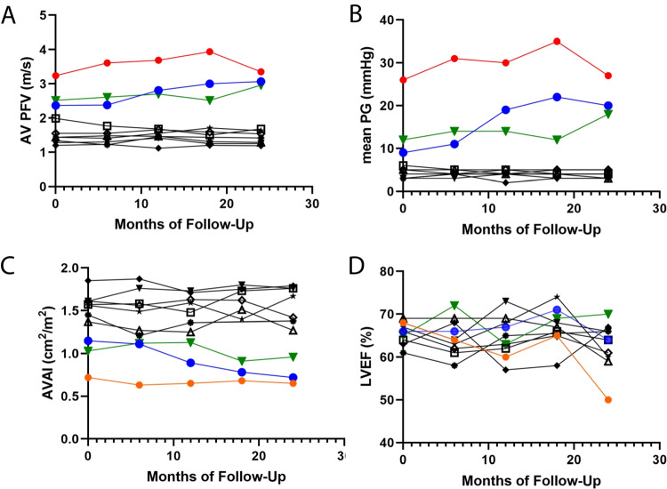
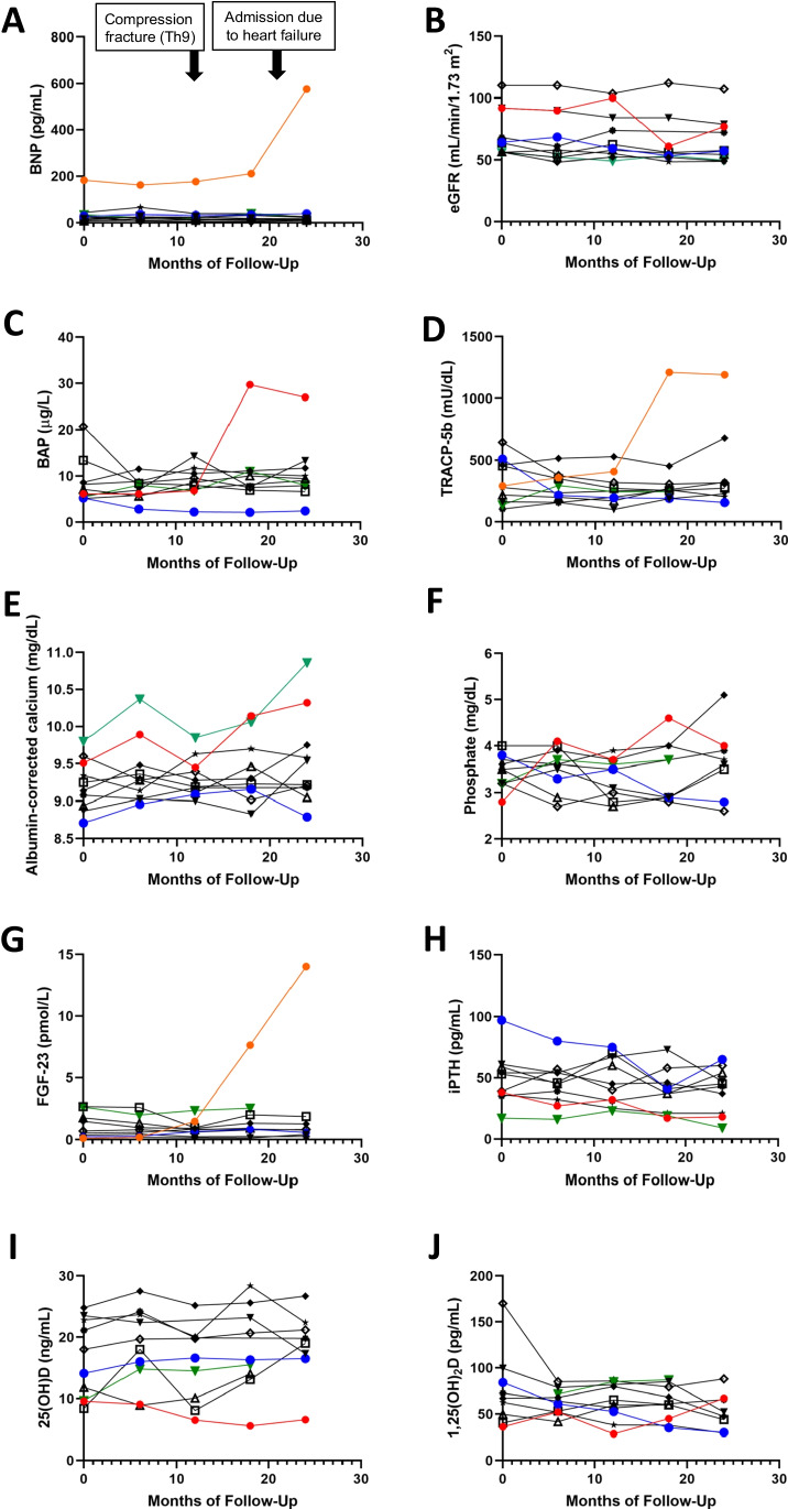
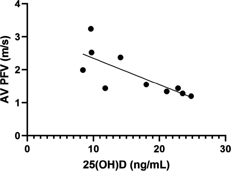
Results: During the 2-year follow-up, three patients with aortic valve peak flow velocity (AV PFV) ≥2 m/s at baseline developed moderate AS, which is defined as AV PFV ≥3 m/s. However, seven patients with AV PFV <2 m/s did not exhibit any progression of AS. There were significant variations in terms of bone mineral density, T-score values, and biomarkers associated with bone turnover (i.e., bone alkaline phosphatase, tartrate-resistance acid phosphatase-5b) among the enrolled patients, but none of these factors were found to be associated with the progression of AS. All patients exhibited low vitamin D status, with a median level of 16.1 ng/mL (25th percentile, 9.7 ng/mL; 75th percentile, 23 ng/mL). The baseline levels of AV PFV values were negatively correlated with 25-hydroxyvitamin D levels, determined by univariate linear regression analysis (beta coefficient = -0.756, 95% confidence interval, -0.136 ̶ -0.023, p = 0.011).
Conclusion: Our data suggest that low vitamin D status might be a potential risk factor for the progression of AS in osteoporosis patients undergoing treatment with anti-resorptive medicines. Elderly patients with osteoporosis patients exhibited a subset of aortic valve stenosis. Our data suggest that the baseline aortic valve peak flow velocity predicts the progression of aortic valve stenosis, and there might be an association between the progression and the co-existing low vitamin D status in these patients.
Keywords: Calcium; Echocardiogram; Osteoporosis; Valvular disease; Vitamin D.
© 2023. The Author(s).
Conflict of interest statement
Toshihiro Tsuruda, Taro Funamoto, Chiyoko Suzuki, Yoshimasa Yamamura, Michikazu Nakai, Etsuo Chosa, and Koichi Kaikita declare that they have no conflict of interest.
Figures
Similar articles
-
Administration of angiotensin-converting enzyme inhibitors is associated with slow progression of mild aortic stenosis in Japanese patients.Heart Vessels. 2011 May;26(3):252-7. doi: 10.1007/s00380-010-0052-x. Epub 2010 Nov 10. Heart Vessels. 2011. PMID: 21063877
-
Association between evolocumab use and slow progression of aortic valve stenosis.Heart Vessels. 2024 Aug;39(8):725-734. doi: 10.1007/s00380-024-02386-6. Epub 2024 Mar 19. Heart Vessels. 2024. PMID: 38499696
-
Progression of aortic valve stenosis is associated with bone remodelling and secondary hyperparathyroidism in elderly patients--the COFRASA study.Eur Heart J. 2013 Jul;34(25):1915-22. doi: 10.1093/eurheartj/ehs450. Epub 2013 Jan 17. Eur Heart J. 2013. PMID: 23329150 Clinical Trial.
-
Asymptomatic Severe Aortic Stenosis in the Elderly.JACC Cardiovasc Imaging. 2017 Jan;10(1):43-50. doi: 10.1016/j.jcmg.2016.05.015. Epub 2016 Sep 14. JACC Cardiovasc Imaging. 2017. PMID: 27639763
-
Aortic Stenosis Progression: A Systematic Review and Meta-Analysis.JACC Cardiovasc Imaging. 2023 Mar;16(3):314-328. doi: 10.1016/j.jcmg.2022.10.009. Epub 2022 Dec 14. JACC Cardiovasc Imaging. 2023. PMID: 36648053
Cited by
-
Vitamin D and Cardiovascular Diseases: From Physiology to Pathophysiology and Outcomes.Biomedicines. 2024 Mar 30;12(4):768. doi: 10.3390/biomedicines12040768. Biomedicines. 2024. PMID: 38672124 Free PMC article. Review.
References
-
- Osnabrugge RLJ, Mylotte D, Head SJ, Van Mieghem NM, Nkomo VT, LeReun CM, Bogers AJJC, Piazza N, Kappetein AP. Aortic Stenosis in the Elderly: Disease Prevalence and Number of Candidates for Transcatheter Aortic Valve Replacement: A Meta-Analysis and Modeling Study. Journal of the American College of Cardiology. 2013;62:1002–1012. - PubMed
-
- Eveborn GW, Schirmer H, Heggelund G, Lunde P, Rasmussen K. The evolving epidemiology of valvular aortic stenosis. the Tromsø study. Heart. 2013;99:396–400. - PubMed
-
- Marquis-Gravel G, Redfors B, Leon MB, Généreux P. Medical Treatment of Aortic Stenosis. Circulation. 2016;134:1766–1784. - PubMed
-
- Otto CM, Prendergast B. Aortic-valve stenosis--from patients at risk to severe valve obstruction. N Engl J Med. 2014;371:744–756. - PubMed
-
- Pawade TA, Newby DE, Dweck MR. Calcification in Aortic Stenosis: The Skeleton Key. J Am Coll Cardiol. 2015;66:561–577. - PubMed
Publication types
MeSH terms
Substances
LinkOut - more resources
Full Text Sources
Medical
Research Materials
