Review
. 2023;40(1):2272065.
doi: 10.1080/02656736.2023.2272065. Epub 2023 Oct 24.

Clinical translation of abdominal histotripsy: a review of preclinical studies in large animal models

Affiliations
Review

Clinical translation of abdominal histotripsy: a review of preclinical studies in large animal models

Katrina L Falk et al. Int J Hyperthermia. 2023.

Abstract

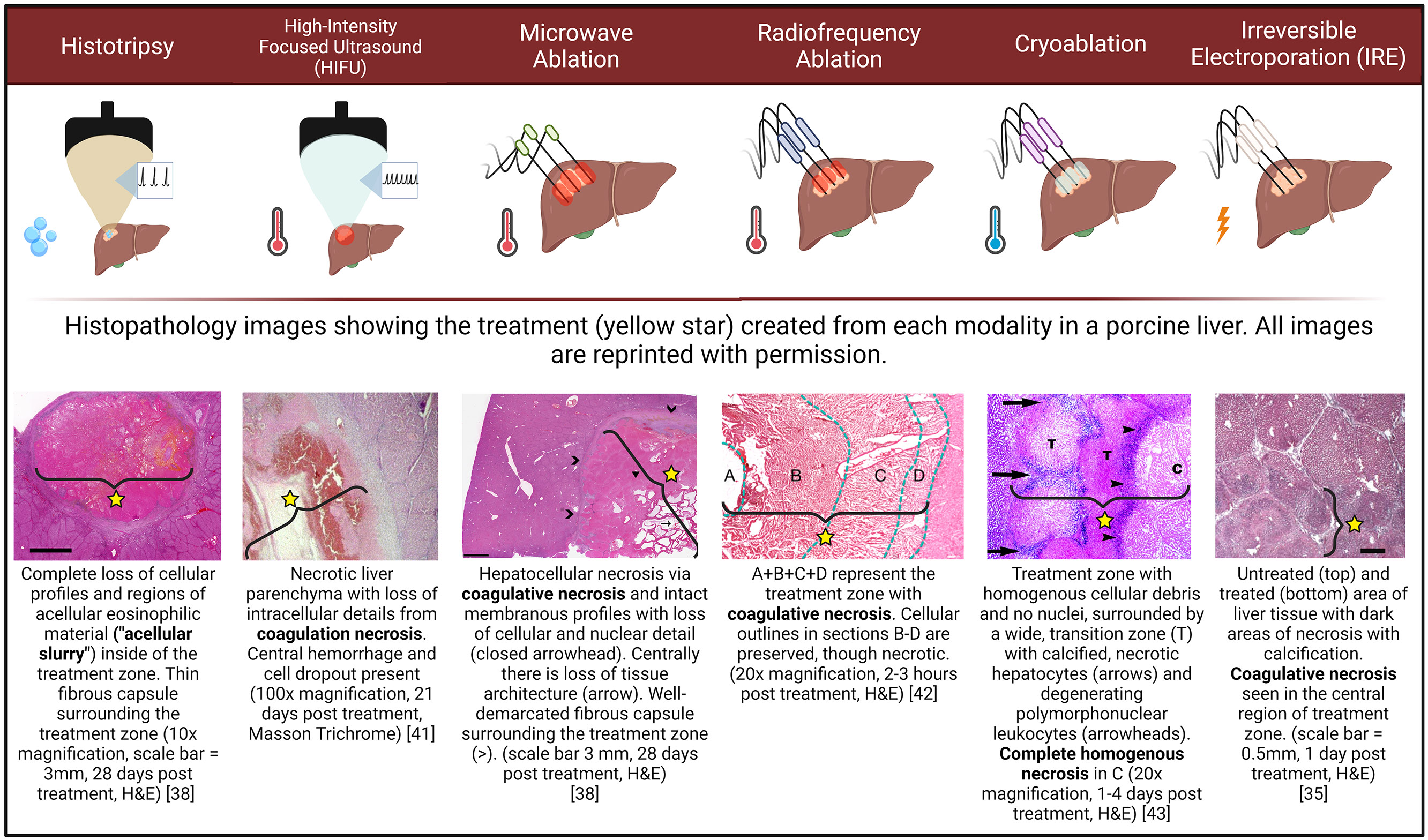
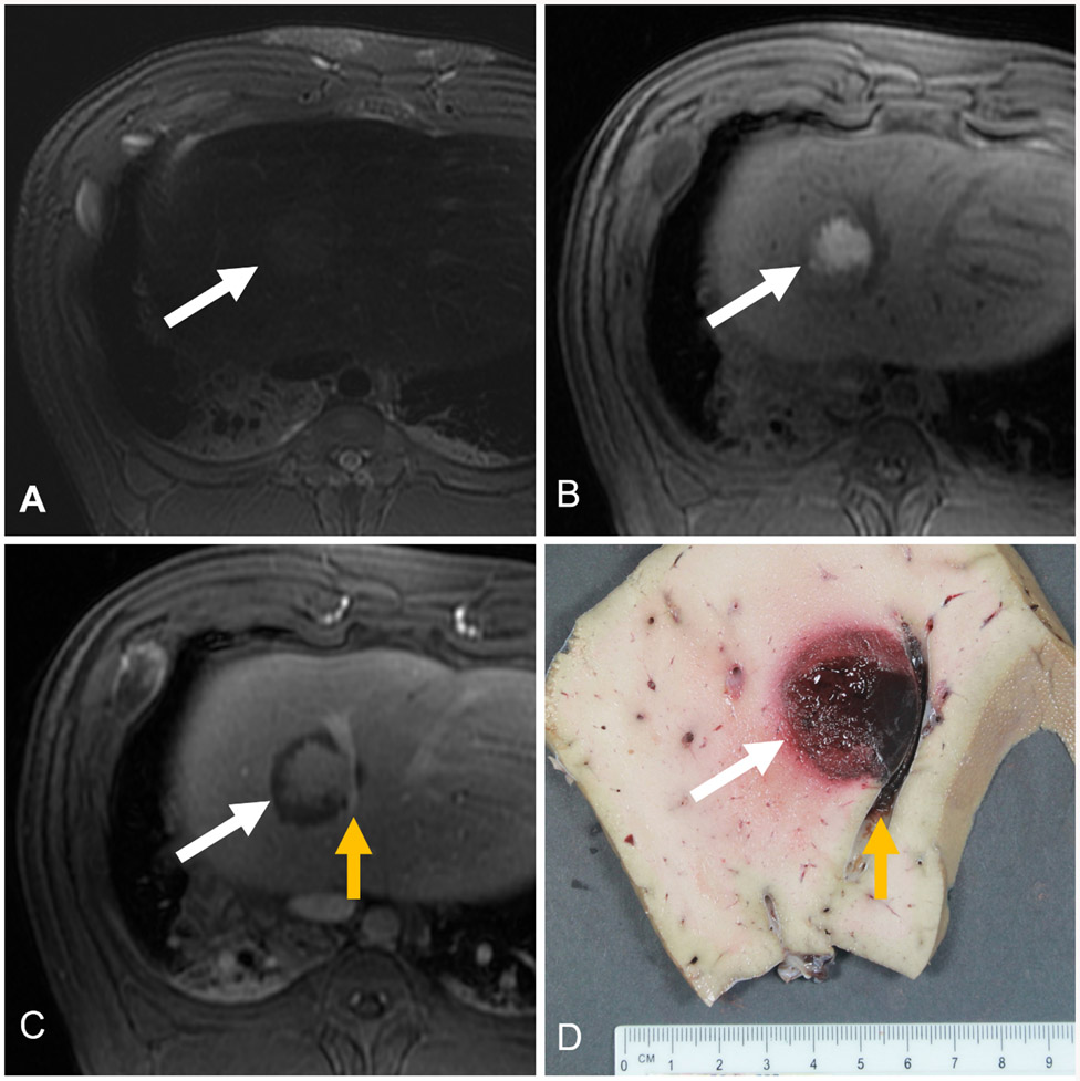
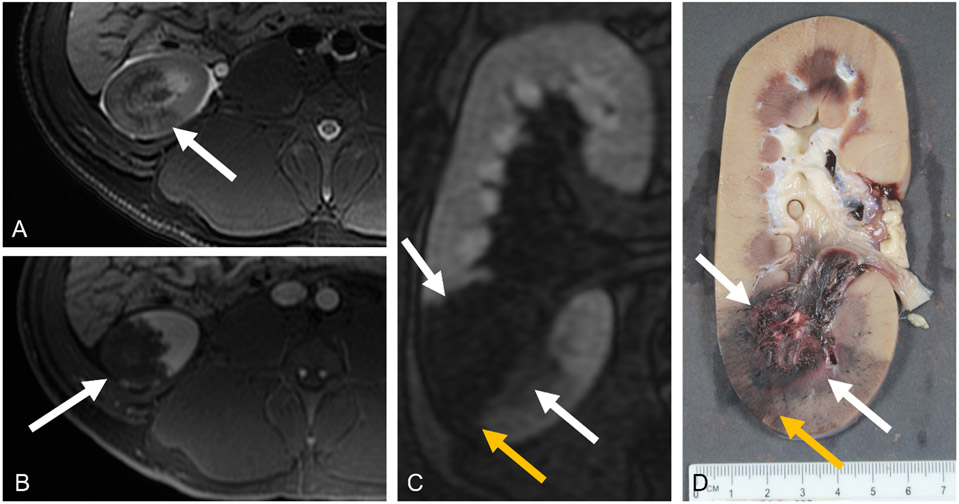
Histotripsy is an emerging noninvasive, non-thermal, and non-ionizing focused ultrasound (US) therapy that can be used to destroy targeted tissue. Histotripsy has evolved from early laboratory prototypes to clinical systems which have been comprehensively evaluated in the preclinical environment to ensure safe translation to human use. This review summarizes the observations and results from preclinical histotripsy studies in the liver, kidney, and pancreas. Key findings from these studies include the ability to make a clinically relevant treatment zone in each organ with maintained collagenous architecture, potentially allowing treatments in areas not currently amenable to thermal ablation. Treatments across organ capsules have proven safe, including in anticoagulated models which may expand patients eligible for treatment or eliminate the risk associated with taking patients off anti-coagulation. Treatment zones are well-defined with imaging and rapidly resorb, which may allow improved evaluation of treatment zones for residual or recurrent tumor. Understanding the effects of histotripsy in animal models will help inform physicians adopting histotripsy for human clinical use.

Keywords: Histotripsy; abdomen; ablation; kidney; liver; pancreas; renal; tumor.


Conflict of interest statement

Disclosure Statement

Conflict of interest. Author PL is a consultant and stockholder with HistoSonics, Inc., a consultant with NeuWave/Ethicon, Inc., and receives research support from Siemens Healthineers. Author AS is a consultant and speaking faculty for NeuWave Medical, on the Advisory Board for AstraZeneca, a consultant for Varian, and advisor and shareholder of HistoSonics, Inc. Author EV is a consultant, stock- holder, and receives research support from HistoSonics, Inc. Author FL is a consultant, stockholder, receives research support, and is on the board of directors at HistoSonics, Inc., is a consultant with Ethicon, Inc., is on the Canon Medical Advisory Board, and has patents and royalties with Medtronic, Inc. Author TZ is a consultant, stockholder, receives research support from HistoSonics, Inc. and is a consultant with Ethicon, Inc.). KF, MK, AZ, EK, CB do not have anything to disclose.

Figures

Figure 1:
Figure 1:
Histopathologic comparison of tissue destruction modalities. Differences in procedural application and mechanism of action between histotripsy, high intensity focused ultrasound (HIFU), microwave ablation (MW), radiofrequency ablation (RF), cryoablation, and irreversible electroporation (IRE) are illustrated in the pictograms under each modality. Histotripsy uses cavitation to mechanically destroy tissue. HIFU, MW, RF and cryoablation use thermal mechanisms to create coagulative necrosis and IRE uses electricity to disrupt the cell membrane potential. Pathologic differences are illustrated below each modality. Images have been reprinted with permission and license numbers can be found in the Acknowledgements section.
Figure 2:
Figure 2:
MRI and gross pathology images of a histotripsy treatment (white arrows) in a porcine liver. A) Axial T2-weighted fat-saturated MRI following histotripsy of a healthy swine liver. B) Axial T1 weighted MRI before the administration of IV contrast. C) Axial T1 weighted MRI after the administration of a gadolinium-based contrast, shown in the portal venous phase. A patent vessel runs through the treatment zone (yellow arrow). D) Corresponding gross pathology of histotripsy treatment with patent vessel.
Figure 3:
Figure 3:
MRI and gross pathology images of a histotripsy treatment (white arrow) in a porcine kidney. A) Axial T2-weighted fat-saturated MRI following histotripsy of a healthy swine kidney. B) Axial T1 weighted MRI with IV contrast (gadobenate dimeglumine) in the corticomedullary phase after treatment (white arrow). C) Coronal MRI with contrast 13 minutes after administering IV contrast. Note the wedge-shaped perfusion defect (yellow arrow) extending peripheral to the treatment zone (white arrows), causing the treatment zone to appear larger. D) Corresponding gross image demonstrating the central area of treatment (white arrows) with adjacent perfusion deficit (yellow arrow).

References

    1. Xu Z, Ludomirsky A, Eun LY, et al. Controlled ultrasound tissue erosion. Proc IEEE Ultrason Symp. 2004;51:726–736. - PMC - PubMed
    1. Vlaisavljevich E, Maxwell A, Mancia L, et al. Visualizing the Histotripsy Process: Bubble Cloud–Cancer Cell Interactions in a Tissue-Mimicking Environment. Ultrasound Med Biol. 2016;42:2466–2477. - PMC - PubMed
    1. Maxwell AD, Yuldashev PV, Kreider W, et al. A Prototype Therapy System for Transcutaneous Application of Boiling Histotripsy HHS Public Access. IEEE Trans Ultrason Ferroelectr Freq Control. 2017;64:1542–1557. - PMC - PubMed
    1. Vlaisavljevich E, Maxwell A, Warnez M, et al. Histotripsy-induced cavitation cloud initiation thresholds in tissues of different mechanical properties. IEEE Trans Ultrason Ferroelectr Freq Control. 2014;61:341–352. - PMC - PubMed
    1. Khokhlova TD, Canney MS, Khokhlova VA, et al. Controlled tissue emulsification produced by high intensity focused ultrasound shock waves and millisecond boiling. J Acoust Soc Am. 2011;130:3498–3510. - PMC - PubMed

Publication types

LinkOut - more resources