. 2023 Oct 24;13(1):18175.
doi: 10.1038/s41598-023-45285-y.

Insight into synthesis and characterisation of Ga0.9Fe2.1O4 superparamagnetic NPs for biomedical applications

Affiliations

Insight into synthesis and characterisation of Ga0.9Fe2.1O4 superparamagnetic NPs for biomedical applications

Amalia Mesaros et al. Sci Rep. .

Abstract

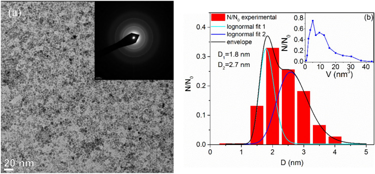
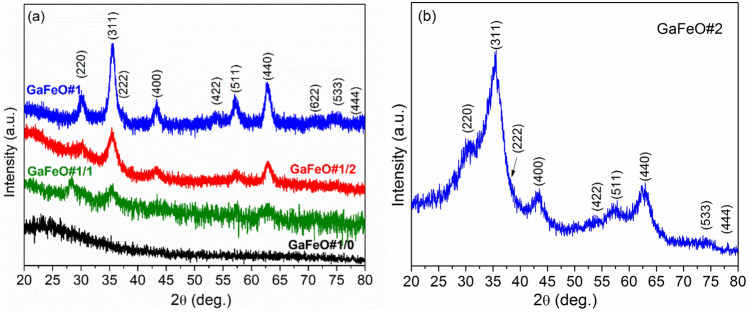
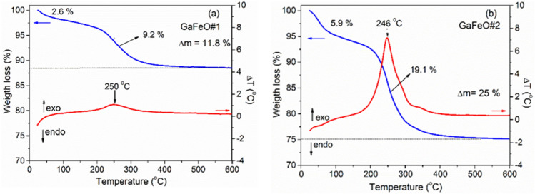
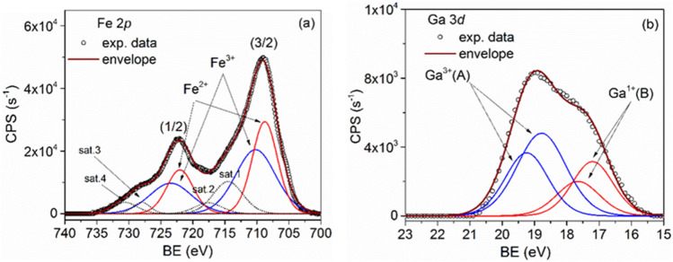
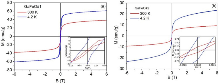
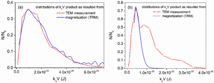
A Ga3+-substituted spinel magnetite nanoparticles (NPs) with the formula Ga0.9Fe2.1O4 were synthesized using both the one-pot solvothermal decomposition method (TD) and the microwave-assisted heating method (MW). Stable colloidal solutions were obtained by using triethylene glycol, which served as a NPs stabilizer and as a reaction medium in both methods. A narrow size distribution of NPs, below 10 nm, was achieved through selected nucleation and growth. The composition, structure, morphology, and magnetic properties of the NPs were investigated using FTIR spectroscopy, thermal analysis (TA), X-ray diffraction (XRD), transmission electron microscopy (TEM), X-ray photoelectron spectroscopy (XPS), and magnetic measurements. NPs with the expected spinel structure were obtained in the case of the TD method, while the MW method produced, additionally, an important amount of gallium suboxide. The NPs, especially those prepared by TD, have superparamagnetic behavior with 2.02 μB/f.u. at 300 K and 3.06 μB/f.u. at 4.2 K. For the MW sample these values are 0.5 μB/f.u. and 0.6 μB/f.u. at 300 K and 4.2 K, respectively. The MW prepared sample contains a secondary phase and very small NPs which affects both the dimensional distribution and the magnetic behavior of NPs. The NPs were tested in vitro on amniotic mesenchymal stem cells. It was shown that the cellular metabolism is active in the presence of Ga0.9Fe2.1O4 NPs and preserves an active biocompatible cytoskeleton.


Conflict of interest statement

The authors declare no competing interests.

Figures

Figure 1
Figure 1
(a) TEM, (b) HRTEM images, and (c) SAED pattern from GaFeO#1 sample. (d) Normalized diameter distribution with, as inset, the corresponding normalized volume distribution.
Figure 2
Figure 2
(a) TEM image and SAED patterns (inset) for GaFeO#2 sample and (b) the bimodal normalized diameter distribution with the corresponding normalized volume distribution in the inset.
Figure 3
Figure 3
XRD patterns of the GaFeO NPs obtained by (a) solvothermal and (b) microwave assisted routes. The reflections have been indexed according to the JCDD PDF 074-2226 for Ga0.9Fe2.1O4 spinel structure. (GaFeO#1/0, GaFeO#1/1, and GaFeO#1/2 dwell time 0, 1, and 2 h).
Figure 4
Figure 4
TG–DTA analysis of the GaFeO NPs: (a) GaFeO#1sample prepared by the solvothermal and (b) GaFeO#2 sample prepared by microwave assisted decomposition.
Figure 5
Figure 5
FT-IR spectra of TEG and GaFeO samples.
Figure 6
Figure 6
XPS spectra of (a) Fe (2p), (b) Ga (3d) core-levels together with the corresponding deconvolutions for the GaFeO#1 sample.
Figure 7
Figure 7
XPS spectra of (a) Fe (2p), (b) Ga (3d) core-levels together with the corresponding deconvolutions for the GaFeO#2 sample.
Figure 8
Figure 8
Magnetization of samples (a) GaFeO#1 and (b) GaFeO#2 as a function of the applied magnetic field, determined at room temperature and 4.2 K.
Figure 9
Figure 9
The magnetization curve of the GaFeO#1 NPs at room temperature (□) together with the best fit obtained by using Eq. 1 (continuous line). The magnetizations were calculated referred to the magnetic content of this sample as given in Table 1.
Figure 10
Figure 10
FC and ZFC curves highlighting changes in magnetization of NPs as a function of temperature for (a) GaFeO#1 and (b) GaFeO#2 samples.
Figure 11
Figure 11
First derivative of TRM magnetizations for samples GaFeO#1 and GaFeO#2.
Figure 12
Figure 12
Compared distributions of the keffV product as resulted from TEM and magnetic measurements for (a) GaFeO#1 and (b) GaFeO#2 samples.
Figure 13
Figure 13
The measured values of the absorbance at 570 nm of the AFSC after 72 h of incubation in the presence of different quantities of GaFeO NPs as compared to the control cells. Here GF1 and GF2 means GaFeO#1 and GaFeO#2 samples respectively.
Figure 14
Figure 14
Biocompatibility assessment of GaFeO#1 and GaFeO#2 NPs through fluorescence microscopic images of AFSC monolayers after 5 days: (a,f) control cells; (b) GaFeO#1—3.3 mg/mL; (c) GaFeO#1—8.3 mg/mL; (d) GaFeO#1—16.66 mg/mL (e) GaFeO#1—33.33 mg/mL; (g) GaFeO#2—3.3 mg/mL; (h) GaFeO#2—8.3 mg/mL; (i) GaFeO#2—16.66 mg/mL; (j) GaFeO#2—33.33 mg/mL. The all scale bars are 20 μm.
Figure 15
Figure 15
Representative fluorescence images of tubulin filaments of AFSC after 72 h of incubation in the presence of (c) GaFeO #1—8.3 mg/mL; (d) GaFeO #2—8.3 mg/mL; (e) GaFeO #1—33.33 mg/mL; (f) GaFeO #2—33.33 mg/mL. Here (a) represents the control cells and (b) is the negative control-lacking primary antibody. The scale bars are 20 μm.

References

    1. Malik R, Sehdev N, Lamba S, Sharma P, Makino A, Annapoorni S. Magnetic memory effects in nickel ferrite/polymer nanocomposites. Appl. Phys. Lett. 2014;104:122407–122407. doi: 10.1063/1.4869724. - DOI
    1. Miller MM, Prinz GA, Cheng SF, Bounnak S. Detection of a micron-sized magnetic sphere using a ring-shaped anisotropic magnetoresistance-based sensor: A model for a magnetoresistance-based biosensor. Appl. Phys. Lett. 2002;81:2211–2213. doi: 10.1063/1.1507832. - DOI
    1. Jain TK, Morales MA, Sahoo SK, Leslie-Pelecky DL, Labhasetwar V. Iron oxide NPs for sustained delivery of anticancer agents. Mol. Pharm. 2005;2:194–205. doi: 10.1021/mp0500014. - DOI - PubMed
    1. Chourpa I, Douziech-Eyrolles L, Okassa LN, Fouquenet JF, Jonathan SC, Souce M, Marchais H, Dubois P. Molecular composition of iron oxide nanoparticles, precursors for magnetic drug targeting, as characterized by confocal Raman microspectroscopy. Analyst. 2005;130(10):1395–1403. doi: 10.1039/b419004a. - DOI - PubMed
    1. Rećko K, Satuła D, Waliszewskia J, Biernacka M, Orzechowska M, Kalska-Szostko B, Soloviov D, Miaskowski A, Beskrovnyy A, Basa A, Szymański K. Magnetism of surface-modified and gallium-doped magnetite particles. J. Surf. Invest. 2020;14:S85–S92. doi: 10.1134/S102745102007040X. - DOI

Publication types

MeSH terms