. 2024 Apr;48(2):705-724.
doi: 10.1007/s11259-023-10237-4. Epub 2023 Oct 24.

Immunomodulatory effect of probiotic exopolysaccharides in a porcine in vitro co-culture model mimicking the intestinal environment on ETEC infection

Affiliations

Immunomodulatory effect of probiotic exopolysaccharides in a porcine in vitro co-culture model mimicking the intestinal environment on ETEC infection

Zuzana Kiššová et al. Vet Res Commun. 2024 Apr.

Abstract

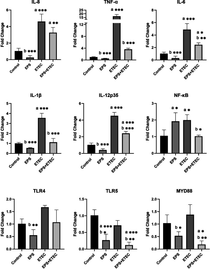
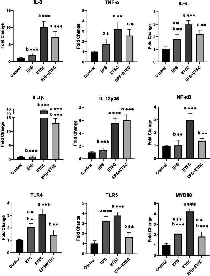
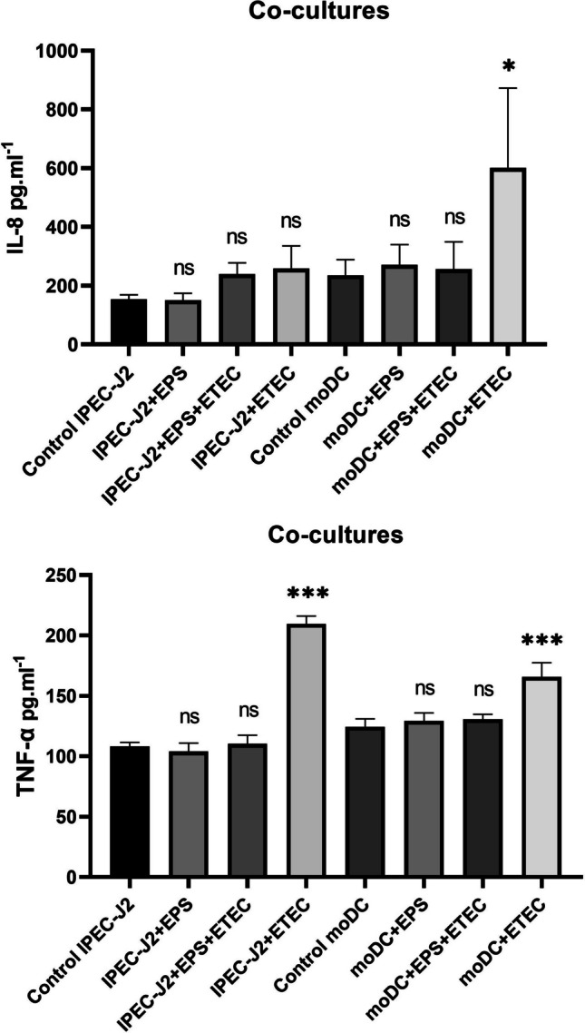
The aim of this study was to evaluate the immunomodulatory effect of EPS-L26 isolated from the probiotic strain Lactobacillus (Limosilactobacillus) reuteri L26 Biocenol™, in a model of infection with an enterotoxigenic E. coli (ETEC) by establishing monocultures consisting of the IPEC-J2 cell line or monocyte-derived dendritic cells (moDCs) and creating a 3D model of cell co-cultures established with IPEC-J2 cells and moDCs. The immunomodulatory and immunoprotective potential of used EPS-L26 was confirmed in monocultures in an experimental group of pretreated cells, where our study showed that pretreatment of cells with EPS-L26 and subsequent exposure to infection resulted in significantly down-regulated mRNA levels of genes encoding inflammatory cytokines compared to ETEC challenge in single cell cultures (in IPEC-J2, decreased mRNA levels for TNF-α, IL-6, IL-1β, IL-12p35; in moDCs, decreased mRNA levels for IL-1β). Similar to monocultures, we also demonstrated the immunostimulatory potential of the ETEC strain in the co-culture model on directly treated IPEC-J2 cells cultivated on insert chambers (apical compartment) and also on indirectly treated moDCs cultivated in the lower chamber (basolateral compartment), however in the co-culture model the expression of inflammatory cytokines was attenuated at the mRNA level compared to monocultures. Pretreatment of the cells on the insert chambers pointed to the immunoprotective properties of EPS-L26, manifested by decreased mRNA levels in both cell lines compared to ETEC challenge (in IPEC-J2 decreased mRNA levels for IL-12p35; in moDCs decreased mRNA levels for IL-1β, IL-6). Our results suggest intercellular communication via humoral signals derived from IPEC-J2 cells by influencing the gene expression of indirectly treated moDC cells located in the basolateral compartment.

Keywords: Co-culture; Cytokines; EPS; ETEC; IPEC-J2; moDCs; qPCR.


Conflict of interest statement

The authors declare no competing interests.

The authors have no pertinent financial or non-financial conflicts of interest to declare.

Figures

Fig. 1
Fig. 1
Schematic diagram showing monoculture and co-culture system. a-b) IPEC-J2 or moDC monocultures seeded onto 12-well culture plates; c) IPEC-J2 cells seeded onto 12-well Transwell®-COL membrane inserts at a density of 2.5 × 105 cells/cm2 and cultured for 15 days. Porcine moDC seeded onto 12-well plates at a cell density of 4,5 × 105 cells/cm2 and cultured for 5 days. IPEC-J2 cell inserts were placed in the wells above the moDC
Fig. 2
Fig. 2
Progression of moDC differentiation by light microscopy (400 x). a) Freshly isolated monocytes on day 0 of culture with a characteristic round shape morphology. b) The presence of the growth factors IL-4 and GM-CSF in the culture media induces moDC differentiation and maturation. Increased elongation of cytoplasmic processes was observed as early as day 3 of culture. The round cell morphology characteristic of monocytes was lost and the cells acquired an elongated shape. c-d) Immature moDCs on day 5 of differentiation; ef) Fully mature moDCs after 48 h of activation with LPS (100 ng/mL) on day 7 of differentiation
Fig. 3
Fig. 3
FACS analysis during moDC differentiation. Legend: APC – allophycocyanin, FITC – fluorescein isothiocyanate, PE – phycoerythrin, SLA II – MHC II
Fig. 4
Fig. 4
Evaluation of transepithelial electrical resistance (TEER). Mean TEER values of a monolayer of IPEC-J2 cells on Transwell®—COL collagen-coated inserts (area 1.12 cm2; pore size 0.4 μm, ± SD n = 2) measured over 22 days of cultivation
Fig. 5
Fig. 5
Macromolecular permeability of the IPEC-J2 monolayer formed on collagen-coated Transwell®—COL inserts. Basolateral HRP concentration was measured during 110 min incubation. Legend: C – inserts without cells; Day 5–20: inserts with IPEC-J2 cells on a corresponding day after planting
Fig. 6
Fig. 6
Immunomodulatory activity of EPS-L26 and ETEC in IPEC-J2 monocultures. Results are expressed as mean ± SD). The statistical significance of the differences was evaluated using Dunnett's multiple comparison test. a—Results significantly differed from the control group of cells without treatment (* p < 0.05; ** p < 0.01; *** p < 0.001). b—Results significantly differed from ETEC (* p < 0.05; ** p < 0.01; *** p < 0.001). Error bars represent standard deviations. Legend: Control – cells treated with IPEC-J2 medium (without FBS and antibiotics); EPS – cells treated with EPS-L26 (100 ug/mL) for 4 h; ETEC – cells challenged with ETEC for 2 h; EPS + ETEC – cells treated with EPS-L26 (100 ug/mL) for 4 h followed by 2 h of ETEC challenge
Fig. 7
Fig. 7
Immunomodulatory activity of EPS-L26 and ETEC in moDC monocultures. Results are expressed as mean ± SD. The statistical significance of the differences was evaluated using Dunnett's multiple comparison test. a—Results significantly different from the control group of untreated cells (* p < 0.05; ** p < 0.01; *** p < 0.001). b—Results significantly different from ETEC (* p < 0.05; ** p < 0.01; *** p < 0.001). Error bars represent standard deviations. Legend: Control – cells treated with moDC medium (without FBS and antibiotics); EPS – cells treated with EPS-L26 (100 ug/mL) for 4 h; ETEC – cells challenged with ETEC for 2 h; EPS + ETEC – cells treated with EPS-L26 (100 ug/mL) for 4 h followed by 2 h of ETEC challenge
Fig. 8
Fig. 8
Immunomodulatory activity of EPS-L26 and ETEC in IPEC-J2/moDC co-cultures. Results are expressed as mean ± SD). The statistical significance of differences was evaluated using Dunnett's multiple comparison test. a – Results significantly different from the control group of untreated cells (* p < 0.05; ** p < 0.01; *** p < 0.001) in each cell culture. b—Results significantly different from ETEC (* p < 0.05; ** p < 0.01; *** p < 0.001) in each cell culture. Error bars represent standard deviations. Legend: Control IPEC-J2 – IPEC-J2 cells only; IPEC-J2 + EPS – IPEC-J2 cells treated with EPS-L26 (100 µg/mL) for 4 h; IPEC-J2 + ETEC – IPEC-J2 cells challenged with ETEC for 2 h; IPEC-J2 + EPS + ETEC – IPEC-J2 cells treated with EPS-L26 (100 µg/mL) for 4 h followed by 2 h of ETEC challenge; Control moDC – moDC cells only; moDC + EPS – moDC cells treated with EPS-L26 (100 µg/mL) for 4 h; moDC + ETEC – moDC cells challenged with ETEC for 2 h; moDC + EPS + ETEC – moDC cells treated with EPS-L26 (100 µg/mL) for 4 h followed by 2 h of ETEC challenge
Fig. 9
Fig. 9
Secretion of IL-8 and TNF-α in the cell supernatant of IPEC-J2 and moDC monocultures. Results are expressed as mean ± SD. The statistical significance of differences was evaluated using Dunnett's multiple comparison test. Results were significantly different from the control group of untreated cells (* p < 0.05; ** p < 0.01; *** p < 0.001). Error bars represent standard deviations. Legend: Control – IPEC-J2 cells, or moDCs only; EPS – IPEC-J2, or moDCs treated with EPS-L26 (100 µg/mL) for 4 h; ETEC – IPEC-J2, or moDCs challenged with ETEC for 2 h; EPS + ETEC – IPEC-J2, or moDCs treated with EPS-L26 (100 µg/mL) for 4 h followed by 2 h of ETEC challenge
Fig. 10
Fig. 10
Secretion of IL-8 and TNF-α into the cell supernatant in IPEC-J2/moDC co-cultures. Results are expressed as mean ± SD. The statistical significance of differences was evaluated using Dunnett's multiple comparison test. Results were significantly different from the control group of untreated cells (* p < 0.05; ** p < 0.01; *** p < 0.001) in the respective cell culture. Error bars represent standard deviations. Legend: Control IPEC-J2 – IPEC-J2 cells only; IPEC-J2 + EPS – IPEC-J2 cells treated with EPS-L26 (100 µg/mL) for 4 h; IPEC-J2 + ETEC – IPEC-J2 cells challenged with ETEC for 2 h; IPEC-J2 + EPS + ETEC – IPEC-J2 cells treated with EPS-L26 (100 µg/mL) for 4 h followed by 2 h of ETEC challenge; Control moDC – moDC cells only; moDC + EPS – moDC cells treated with EPS-L26 (100 µg/mL) for 4 h; moDC + ETEC – moDC cells challenged with ETEC for 2 h; moDC + EPS + ETEC – moDC cells treated with EPS-L26 (100 µg/mL) for 4 h followed by 2 h of ETEC challenge

References

    1. Allaire JM, Crowley SM, Law HT, Chang S-Y, Ko H-J, Vallance BA. The Intestinal Epithelium: Central Coordinator of Mucosal Immunity. Trends Immunol. 2018;39:677–696. doi: 10.1016/j.it.2018.04.0021. - DOI - PubMed
    1. Bernardini C, Algieri C, Mantia LD, Zannoni A, Salaroli R, Trombetti F, et al. Relationship between serum concentration, functional parameters and cell bioenergetics in IPEC-J2 cell line. Histochem Cell Biol. 2021;156:59–67. doi: 10.1007/s00418-021-01981-2. - DOI - PubMed
    1. Bleau C, Monges A, Rashidan K, Laverdure J-P, Lacroix M, Van Calsteren M-R, et al. Intermediate chains of exopolysaccharides from Lactobacillus rhamnosus RW-9595M increase IL-10 production by macrophages. J Appl Microbiol. 2010;108:666–675. doi: 10.1111/j.1365-2672.2009.04450.x. - DOI - PubMed
    1. Carrasco CP, Rigden RC, Schaffner R, Gerber H, Neuhaus V, Inumaru S, et al. Porcine dendritic cells generated in vitro: Morphological, phenotypic and functional properties. Immunology. 2001;104:175–184. doi: 10.1046/j.1365-2567.2001.01299.x. - DOI - PMC - PubMed
    1. Castro-Bravo N, Wells JM, Margolles A, Ruas-Madiedo P. Interactions of surface exopolysaccharides from bifidobacterium and lactobacillus within the intestinal environment. Front Microbiol. 2018;9:1–15. doi: 10.3389/fmicb.2018.02426. - DOI - PMC - PubMed

MeSH terms