. 2023 Oct 24;56(1):54.
doi: 10.1186/s40659-023-00469-8.

Two murine models of sepsis: immunopathological differences between the sexes-possible role of TGFβ1 in female resistance to endotoxemia

Affiliations

Two murine models of sepsis: immunopathological differences between the sexes-possible role of TGFβ1 in female resistance to endotoxemia

Rafael Bojalil et al. Biol Res. .

Abstract

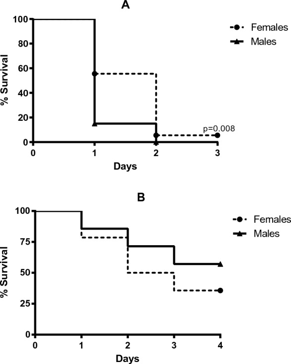
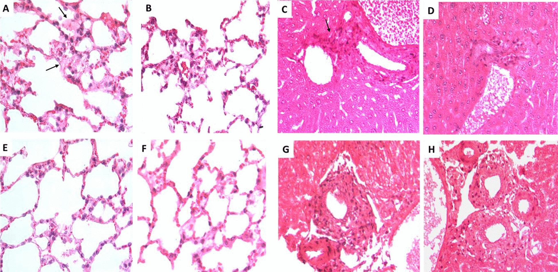
Endotoxic shock (ExSh) and cecal ligature and puncture (CLP) are models that induce sepsis. In this work, we investigated early immunologic and histopathologic changes induced by ExSh or CLP models in female and male mice. Remarkable results showed that females supported twice the LD100 of LPS for males, CLP survival and CFU counts were similar between genders, high circulating LPS levels in ExSh mice and low levels of IgM anti-LPS in males. In the serum of ExSh males, TNF and IL-6 increased in the first 6 h, in CLP males at 12 h. In the liver of ExSh mice, TNF increased at 1.5 and 12 h, IL-1 at 6 h. TGFβ1 increased in females throughout the study and at 12 h in males. In CLP mice, IL-6 decreased at 12 h, TGFβ1 increased at 6-12 h in males and at 12 h in females. In the lungs of ExSh males, IL-1β increased at 1.5-6 h and TGFβ1 at 12 h; in females, TNF decrease at 6 h and TGFβ1 increased from 6 h; in CLP females, TNF and IL-1β decreased at 12 h and 1.5 h, respectively, and TGFβ1 increased from 6 h; in males, TGFβ1 increased at 12 h. In the livers of ExSh mice, signs of inflammation were more common in males; in the CLP groups, inflammation was similar but less pronounced. ExSh females had leucocytes with TGFβ1. The lungs of ExSh males showed patches of hyaline membranes and some areas of inflammatory cells, similar but fewer and smaller lesions were seen in male mice with CLP. In ExSh females, injuries were less extent than in males, similar pulmonary lesions were seen in female mice with CLP. ExSh males had lower levels of TGFβ1 than females, and even lower levels were seen in CLP males. We conclude that the ExSh was the most lethal model in males, associated with high levels of free LPS, low IgM anti-LPS, exacerbated inflammation and target organ injury, while females showed early TGFβ1 production in the lungs and less tissue damage. We didn't see any differences between CLP mice.


Conflict of interest statement

The authors declare that they have no competing interest.

Figures

Fig. 1
Fig. 1
Survival. Survival in A endotoxic shock model and B CLP model was evaluated one week after challenge, the N in each group was 10 animals. Dotted lines represent female mice, while solid lines represent male mice. Comparisons of survival curves were performed using the Mantel-Haenzel log-rank test. Differences were considered when p < 0.05
Fig.  2
Fig.  2
Inflammation of lung and liver from mice treated with LPS or CLP. A Lung section from male mice treated with LPS by the ip route, mildly sized patches of lung tissue whose alveolar lumen is occupied by acellular fibrillary acidophilic material (arrows) with few inflammatory cells. B Similar but fewer and smaller lesions are seen in a male mouse with CLP. C Liver section from male mice treated with LPS shows some portal areas with mild inflammatory infiltrate (arrow). D Similar lesions are seen in male mouse with CLP. E Compared with male mice, female mice treated with LPS show fewer and smaller areas of inflammatory infiltrate in the alveolar-capillary interstitium and scarce eosinophilic fibrillar material on the surface of the alveolar epithelium. F Similar lung lesions are seen in a female mouse with CLP. G Some portal areas show a scar-like inflammatory infiltrate in the liver of female mice treated with LPS. H Similar liver lesions are seen in female mouse with CLP (all sections were stained with hematoxylin and eosin, original magnification × 400). This is a representative photomicrograph of three
Fig. 3
Fig. 3
TGF detection by immunohistochemistry in the lungs of LPS- or CLP-treated mice. A Occasional alveolar macrophages and alveolar cells show TGF immunostaining in the lung of a control female mouse. B In contrast, numerous TGF-immunostained cells are present in the alveolar capillary interstitium of the lungs of female mice treated with LPS. C Numerous TGF-immunostained cells are also present in the lumen and alveolar capillary interstitium of the lungs of CLP-treated female mice. D The lungs of male mice do not show TGF-immunostained cells. E The alveolar space and walls of the lungs of male mice treated with LPS show some TGF-immunostained cells. F Similar TGF immunostaining pattern is seen in the lungs of mice after CLP procedure. (all photomicrographs × 400 magnification). This is a representative micrograph of three

References

    1. Singer M, Deutschman C, Warren C, Shankar-Hari M, Annane D, Bauer M, et al. The Third International Consensus Definitions for Sepsis and Septic Shock (Sepsis-3) JAMA. 2016;315:801. - PMC - PubMed
    1. Deutschman C, Tracey K. Sepsis: current dogma and new perspectives. Immunity. 2014;40:463. - PubMed
    1. Rudd KE, Johnson SC, Agesa KM, Shackelford KA, Tsoi D, Kievlan DR, Colombara DV, et al. Global, regional, and national sepsis incidence and mortality, 1990–2017: analysis for the Global Burden of Disease Study. Lancet. 2020;18(395):200. - PMC - PubMed
    1. Paoli CJ, Reynolds MA, Sinha M, Gitlin M, Crouser E. Epidemiology and costs of sepsis in the United States—an analysis based on timing of diagnosis and severity level. Crit Care Med. 2018;46:1889. - PMC - PubMed
    1. Gyawali B, Ramakrishna K, Dhamoon AS. Sepsis: the evolution in definition, pathophysiology, and management. SAGE Open Med. 2019;7:1. - PMC - PubMed