. 2023 Oct 24;20(1):245.
doi: 10.1186/s12974-023-02920-9.

Complement-membrane regulatory proteins are absent from the nodes of Ranvier in the peripheral nervous system

Affiliations

Complement-membrane regulatory proteins are absent from the nodes of Ranvier in the peripheral nervous system

Netanel Karbian et al. J Neuroinflammation. .

Abstract

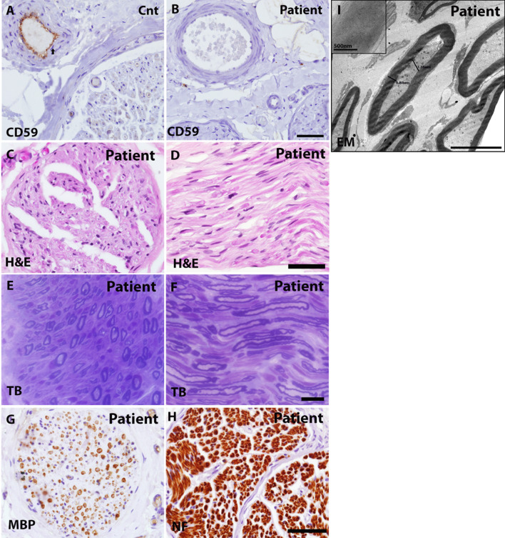
Background: Homozygous CD59-deficient patients manifest with recurrent peripheral neuropathy resembling Guillain-Barré syndrome (GBS), hemolytic anemia and recurrent strokes. Variable mutations in CD59 leading to loss of function have been described and, overall, 17/18 of patients with any mutation presented with recurrent GBS. Here we determine the localization and possible role of membrane-bound complement regulators, including CD59, in the peripheral nervous systems (PNS) of mice and humans.

Methods: We examined the localization of membrane-bound complement regulators in the peripheral nerves of healthy humans and a CD59-deficient patient, as well as in wild-type (WT) and CD59a-deficient mice. Cross sections of teased sciatic nerves and myelinating dorsal root ganglia (DRG) neuron/Schwann cell cultures were examined by confocal and electron microscopy.

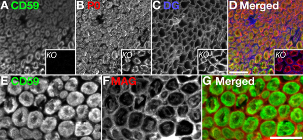
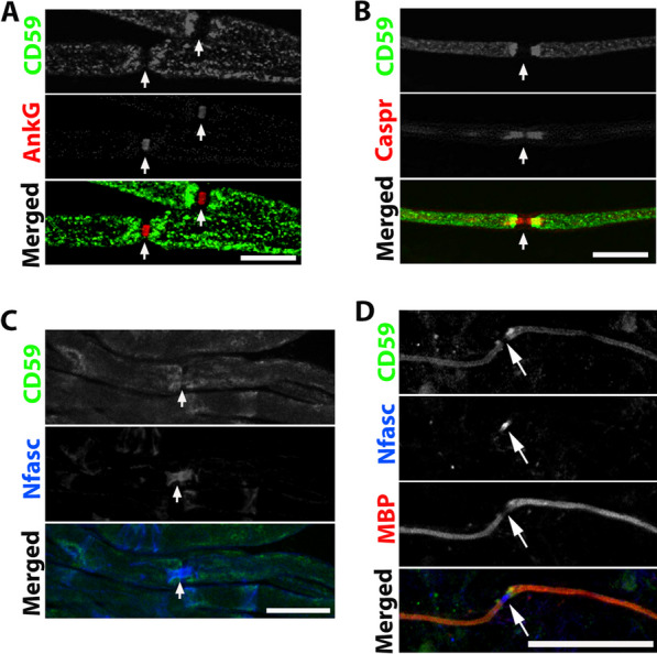
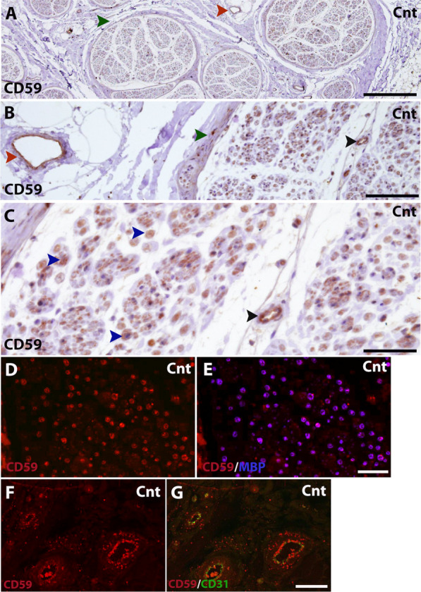
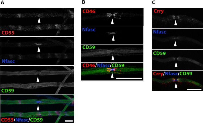
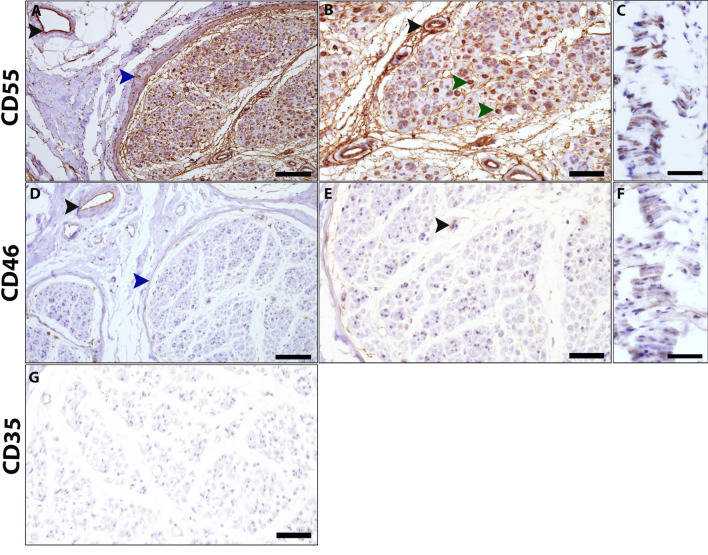
Results: We demonstrate that CD59a-deficient mice display normal peripheral nerve morphology but develop myelin abnormalities in older age. They normally express myelin protein zero (P0), ankyrin G (AnkG), Caspr, dystroglycan, and neurofascin. Immunolabeling of WT nerves using antibodies to CD59 and myelin basic protein (MBP), P0, and AnkG revealed that CD59 was localized along the internode but was absent from the nodes of Ranvier. CD59 was also detected in blood vessels within the nerve. Finally, we show that the nodes of Ranvier lack other complement-membrane regulatory proteins, including CD46, CD55, CD35, and CR1-related gene-y (Crry), rendering this area highly exposed to complement attack.

Conclusion: The Nodes of Ranvier lack CD59 and are hence not protected from complement terminal attack. The myelin unit in human PNS is protected by CD59 and CD55, but not by CD46 or CD35. This renders the nodes and myelin in the PNS vulnerable to complement attack and demyelination in autoinflammatory Guillain-Barré syndrome, as seen in CD59 deficiency.

Keywords: CD59; Complement; Guillain-Barré syndrome; Myelin; Nodes of Ranvier; Nodopathies; Peripheral nervous system.


Conflict of interest statement

The authors declare no competing interests.

Figures

Fig. 1
Fig. 1
Localization of CD59 in peripheral nerve and murine sciatic nerve cross-sections. AD CD59 (green), P0 (red) and beta dystroglycan (DG, blue) staining of murine wild-type (WT) and knock-out (KO) cross section fibers (A-D small squares). EG CD59 (green) and myelin-associated glycoprotein (MAG, red) staining of fibers from WT mice. Staining shows that CD59 and P0, which represent compact myelin, have identical patterns in WT mice. Sections were taken from 4-month-old mice. Immunolabeling was performed with methanol and 0.1% triton. Scale bars 20 μm
Fig. 2
Fig. 2
Localization of CD59 in murine teased nerve and myelinated cultures. A CD59 (green) and ankyrin-G (AnkG, red) staining of WT teased fibers. B CD59 (green) and Caspr (red) staining of WT teased fibers. C CD59 (green) and neurofascin (Nfasc, blue) staining of WT teased fibers. D CD59 (green), myelin basic protein (MBP, red), and neurofascin (Nfasc, blue) staining of WT ICR DRG. CD59 is co-localized with MBP and localized in the internodes and paranodes. There is no CD59 localization in the nodes of Ranvier. AC Sections taken from a 4-month-old mouse. Immunolabeling procedure handled with methanol and 0.1% triton. Scale bars 20 μm and in D 50 μm
Fig. 3
Fig. 3
Localization of CD59 in human sural nerve. AC Immunohistochemical staining for CD59 (DAB, red) in sural nerves of a healthy control. Healthy control staining shows compact myelin localization (C, blue arrow heads). CD59 was also detected in endothelial blood vessels of the epineurium (A and B, red arrow heads) and endoneurium (B and C, black arrow heads), as well as the perineurium (A and B, green arrow heads). D, E Immunofluorescence staining for CD59 (red) and MBP (blue). F, G Immunofluorescence staining for CD59 (red) and CD31 (green). Scale bars: A 500 μm, B 100 μm, CG 50 μm. Magnifications: A ×4, B, DG and ×20, C ×40. AC 1:400 dilution
Fig. 4
Fig. 4
Electron microscopy (EM) and immunofluorescence labeling of murine sciatic nerve. EM pictures of cross sections from WT (AD (and CD59a-deficient (EH) murine sciatic nerve at different ages. A and E are low-resolution toluidine blue pictures from a 6-month-old mouse. Scale bars: A and E 50 μm, B and F 10 μm, C, D, G and H 5 μm. IL EM (higher magnification) showing accumulation of axoplasmatic organelles at an older age (18 months) in longitudinal sections from the paranodal regions (I, J, scale bars: I 2 μm, J 5 μm), and mitochondria and dense bodies in cross sections at regions with of noncompact myelin (K, scale bar = 500 nm; IK are from CD59 KO mice). WT vs CD59 KO mice, comparison of the percentage of nodes of Ranvier with accumulation of axoplasmic organelles is shown in L (WT: n = 5, 82 nodes. KO: n = 5, 117 nodes; (p = 0.01, unpaired two-tailed student’s t-tests). MP Immunofluorescence labeling of WT (left panels) and CD59 KO (right panels) teased fibers show normal node structure by CD59 (green) and AnkG (M, red), Caspr (N, red), neurofascin (O, Nfasc, blue), and myelin-associated glycoprotein (P, MAG, red). Sections taken from a 4-month-old mouse. Immunolabeling procedure handled with methanol and 0.1% triton. Scale bars 20 μm
Fig. 5
Fig. 5
CD59 patient sural nerve biopsy. A, B Immunohistochemical staining of CD59 (DAB, red) from a healthy control subject (A) and a CD59 patient (B). CD59-deficient patient staining of CD59 was completely absent in comparison to a healthy control subject where CD59 appears to localize in compact myelin. Scale bars = 50 μm, magnifications × 40, 1:400 dilution. CH Hematoxylin and eosin (H&E) (C and D), epoxy-resin embedded semi-thin sections stained with toluidine-blue (E and F), immunohistochemical staining for MBP (G) and neurofilament (NF) (H). Staining displayed normal nerve fiber density without evidence of active axonal or myelin damage. Scale bars: C, D, G and H 50 μm; E and F 20 μm; magnifications: C, D, G and H ×40; E and F ×60. I Electron microscopy image, tissue from a CD59-deficient patient showing thin myelin sheaths relative to axonal diameter with a relatively high g-ratio, suggestive of remyelination. Scale bar 5 μm. small squares, myelin sheath lamellar structure. Scale bar 500 nm
Fig. 6
Fig. 6
Localization of complement membrane regulatory proteins in murine teased fibers of sciatic nerve. Staining of WT teased fibers. CD55 (red), neurofascin (Nfasc, blue) and CD59 (green) (A), CD46 (red), neurofascin (Nfasc, blue) and CD59 (green) (B), Crry (red), neurofascin (Nfasc, blue) and CD59 (green) (C). Sections taken from a 4-month-old mouse. Immunolabeling procedure performed with the TSA method (A, C) and methanol and 0.1% triton (B). Scale bars 20 μm. CD55 staining was localized in Schmidt Lanterman incisures. CD46 staining was localized in the paranodes-juxtaparanodes, and Crry staining was localized in the paranodes and the internodes corresponding to compact myelin. White arrowheads indicate the nodes of Ranvier
Fig. 7
Fig. 7
Complement membrane regulatory proteins in human peripheral sural nerve. AG. Human sural nerve from a healthy control. Cross and longitudinal section immunohistochemical staining for CD55 (DAB, red) (AC), CD46 (DAB, red) (DF) and CD35 (G). CD55 staining corresponds to compact myelin (B, green arrow heads) and endothelial blood vessels of the epineurium (A, black arrowhead) and endoneurium (B, black arrowhead), as well as the perineurium (A, blue arrowhead). CD46 was detected in endothelial blood vessels of the epineurium (D, black arrowhead) and endoneurium (E, black arrowhead), as well as the perineurium (D, blue arrowhead). CD35 was not detected anywhere. Scale bars: A, D 100 μm; B, C, EG 50 μm. Magnifications: A, D ×20; B, C, EG ×40

References

    1. Yuki N, Hartung HP. Guillain-Barre syndrome. N Engl J Med. 2012;366:2294–2304. - PubMed
    1. Karbian N, Eshed-Eisenbach Y, Tabib A, Hoizman H, Morgan BP, Schueler-Furman O, et al. Molecular pathogenesis of human CD59 deficiency. Neurol Genet. 2018;4:e280. - PMC - PubMed
    1. Mevorach D, Reiner I, Grau A, Ilan U, Berkun Y, Ta-Shma A, et al. Therapy with eculizumab for patients with CD59 p.Cys89Tyr mutation. Ann Neurol. 2016;80:708–717. - PubMed
    1. Tabib A, Karbian N, Mevorach D. Demyelination, strokes, and eculizumab: lessons from the congenital CD59 gene mutations. Mol Immunol. 2017;89:69–72. - PubMed
    1. Ardicli D, Taskiran EZ, Kosukcu C, Temucin C, Oguz KK, Haliloglu G, et al. Neonatal-onset recurrent Guillain-Barre syndrome-like disease: clues for inherited CD59 deficiency. Neuropediatrics. 2017;48:477–481. - PubMed