Review
. 2023 Aug 8;3(5):389-417.
doi: 10.1021/acsbiomedchemau.3c00021. eCollection 2023 Oct 18.

Functional Diversity in Radiolabeled Nanoceramics and Related Biomaterials for the Multimodal Imaging of Tumors

Affiliations
Review

Functional Diversity in Radiolabeled Nanoceramics and Related Biomaterials for the Multimodal Imaging of Tumors

David G Calatayud et al. ACS Bio Med Chem Au. .

Abstract

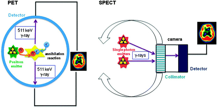
Nanotechnology advances have the potential to assist toward the earlier detection of diseases, giving increased accuracy for diagnosis and helping to personalize treatments, especially in the case of noncommunicative diseases (NCDs) such as cancer. The main advantage of nanoparticles, the scaffolds underpinning nanomedicine, is their potential to present multifunctionality: synthetic nanoplatforms for nanomedicines can be tailored to support a range of biomedical imaging modalities of relevance for clinical practice, such as, for example, optical imaging, computed tomography (CT), magnetic resonance imaging (MRI), single photon emission computed tomography (SPECT), and positron emission tomography (PET). A single nanoparticle has the potential to incorporate myriads of contrast agent units or imaging tracers, encapsulate, and/or be conjugated to different combinations of imaging tags, thus providing the means for multimodality diagnostic methods. These arrangements have been shown to provide significant improvements to the signal-to-noise ratios that may be obtained by molecular imaging techniques, for example, in PET diagnostic imaging with nanomaterials versus the cases when molecular species are involved as radiotracers. We surveyed some of the main discoveries in the simultaneous incorporation of nanoparticulate materials and imaging agents within highly kinetically stable radio-nanomaterials as potential tracers with (pre)clinical potential. Diversity in function and new developments toward synthesis, radiolabeling, and microscopy investigations are explored, and preclinical applications in molecular imaging are highlighted. The emphasis is on the biocompatible materials at the forefront of the main preclinical developments, e.g., nanoceramics and liposome-based constructs, which have driven the evolution of diagnostic radio-nanomedicines over the past decade.


Conflict of interest statement

The authors declare no competing financial interest.

Figures

Figure 1
Figure 1
An overview of the main techniques used for molecular imaging in clinical practice, which will be the focus of this review. Adapted with permission from ref (3). Copyright 2008 Springer Nature.
Figure 2
Figure 2
Early example focused on preclinical investigations indicative of a feasible way to combine molecular imaging modalities. Reproduced with permission under a Creative Commons CC-BY License from ref (27). Copyright 2003 CSH press.
Figure 3
Figure 3
(a) Structural representations of the small-molecular tags 68Ga-DOTA-CHCO-Gly-4-aminobenzyl bombesin (1), 89Zr-5A10 monoclonal antibody (2), and 1-(2′-deoxy-2′-fluoro-b-d-arabinofuranosyl) thymidine (3). (b) Structural representation of a multifunctionalized nanoparticle and its constituents: Gadolinium diethylenetriaminepentaacetate-di(stearylamide) (Gd-DTPA-DSA, yellow dot) as MRI contrast agent, 1,1′-dioctadecyl-3,3,3′,3′-tetramethylindotricarbocyanine iodide (DiR, green dot) as NIR dye, and the cyclic RGD-containing pentapeptide (c(RGDf(S-acetylthioacetyl) K) (RGD) as specific targeting agent. (c) Structural representations of clinically relevant small molecular PET radiotracers. 18F-fluoromisonidazole (FMISO), 18F-fluoroazomycin-arabinofuranoside (FAZA), 18F-fluoroerythronitroimidazole (FETNIM), [18F]-2-(2-Nitro-1H-imidazol-1-yl)-N-(2,2,3,3,3-pentafluoropropyl) acetamide (EF5), EF3, RP-170 (1-(2-1-(1H-methyl) ethoxy)-methyl-2-nitroimidazole) (FRP-170), 3-[18F]-2-(4-((2-nitro-1H-imidazol-1-yl) methyl)-1H-1,2,3,-triazol-1-yl)-propan-1-ol (HX4), copper-labeled diacetyl-bis(N-methylthiosemicarbazone) (Cu-ATSM).
Figure 4
Figure 4
Schematic representations for PET and SPECT applications. In PET, the emitted positron undergoes an annihilation process with an electron, thus giving rise two γ-rays situated at 180° from each other. Their emergence is detected, and a 3D image of the tracer concentration is obtained by software reconstruction.
Figure 5
Figure 5
Representation of a nanodimensional synthetic platform suggestive of the multifunctional possibility of nanomedicines. Image reproduced with permission from ref (70). Copyright 2009 John Wiley and Sons.
Figure 6
Figure 6
Representations of a malignant solid tumor with its different areas depicting deregulated pharmacology. Image reprinted with permission under a Creative Commons [CC-BY 4.0] from ref (99), an Open Access article distributed under the terms and conditions of the Creative Commons Attribution (CC BY) license. Copyright 2018. [2018 by the authors].
Figure 7
Figure 7
Overview of different categories of nanocarriers relevant as synthetic materials scaffold for radionanoparticulate drug delivery and their relative size.
Figure 8
Figure 8
Trends in the number of published articles with keyword “nanomedicine” as emerging from PubMed and SciFinder overview of the decades 2000–2022.
Figure 9
Figure 9
Overview of selected nanoparticles and their main properties and characteristics applicable to theranostics design.
Figure 10
Figure 10
Polyethylene glycol prevents uptake by the reticuloendothelial system. (A) Nanoparticles (NP) (A1) are coated with opsonin proteins (A2) and associate with macrophages (A3) for their transit to the liver (A4). Macrophages stationary in the liver, known as Kupffer cells, also participate in nanoparticle scavenging. (B) Nanoparticles coated with PEG coating (B1) prevents this opsonization (B2), resulting in decreased liver accumulation (B3) and increased availability of the NP for imaging or therapy. NP: Nanoparticle; PEG: Polyethylene glycol. Reproduced with permission from ref (112). Copyright 2018 Elsevier.
Figure 11
Figure 11
Nanoparticle interactions with cell membrane receptors, ultimately influencing delivery, mediated by size, shape charge of NPs, ligand density, receptor expression levels, internalization mechanism, and cell properties (phenotype, location, etc.). Image reproduced from ref (108).
Figure 12
Figure 12
(a) Mechanisms of nanoparticle passive targeting: Nanoparticles smaller than 3 nm in diameter could extravasate different tissues nonspecifically. Nanoparticles with large negative surface charge or larger than 150 nm in diameter could be captured by Kupffer cells. Nanoparticles less than 200 nm in diameter could pass through sinusoidal fenestrations after intravenous administration and (b) gold–dendrimer nanoparticles and their biodistibution in vivo. Dendrimers are branched molecules that can be used as scaffolds for metals such as gold to attach to, enabling nanoparticles with different diameters and surfaces charges (left and right; – is negative charge, + is positive charge, and n is neutral) to be produced. Recent experiments show that the size and charge of the nanoparticles influence their biodistribution in mice. (Figure adapted with permission from ref (115). Copyright 2012 Royal Society of Chemistry.)
Figure 13
Figure 13
Active and passive targeting of nanoparticles (liposomes) to target cancer cells in chemotherapy. Reproduced with permission from ref (118). Copyright 2009 Elsevier.
Figure 14
Figure 14
Various magnetic nanoparticles coated with silica shells: Backscattered electron images (a) and TEM images (b) of Fe3O4-core/SiO2-mesoporous-shell magnetic nanoparticles. TEM image (c) of Fe3O4-core/SiO2-mesoporous-shell magnetic nanoparticles. TEM image (d) of Fe3O4-core/SiO2-shell magnetic nanoparticles. Reproduced with permission under a Creative Commons CC-BY license from ref (223). Copyright 2019 The Author.
Figure 15
Figure 15
(a–f) PET/MR images of SLNs in a rat at 1 h post injection of 124I-SA-MnMEIO into the right forepaw (I = nanoprobe injection site). Coronal (a) MR and (b) PET images in which a brachial LN (white circle) is detected. (c) The position of the brachial LN is well-matched in a PET/MR fusion image. Four small pipet tips containing Na124I solution are used as a fiducial marker (white arrowheads) for the concordant alignment in PET/MR images. In the transverse images, axillary (red circle) and brachial LNs (white circle) are detected in the (d) MR and (e) PET images, and images of each node are nicely overlapped in the corresponding PET/MR fusion image (f). (g) The explanted brachial LN also shows consistent results with in vivo images by PET and MR. Only the LN from the right-hand side of the rat containing 124I-SA-MnMEIO shows strong PET and dark MR images. The schematics of the rat in the (h) coronal and (i) transverse directions show the locations of the LNs. Reproduced with permission from ref (143). Copyright 2008 John Wiley and Sons.
Figure 16
Figure 16
General overview for silica-coated nanoparticles functionalized with fluorescent quantum dots and a chelator from the Lledos, Calatayud, and Pascu state-of-the-art: Cd0.1Zn0.9Se QDs modified silica-coated magnetic IONPs; Fe3O4@SiO2@68Ga@SiO2 (RCY 70%) and Fe3O4/Cd0.1Zn0.9Se@SiO2@68Ga@SiO2 (66%), Fe3O4@SiO2@Zn(ATSM/A)@68Ga (RCY > 99%).

References

    1. Jennings L. E.; Long N. J. ‘Two Is Better than One’—Probes for Dual-Modality Molecular Imaging. Chem. Commun. 2009, (24), 3511. 10.1039/b821903f. - DOI - PubMed
    1. Smith S. V. Molecular Imaging with Copper-64. J. Inorg. Biochem. 2004, 98 (11), 1874–1901. 10.1016/j.jinorgbio.2004.06.009. - DOI - PubMed
    1. Willmann J. K.; van Bruggen N.; Dinkelborg L. M.; Gambhir S. S. Molecular Imaging in Drug Development. Nat. Rev. Drug Discovery 2008, 7 (7), 591–607. 10.1038/nrd2290. - DOI - PubMed
    1. Sausville E. A.; Longo D. L. Principles of Cancer Treatment: Surgery, Chemotherapy, and Biologic Therapy. Harrisons Princ. Int. Med. 2005, 16 (1), 464.
    1. Wagner V.; Dullaart A.; Bock A.-K.; Zweck A. The Emerging Nanomedicine Landscape. Nat. Biotechnol. 2006, 24 (10), 1211–1217. 10.1038/nbt1006-1211. - DOI - PubMed