. 2023 Sep 25;26(11):108050.
doi: 10.1016/j.isci.2023.108050. eCollection 2023 Nov 17.

Pre- and postsynaptic N-methyl-D-aspartate receptors are required for sequential printing of fear memory engrams

Affiliations

Pre- and postsynaptic N-methyl-D-aspartate receptors are required for sequential printing of fear memory engrams

Ilaria Bertocchi et al. iScience. .

Abstract

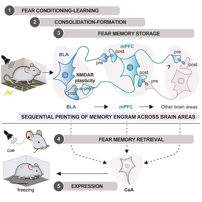
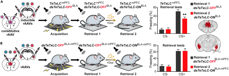
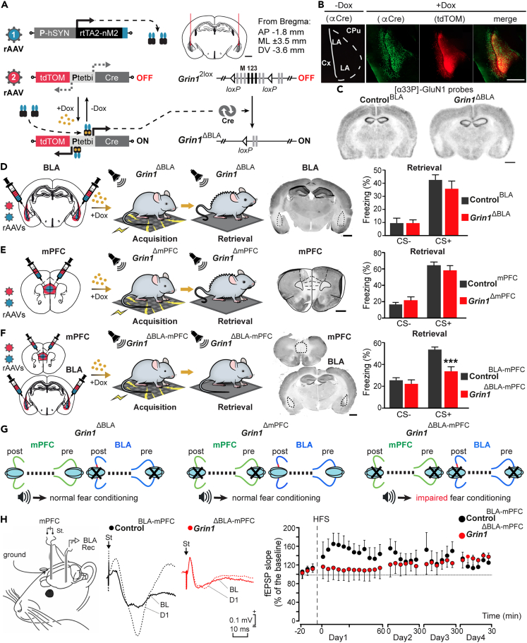
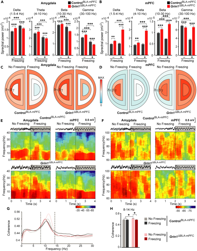
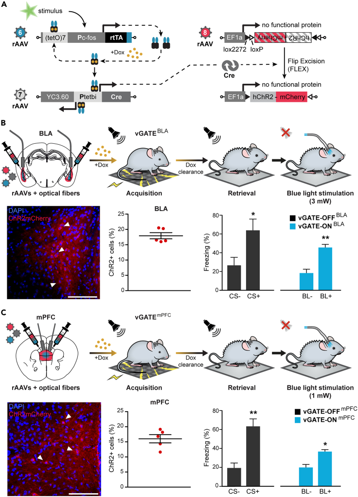
The organization of fear memory involves the participation of multiple brain regions. However, it is largely unknown how fear memory is formed, which circuit pathways are used for "printing" memory engrams across brain regions, and the role of identified brain circuits in memory retrieval. With advanced genetic methods, we combinatorially blocked presynaptic output and manipulated N-methyl-D-aspartate receptor (NMDAR) in the basolateral amygdala (BLA) and medial prefrontal cortex (mPFC) before and after cued fear conditioning. Further, we tagged fear-activated neurons during associative learning for optogenetic memory recall. We found that presynaptic mPFC and postsynaptic BLA NMDARs are required for fear memory formation, but not expression. Our results provide strong evidence that NMDAR-dependent synaptic plasticity drives multi-trace systems consolidation for the sequential printing of fear memory engrams from BLA to mPFC and, subsequently, to the other regions, for flexible memory retrieval.

Keywords: Behavioral neuroscience; Cellular neuroscience.


Conflict of interest statement

The authors declare no competing interests.

Figures

None
Graphical abstract
Figure 1
Figure 1
Inducible silencing of the amygdala and prefrontal output (A) Schematic showing the components of the second-generation virus-delivered INducible SIlencing of Synaptic Transmission (“vINSIST-2”) system and their operation. Abbreviations: Dox, doxycycline; dsTeTxLC, destabilized tetanus toxin light chain protein; PhSYN, human synapsin specific promoter; Ptetbi, bidirectional tet responder promoter; rAAV, recombinant adenovirus serotype 1/2; tdTOM, tdTomato red fluorescent protein; rtTA2-nM2, Dox-sensitive recombinant transactivator; Syb-2, synaptobrevin 2 vesicle protein. (B) Silencing of BLA output by dsTeTxLC in dsTeTxLC-ONBLA mice before conditioning induces a significant decrease of freezing during the cued test (N = 7,8; two-way RM ANOVA; significant main effect of genotype F(1,13) = 4.98, p = 0.044; significant effect of cue/genotype interaction F(1,13) = 10.04, p = 0.007) ∗∗p < 0.01 by Bonferroni post-test. Scale bar, 1mm. (C) Silencing of BLA output induced after fear conditioning does not affect freezing in the cued test (retrieval-2/dsTeTxLC-ONBLA compared to retrieval-1/dsTeTxLC-OFFBLA) (N = 10; two-way RM ANOVA; no significant effect of treatment F(1,18) = 0.099, p = 0.76). (D) Silencing of mPFC output by constitutive TeTxLC expression has no significant effect on fear learning and expression (N = 11, 8; Freezing: two-way RM ANOVA, no significant effect of genotype F(1,17) = 0.96, p = 0.34). (E) Silencing of mPFC outputs induced after fear conditioning has no significant effect on freezing in the cued test (retrieval-2/dsTeTxLC-ONmPFC compared to retrieval-1/dsTeTxLC-OFFmPFC) (N = 9; two-way RM ANOVA; no significant impact of treatment F(1,16) = 0.01, p = 0.91). Scale bar, 1mm. Data are presented as mean ± s.e.m.
Figure 2
Figure 2
Fear memory is distributed between the medial prefrontal cortex and the amygdala (A) When mPFC outputs are inhibited since acquisition (TeTxLCmPFC), silencing of BLA output induced after conditioning (dsTeTxLC-ONBLA) has a significant effect on freezing in the cued test for memory recall (N = 8, two-way RM ANOVA; cue × treatment interaction, F(1,14) = 10.44, p = 0.006) ∗∗p < 0.01 by Bonferroni post-test. (B) Silencing of both mPFC and BLA outputs after conditioning has no significant effect on freezing in the cued test (retrieval-2/dsTeTxLC-ONBLA-mPFCmPFC compared to retrieval-1/dsTeTxLC-OFFBLA-mPFCmPFC) (N = 6; two-way RM ANOVA; no significant effect of genotype F(1,10) = 0.02, p = 0.87). Data are presented as mean ± s.e.m. Scale bars, 1mm. All injection sites were validated by a tracer rAAV expressing tdTOM.
Figure 3
Figure 3
Role of NMDARs in fear memory conditioning (A) rAAVs were bilaterally delivered into the BLA of Grin12lox mice to induce Cre/loxP mediated deletion of the Grin1 gene and the generation of BLA-specific Grin1 gene knockout mice (Grin1ΔBLA). Scale bar, 1 mm. (B) tdTOM fluorescence and Cre immunostaining in the BLA are overlapping and evident in Dox-treated (Grin1ΔBLA) but not in untreated mice (Grin12lox mice, also called ControlBLA). Scale bar, 500 μM. Abbreviations: BA, basal amygdaloid nucleus; CpU, caudate-putamen; Cx, cortex; LA, lateral amygdaloid nucleus. (C) in situ hybridization for Grin1 mRNA of coronal brain slices from ControlBLA and Grin1ΔBLA mice. Scale bar, 1 mm. (D) Genetic deletion of Grin1 bilaterally in the BLA before fear conditioning had no effects on freezing during the cued test for memory recall in Grin1ΔBLA compared to ControlBLA mice (N = 8, 6; two-way RM ANOVA; no significant effect of genotype F(1,12) = 0.15; p = 0,7). (E) Genetic deletion of Grin1 bilaterally in the mPFC before fear conditioning had no effects on freezing during the cued test in Grin1ΔmPFC compared to ControlmPFC mice (N = 8, 12; two-way RM ANOVA; no significant effect of genotype; F(1,18) = 0.004; p = 0.9). (F) Genetic deletion of Grin1 bilaterally in both the BLA and mPFC before fear conditioning significantly reduced the percentage of freezing during the cued test in Grin1ΔBLA-mPFC compared to ControlBLA-mPFC mice (N = 8, 8; two-way RM ANOVA followed by Bonferroni post-test; significant main effect of genotype F(1,14) = 7.6; p < 0.001 during tone). (G) Schematics reproducing the effects of targeted NMDAR removal in the BLA, in the mPFC, and in both areas. (H) Schematic depicting in vivo electrodes implanted for stimulating neurons in the mPFC and recording in BLA. In the middle, examples of fEPSPs (averaged 5 times) were collected from representative ControlBLA-mPFC and Grin1ΔBLA-mPFC mice (n = 6 and n = 5, respectively). On the right, illustrated data correspond to fEPSPs evoked by the second pulse. While control mice presented a significant LTP (F(32,96) = 1.787, p = 0.016), Grin1ΔBLA-mPFC mice did not reach significant values (F(32,160) = 0.765, p = 0.812).
Figure 4
Figure 4
Spectral analyses of LFPs recorded in the amygdalar complex and mPFC of Grin1ΔBLA-mPFC and ControlBLA-mPFC mice (A) Spectral power values during no freezing and freezing for the four selected bands in the amygdala of control (ControlBLA-mPFC) and double knockout mice (Grin1ΔBLA-mPFC). (B) Spectral power values during no freezing and freezing for the four selected bands in the mPFC of control (ControlBLA-mPFC) and double knockout mice (Grin1ΔBLA-mPFC). ∗∗∗p < 0.001 and ∗∗p ≤ 0.01. Data are presented as mean ± s.e.m. (C and D) Schematic diagrams representing the spectral power differences between Grin1ΔBLA-mPFC and ControlBLA-mPFC mice for the 4 selected bands in the BLA (C) and the mPFC (D). The color scale is illustrated in the middle. (E) Time-frequency power analysis for delta, theta, and beta bands (0–40 Hz) of representative LFP signals recorded in the mPFC (left) and the amygdala (right) of ControlBLA-mPFC mice (upper panel) and Grin1ΔBLA-mPFC mice (lower panel) during a non-freezing period of 2 s (0–2 s) and a freezing period of 2 s (2–4 s). (F) Time-frequency power analysis for the gamma band (60–100 Hz) of representative LFP signals recorded in the mPFC (left) and the amygdala (right) of ControlBLA-mPFC mice (upper panel) and Grin1ΔBLA-mPFC mice (lower panel) during a non-freezing period of 2 s (0–2 s) and a freezing period of 2 s (2–4 s). The color scale is illustrated at the bottom right. (G) Coherence analysis between mPFC and amygdala during no freezing and freezing behavior. Coherence spectra of Grin1ΔBLA-mPFC and ControlBLA-mPFC mice during freezing (dark colors) and no freezing (light colors) behaviors (N = 100 epochs of 1 s per behavior). The dashed lines correspond to 8 Hz and 14 Hz. Mean ± s.e.m. of coherence values in the 8–14 Hz band. (H) Grin1ΔBLA-mPFC mice presented more significant coherence at the indicated band during the non-freezing period (N = 5, 5; ∗p < 0.05).
Figure 5
Figure 5
Optogenetic reactivation of mPFC and BLA engram cells (A) A schematic of the vGATE method (virus-delivered Genetic Activity-induced Tagging of Ensembles). (B) Blue light stimulation of the BLA (BL+, 3mW) allowed the recruitment of the same ensemble of cells activated during conditioning. It produced significant freezing (vGATE-ONBLA), comparable to the fear-conditioned response observed in the cued test (vGATE-OFFBLA) for memory recall (N = 5, Retrieval: two-tailed paired t-test (no tone versus tone) p = 0.017, t = 3.92 df = 4. BL stimulation: two-tailed paired t-test (no BL versus BL) p = 0.005, t = 5.53 df = 4.). (C) Blue light stimulation of the mPFC (BL+, 1mW) allows the recruitment of the same ensemble of cells activated during conditioning. It produces significant freezing (vGATE-ONmPFC), comparable to the fear-conditioned response observed in the cued test (vGATE-OFFmPFC) for memory recall (N = 5, Retrieval: two-tailed paired t-test (no tone versus tone) p = 0.0014, t = 7.95 df = 4; BL stimulation: two-tailed paired t-test (no BL versus BL) p = 0.025, t = 3.47 df = 4). ∗p < 0.05, ∗∗p < 0.01 by two-tailed paired t-test. Data are presented as mean ± s.e.m. Scale bars, 100 mm.

References

    1. Gale G.D., Anagnostaras S.G., Godsil B.P., Mitchell S., Nozawa T., Sage J.R., Wiltgen B., Fanselow M.S. Role of the basolateral amygdala in the storage of fear memories across the adult lifetime of rats. J. Neurosci. 2004;24:3810–3815. doi: 10.1523/JNEUROSCI.4100-03.2004. - DOI - PMC - PubMed
    1. Do Monte F.H., Quirk G.J., Li B., Penzo M.A. Retrieving fear memories, as time goes by. Mol. Psychiatr. 2016;21:1027–1036. doi: 10.1038/mp.2016.78. - DOI - PMC - PubMed
    1. Kandel E.R., Dudai Y., Mayford M.R. The molecular and systems biology of memory. Cell. 2014;157:163–186. doi: 10.1016/j.cell.2014.03.001. - DOI - PubMed
    1. Squire L.R., Bayley P.J. The neuroscience of remote memory. Curr. Opin. Neurobiol. 2007;17:185–196. doi: 10.1016/j.conb.2007.02.006. - DOI - PMC - PubMed
    1. Tulving E. Multiple memory systems and consciousness. Hum. Neurobiol. 1987;6:67–80. - PubMed