Longitudinal Choroidal Development in Preterm Infants
- PMID: 37877004
- PMCID: PMC10591002
- DOI: 10.1016/j.xops.2023.100359
Longitudinal Choroidal Development in Preterm Infants
Abstract
Purpose: To characterize changes in subfoveal choroidal thickness in preterm infants from 30 to 60 weeks' postmenstrual age (PMA).
Design: The prospective, observational Study of Eye Imaging in Preterm infantS (BabySTEPS) enrolled infants eligible for retinopathy of prematurity screening per the American Association of Pediatrics guidelines.
Subjects: Infants imaged with an investigational, handheld OCT at ≥ 4 distinct imaging sessions between 30 to 60 weeks' PMA as part of BabySTEPS.
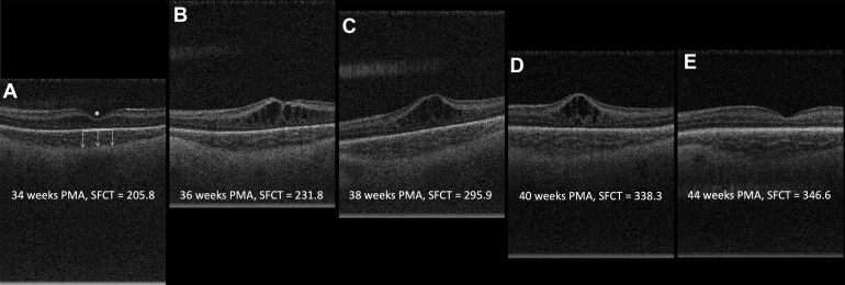
Methods: Average choroidal thickness across the central subfoveal 1 mm in each eye at each time point was measured using custom segmentation software, and errors were manually corrected by a trained grader. We prospectively collected birth history data. A segmented mixed model was used to analyze the change in choroidal thickness as a function of PMA, birth weight, and gestational age (GA).
Main outcome measures: Characterization of normative subfoveal choroidal thickness values and choroidal growth rate between 30 to 60 weeks' PMA.
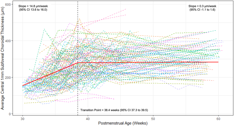
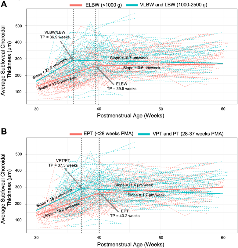
Results: We included 592 imaging sessions of 79 preterm infants (152 eyes). Mean (± standard deviation) GA was 27.5 ± 2.5 weeks. Mean choroidal thickness was 141.4 ± 34.5 μm at 30 weeks, 272.2 ± 83.9 μm at 38 weeks, and 306.2 ± 77.4 μm between 56 and 60 weeks. Between 30 and 60 weeks' PMA, choroidal growth followed a biphasic model, with a linear growth rate of 14.8 μm per week (95% confidence interval [CI], 13.6-16.0) from 30 until 38.4 weeks, then cessation of growth, with a growth rate of 0.3 μm per week (95% CI, -1.1 to 1.6) from 38.4 to 60 weeks. Infants with extremely low birth weight (ELBW; < 1000 g) and extremely preterm (GA < 28 weeks) infants had significantly slower initial growth rates compared with very low and low birth weight and very preterm and preterm infants (ELBW 13.0 vs. 21.0 μm per week; P < 0.0001 and extremely preterm 13.2 vs. 18.0 μm per week; P = 0.003).
Conclusions: Preterm infant choroidal thickness experiences rapid linear growth from 30 to 38 weeks' PMA, at which time growth nearly stops. These foundational measurements and identification of the impact of extremes of low birth weight and prematurity on choroidal development will be essential as researchers begin to understand the role of choroidal development in ocular and retinal health in human infants.
Financial disclosures: Proprietary or commercial disclosure may be found in the Footnotes and Disclosures at the end of this article.
Keywords: Choroid; Optical coherence; Premature infant; Tomography.
© 2023 by the American Academy of Ophthalmology.
Figures