. 2023 Nov;29(11):2275-2284.
doi: 10.3201/eid2911.230804.

Neurologic Effects of SARS-CoV-2 Transmitted among Dogs

Neurologic Effects of SARS-CoV-2 Transmitted among Dogs

Dong-Hwi Kim et al. Emerg Infect Dis. 2023 Nov.

Abstract

SARS-CoV-2 induces illness and death in humans by causing systemic infections. Evidence suggests that SARS-CoV-2 can induce brain pathology in humans and other hosts. In this study, we used a canine transmission model to examine histopathologic changes in the brains of dogs infected with SARS-CoV-2. We observed substantial brain pathology in SARS-CoV-2-infected dogs, particularly involving blood-brain barrier damage resembling small vessel disease, including changes in tight junction proteins, reduced laminin levels, and decreased pericyte coverage. Furthermore, we detected phosphorylated tau, a marker of neurodegenerative disease, indicating a potential link between SARS-CoV-2-associated small vessel disease and neurodegeneration. Our findings of degenerative changes in the dog brain during SARS-CoV-2 infection emphasize the potential for transmission to other hosts and induction of similar signs and symptoms. The dynamic brain changes in dogs highlight that even asymptomatic individuals infected with SARS-CoV-2 may develop neuropathologic changes in the brain.

Keywords: 2019 novel coronavirus disease; COVID-19; SARS-CoV-2; South Korea; blood–brain barrier; coronavirus disease; dogs; infection transmission; neurodegenerative diseases; neurological model; respiratory infections; severe acute respiratory syndrome coronavirus 2; viruses; zoonoses.


Figures

Figure 1
Figure 1
Schematic diagram representing susceptibility to SARS-CoV-2 infection in the lungs and brain of animals with potential for human transmission and homology of ACE2 amino acid sequences in study of the neurologic effects of SARS-CoV-2. *Number of mutations among the key 20 residues involved in interacting with the SARS-CoV-2 RBD. ACE2, angiotensin-converting enzyme 2; RBD, receptor-binding domain.
Figure 2
Figure 2
SARS-CoV-2 in the brain of dogs in study of the neurologic effects of SARS-CoV-2, showing transmission at an early stage of infection. A) Quantitative real-time PCR validation of the SARS-CoV-2 gene in the SARS-CoV-2–infected and contact groups of dogs, at each postinfection day. The dashed line indicates the regions where gene copy numbers of SARS-CoV-2 were considered negative. B) Representative fluorescent images of S1 protein (a marker of virus infection) and NeuN (a specific marker of neuronal cells) demonstrated SARS-CoV-2 infection in the canine brain sections of SARS-CoV-2–infected and contact dogs, at early and late days after infection. SARS-CoV-2–infected neuronal cells at the early stage of infection. At the late stage of infection, the presence of the virus appeared to diminish. Scale bars indicate 100 μm.
Figure 3
Figure 3
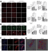
SARS-CoV-2 infection disrupting the blood–brain barrier (BBB) and immune cells infiltrating the brains of infected dogs in study of the neurologic effects of SARS-CoV-2 transmitted among dogs. A) Representative fluorescent images and statistical results of vascular markers including laminin, claudin 5, collagen IV, and PDGFR-β staining of canine brain sections derived from SARS-CoV-2–infected and contact dogs at early and late days after infection. Scale bar indicates 200 μm. B) Brain sections from SARS-CoV-2–infected and contact dogs stained with fibrinogen (green) and IgG (red), which represent BBB leakage staining, at early and late days after infection. Scale bar indicates 200 μm. (C) Representative fluorescent images of the brain sections stained with the CD4 (green) and aquaporin 4 (red), which are markers of CD4-positive T cells and astrocytic end foot, respectively. The CD4-positive cells were infiltrated in the brain parenchyma (white arrows) in the SARS-CoV-2–infected group. Scale bar indicates 50 μm. (D) Representative 3D images of CD4 (green) and aquaporin 4 (red). Statistical significance was determined using a 1-way analysis of variance. Data are presented as mean ±SEM.
Figure 4
Figure 4
SARS-CoV-2 induces activation of microglial cells in the brain white matter in a region-specific manner in SARS-CoV-2–infected and contact dogs in study of the neurologic effects of SARS-CoV-2 transmitted among dogs. A) Representative fluorescent images of glial fibrillary acidic protein (activation astrocyte marker, green) staining of canine brain sections derived from SARS-CoV-2–infected and contact groups at early and late days after infection. Scale bars indicate 200 μm; in insets, 50 μm. B) Representative fluorescent images and statistical results of Iba-1 (a marker of microglia; green) staining of canine brain white matter sections derived from SARS-CoV-2–infected and contact dogs at early and late dpi. Scale bars indicate 200 μm; in insets, 50 μm. C) Representative fluorescent images and statistical results of Iba-1 (a marker of microglia, green) staining of canine brain gray matter sections derived from SARS-CoV-2–infected and contact dogs at early and late dpi. Scale bars indicate 200 μm; in insets, 50 μm. Statistical significance was determined using a 1-way analysis of variance. Data in graphs are presented as means ±SEM.
Figure 5
Figure 5
SARS-CoV-2 infection causing perivascular demyelination in the brain in dogs in study of the neurologic effects of SARS-CoV-2 transmitted among dogs. A, B) Representative fluorescent images and statistical analysis results of NFL (a marker of neurofilament light chain; gray) staining of canine brain white matter sections derived from SARS-CoV-2 infected and contact dogs at early and late dpi. Scale bar indicates 200 μm. Overall images from infected dogs demonstrate irregular axonal morphology compared with that of control dogs. Single-layer slice images. Scale bar indicates 20 μm. C, D) Representative fluorescent images and statistical analysis results of myelin (fluomyelin; green) and DAPI (blue) staining of canine brain white matter sections derived from SARS-CoV-2–infected and contact dogs at early and late dpi. Scale bar indicates 200 μm. E) Representative fluorescent images of p-tau (red) staining of canine brain gray matter sections derived from SARS-CoV-2–infected and contact dogs at early and late dpi. Scale bar indicates 200 μm. F–H) Representative fluorescent images (F) and statistical analysis results (G, H) of NeuN (a marker of neuron; green) staining of canine brain gray matter sections derived from SARS-CoV-2–infected and contact dogs at early (G) and late (H) infection. Scale bar indicates 200 μm. Statistical significance was determined using a 1-way analysis of variance. Data are presented as mean ±SEM. NS, not significant.

References

    1. Abdel-Moneim AS, Abdelwhab EM. Evidence for SARS-CoV-2 infection of animal hosts. Pathogens. 2020;9:529. 10.3390/pathogens9070529 - DOI - PMC - PubMed
    1. Mahdy MAA, Younis W, Ewaida Z. An overview of SARS-CoV-2 and animal infection. Front Vet Sci. 2020;7:596391. 10.3389/fvets.2020.596391 - DOI - PMC - PubMed
    1. Lee DH, Helal ZH, Kim J, Hunt A, Barbieri A, Tocco N, et al. Severe acute respiratory syndrome coronavirus 2 (SARS-CoV-2) in a dog in Connecticut in February 2021. Viruses. 2021;13:2141. 10.3390/v13112141 - DOI - PMC - PubMed
    1. Sit THC, Brackman CJ, Ip SM, Tam KWS, Law PYT, To EMW, et al. Infection of dogs with SARS-CoV-2. Nature. 2020;586:776–8. 10.1038/s41586-020-2334-5 - DOI - PMC - PubMed
    1. Bosco-Lauth AM, Hartwig AE, Porter SM, Gordy PW, Nehring M, Byas AD, et al. Experimental infection of domestic dogs and cats with SARS-CoV-2: Pathogenesis, transmission, and response to reexposure in cats. Proc Natl Acad Sci U S A. 2020;117:26382–8. 10.1073/pnas.2013102117 - DOI - PMC - PubMed

Publication types