. 2023 Nov 23;12(6):e220231.
doi: 10.1530/ETJ-22-0231. Print 2023 Dec 1.

Decreased hepatic thyroid hormone signaling in systemic and liver-specific but not brain-specific accelerated aging due to DNA repair deficiency in mice

Affiliations

Decreased hepatic thyroid hormone signaling in systemic and liver-specific but not brain-specific accelerated aging due to DNA repair deficiency in mice

Sander Barnhoorn et al. Eur Thyroid J. .

Abstract

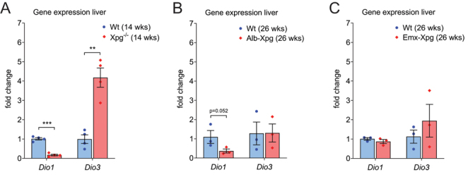
Background: Thyroid hormone signaling is essential for development, metabolism, and response to stress but declines during aging, the cause of which is unknown. DNA damage accumulating with time is a main cause of aging, driving many age-related diseases. Previous studies in normal and premature aging mice, due to defective DNA repair, indicated reduced hepatic thyroid hormone signaling accompanied by decreased type 1 deiodinase (DIO1) and increased DIO3 activities. We investigated whether aging-related changes in deiodinase activity are driven by systemic signals or represent cell- or organ-autonomous changes.

Methods: We quantified liver and plasma thyroid hormone concentrations, deiodinase activities and expression of T3-responsive genes in mice with a global, liver-specific and for comparison brain-specific inactivation of Xpg, one of the endonucleases critically involved in multiple DNA repair pathways.

Results: Both in global and liver-specific Xpg knockout mice, hepatic DIO1 activity was decreased. Interestingly, hepatic DIO3 activity was increased in global, but not in liver-specific Xpg mutants. Selective Xpg deficiency and premature aging in the brain did not affect liver or systemic thyroid signaling. Concomitant with DIO1 inhibition, Xpg -/- and Alb-Xpg mice displayed reduced thyroid hormone-related gene expression changes, correlating with markers of liver damage and cellular senescence.

Conclusions: Our findings suggest that DIO1 activity during aging is predominantly modified in a tissue-autonomous manner driven by organ/cell-intrinsic accumulating DNA damage. The increase in hepatic DIO3 activity during aging largely depends on systemic signals, possibly reflecting the presence of circulating cells rather than activity in hepatocytes.

Keywords: DNA damage; aging; deiodinase; liver; nucleotide excision repair; progeria; thyroid hormone.


Conflict of interest statement

The authors declare that there is no conflict of interest that could prejudice the impartiality of the research reported. Edward Visser is on the editorial board of European Thyroid Journal. Edward Visser was not involved in the review or editorial process for this paper, on which he is listed as an author.

Figures

Figure 1
Figure 1
Research question and experimental design. (A) Life span in mice with reduced somato-, lacto-, and/or thyrotropic signaling. The relationship between changes in percentage survival (x-axis) and log-hazard ratio effect size (y-axis) for median life span. Data were obtained from various life span cohorts (1) and separated by mutation. Mean values ± s.d.of the different cohorts are depicted for the various long-lived dwarf mutant mouse lines. (B) Schematic representation of the research question and experimental design.
Figure 2
Figure 2
Deiodinase activity in liver. Deiodinase 1 (DIO1) and 3 (DIO3) activity in livers of DNA repair-deficient (indicated in red) 4- and 14-week-old male Xpg−/− mice (A, B), 26-week-old liver-specific male Alb-Xpg mice (C, D) and 26-week-old brain-specific male and female Emx-Xpg mice (E, F). n = 3–4 animals/group. Wild-type (Wt) littermate controls are indicated in blue. Error bars denote mean ± s.e. *P < 0.05, ***P < 0.001.
Figure 3
Figure 3
Deiodinase gene expression in liver. Dio1 and Dio3 gene expression in livers of 4- and 14-week-old male Xpg−/− mice (A), 26-week-old liver-specific male Alb-Xpg mice (B), and 26-week-old brain-specific male and female Emx-Xpg mice (C). n = 3–4 animals/group. Error bars denote mean ± s.e. **P <0.01, ***P < 0.001.
Figure 4
Figure 4
Thyroid hormone concentrations in liver and plasma. Liver T3 and T4 concentrations in 4- and 14-week-old male and female Xpg−/− mice (A, D), 26-week-old liver-specific male Alb-Xpg mice (B, E), and 26-week-old brain-specific male and female Emx-Xpg mice (C, F). Plasma T3 and T4 concentrations in 4- and 14-week-old male and female Xpg−/− mice (G, J), 26-week-old liver-specific male Alb-Xpg mice (H, K), and 26-week-old male and female brain-specific Emx-Xpg mice (I, L). n = 3 animals/group. Error bars denote mean ± s.e. *P < 0.05.
Figure 5
Figure 5
Expression of thyroid hormone responsive genes and thyroid hormone-binding proteins in liver. Gene expression in livers of 4- and 14-week-old male Xpg−/− mice (A, B), 26-week-old liver-specific male Alb-Xpg mice (C), and 26-week-old brain-specific male and female Emx-Xpg mice (D). n = 3–4 animals/group. The dotted line separates genes that are upregulated (left) or downregulated (right) in hypothyroidism. Alb, Tbg, and Ttr gene expression in livers of 4-week-old male Xpg−/− mice (E), 14-week-old male Xpg−/− mice (F), 26-week-old male Alb-Xpg mice (G) and 26-week-old male and female Emx-Xpg mice (H). n = 3–4 animals/group. Error bars denote mean ± s.e. *P < 0.05, **P < 0.01.
Figure 6
Figure 6
Alanine transferase and albumin concentrations in plasma. Alanine amino transferase (ALAT) concentration in plasma of 4- and 14-week-old male Xpg−/− mice (A), and 26-week-old liver-specific male Alb-Xpg mice (B). Albumin concentration in plasma of 4- and 14-week-old male Xpg−/− mice (C), and 26-week-old, liver-specific male Alb-Xpg mice (D). n = 2–3 animals/group. Error bars denote mean ± s.e. **P < 0.01.
Figure 7
Figure 7
Expression of senescence-associated factors in liver. P21, IL-6, Mmp12, and Timp1 expression in livers of 14-week-old male Xpg−/− mice (A), 26-week-old liver-specific male Alb-Xpg mice (B), and 26-week-old brain-specific male and female Emx-Xpg mice (C). n = 3–4 animals/group. Error bars denote mean ± s.e. **P < 0.01, ***P < 0.001.

References

    1. Lopez-Otin C Galluzzi L Freije JMP Madeo F & Kroemer G. Metabolic control of longevity. Cell 2016166802–821. (10.1016/j.cell.2016.07.031) - DOI - PubMed
    1. Franceschi C Ostan R Mariotti S Monti D & Vitale G. The aging thyroid: a reappraisal within the geroscience integrated perspective. Endocrine Reviews 2019401250–1270. (10.1210/er.2018-00170) - DOI - PubMed
    1. Bowers J Terrien J Clerget-Froidevaux MS Gothié JD Rozing MP Westendorp RG van Heemst D & Demeneix BA. Thyroid hormone signaling and homeostasis during aging. Endocrine Reviews 201334556–589. (10.1210/er.2012-1056) - DOI - PubMed
    1. Engels K Rakov H Hönes GS Brix K Köhrle J Zwanziger D Moeller LC & Führer D. Aging alters phenotypic traits of thyroid dysfunction in male mice with divergent effects on complex systems but preserved thyroid hormone action in target organs. Journals of Gerontology. Series A, Biological Sciences and Medical Sciences 2019741162–1169. (10.1093/gerona/glz040) - DOI - PubMed
    1. Visser WE Visser TJ & Peeters RP. Thyroid disorders in older adults. Endocrinology and Metabolism Clinics of North America 201342287–303. (10.1016/j.ecl.2013.02.008) - DOI - PubMed

Publication types