. 2023 Oct 27;9(43):eadg6874.
doi: 10.1126/sciadv.adg6874. Epub 2023 Oct 25.

Human interleukin-12α and EBI3 are cytokines with anti-inflammatory functions

Affiliations

Human interleukin-12α and EBI3 are cytokines with anti-inflammatory functions

Karen Hildenbrand et al. Sci Adv. .

Abstract

Interleukins are secreted proteins that regulate immune responses. Among these, the interleukin 12 (IL-12) family holds a central position in inflammatory and infectious diseases. Each family member consists of an α and a β subunit that together form a composite cytokine. Within the IL-12 family, IL-35 remains particularly ill-characterized on a molecular level despite its key role in autoimmune diseases and cancer. Here we show that both IL-35 subunits, IL-12α and EBI3, mutually promote their secretion from cells but are not necessarily secreted as a heterodimer. Our data demonstrate that IL-12α and EBI3 are stable proteins in isolation that act as anti-inflammatory molecules. Both reduce secretion of proinflammatory cytokines and induce the development of regulatory T cells. Together, our study reveals IL-12α and EBI3, the subunits of IL-35, to be functionally active anti-inflammatory immune molecules on their own. This extends our understanding of the human cytokine repertoire as a basis for immunotherapeutic approaches.


Figures
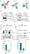
Fig. 1.
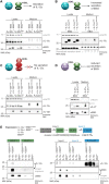
Fig. 1.. IL-12α and EBI3 mutually promote their secretion and form IL-35.
(A) IL-35 (alphaFold2 docked model) shares its α subunit IL-12α with IL-12 [Protein Data Bank (PDB): 3HMX], and its β subunit EBI3 with IL-27 (PDB: 7u7n). To assess mutually induced secretion of IL-12α and EBI3, constant DNA amounts of IL-12α were cotransfected with increasing DNA amounts of EBI3 (B) or vice versa (C). Both IL-12α and EBI3 show a reduced mobility in the medium, indicating that both subunits are retained in the endoplasmic reticulum in isolation and traverse the Golgi during secretion, as indicated by modification of their glycans. (D) Co-IP of FLAG-tagged IL-12α coexpressed with EBI3 in the cell medium verifies assembly for these two proteins. Quantification of the IL-12α IP efficiency and the fraction of EBI3 that is found in complex with IL-12α is shown (n = 3 ± SD). (E) Co-IP of secreted HA-tagged EBI3 coexpressed with IL-12αFLAG verifies assembly for these two proteins in the medium. Quantification of the EBI3HA IP efficiency and the fraction of IL-12αFLAG that is found in complex with EBI3HA is shown (n = 5 ± SD). Constructs were expressed in human embryonic kidney (HEK) 293T cells. One representative immunoblot is shown in each case.
Fig. 2.
Fig. 2.. IL-35 subunits can be secreted as nonheterodimers in contrast to IL-12 and IL-27.
(A) EBI3 induces the secretion of IL-12α even when it is retained in the ER (via a C-terminal KDEL sequence). EBI3-induced secretion is observed for wild-type IL-12α and a variant lacking the cysteine that forms an interchain disulfide bond in IL-12 (C96S). (B) The same as in (A), only that IL-12α was furnished with a KDEL ER-retention sequence and secretion of EBI3 was monitored. EBI3 secretion was slightly increased by coexpression with both IL-12α and the C96S variant. Quantifications of EBI3 secretion are shown below the blot. (C) A similar analysis for IL-12 reveals that IL-12βC199S, with an ER retention sequence, does not induce secretion of free IL-12α but instead co-retains it in the cell. The same is observed for the combination of IL-12αC96S with IL-12βC199S, with both proteins lacking the cysteines that form the interchain disulfide bond in IL-12. (D) Coexpression of wild-type EBI3 and IL-27α leads to the secretion of IL-27. When IL-27α was ER-retained (IL-27αKDEL), EBI3 secretion was reduced, in contrast to (B). (E) Free IL-12α can be detected in medium samples after pulldown of EBI3. IL-12α and EBI3 are cotransfected, and cell supernatants underwent two consecutive HA-IPs to isolate EBI3HA-containing complexes. IL-12α is also coimmunoprecipitated as can be seen in the IL-12α blot after HA-IP I and II, indicating pulldown of IL-35. The final IL-12α–IP reveals remaining IL-12α in the medium that is not interacting with EBI3. Band intensities show a significantly higher amount of free IL-12α when coexpressed with EBI3 (or EBI3KDEL) compared to transfection in isolation without EBI3.
Fig. 3.
Fig. 3.. Recombinant human IL-12αC96S and EBI3 are stable and well-structured proteins.
(A) Analysis of purified IL-12αC96S and EBI3 by reducing and nonreducing SDS–polyacrylamide gel electrophoresis (SDS-PAGE). Faster migration on nonreducing SDS-PAGE indicates the presence of disulfide bonds. Positions of intramolecular disulfide bonds are indicated in each subunit structure. (B) Reconstituted IL-12 and IL-27 are able to induce receptor heterodimerization. COS-7 cells were cotransfected with the indicated receptor chains equipped with the NanoBRET reporter system. Cells were stimulated with purified IL-12αC96S, which was previously incubated with recombinant human IL-12βC199S, or EBI3, previously incubated with murine IL-27α or the isolated subunits as indicated (10 nM final concentrations). Graphs represent the normalized NanoBRET signal (n = 3 ± SD). Statistical significance was calculated by one way analysis of variance (ANOVA) followed by Dunnett’s multiple comparisons test; ****P < 0.0001 compared with the corresponding phosphate-buffered saline (PBS) control. (C) NK-92 or BL-2 cells were stimulated with preincubated IL-12αC96S + IL-12βC199S or EBI3 + mIL-27α or the heterodimeric cytokines (10 ng/ml final concentrations). Downstream signaling was detected by STAT-phosphorylation via immunoblot. (D) Far-UV CD spectra for IL-12αC96S and EBI3. (E) IL-12αC96S unfolds cooperatively with an apparent melting temperature of 47 ± 0.2°C and EBI3 with an apparent melting temperature of 50 ± 0.2°C (green line, experimental data; black line, Boltzmann sigmoidal nonlinear curve fit; transitions were not reversible).
Fig. 4.
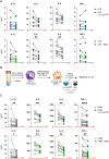
Fig. 4.. IL-12aC96S and EBI3 act as immunosuppressors.
(A) Concentrations of secreted IL-1β, IL-6, IL-8, and TNF-α [enzyme-linked immunosorbent assay (ELISA)] in supernatants from LPS stimulated human PBMCs after stimulation with IL-12aC96S or EBI3 (n = 10 donors). (B) Scheme of the experimental workflow to assess IL-12aC96S and EBI3 effects on alveolar like monocyte-derived macrophages (MDMs). (C) Amounts of IL-1β, IL-6, IL-8, and TNF-α (ELISA) produced by human MDMs (n = 10 to 17 donors) in supernatants after stimulation with HDM and IL-12aC96S or EBI3. Dotted line indicates mean secretion of PBS-treated PBMCs or MDMs. Data are presented as individual values. Donor-dependent effect is shown by connecting line. Statistical significance was determined by Wilcoxon test. *P < 0.05, **P < 0.01, ***P < 0.001, and ****P < 0.0001.
Fig. 5.
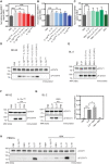
Fig. 5.. IL-12αC96S and EBI3 show different signaling characteristics.
(A) The NanoBRET reporter system indicates IL-12 receptor heterodimerization after stimulation with 1 nM IL-12, which can be blocked by a 30-min pretreatment with IL-12βC199S (1, 10, or 100 nM). (B) In the same assay, a 30-min pretreatment with IL-12αC96S (1, 10, or 100 nM) shows reduced IL-12–induced receptor heterodimerization at a 10-fold excess of IL-12αC96S. (C) A preincubation with increasing concentration of EBI3 did not have any effect on IL-27 activity. Graphs represent the normalized NanoBRET signal (n = 3 ± SD). (D) STAT4 phosphorylation in NK-92 cells after treatment with IL-12 (1 ng/ml) is blocked by a 30-min pretreatment with IL-12βC199S (10 and 100 ng/ml) or IL-12αC96S (10 ng/ml). Representative immunoblots from one of three independent experiments are shown. (E) BL-2 cells were used to monitor IL-27 (10 ng/ml)–mediated STAT1 phosphorylation, which is not blocked by pretreatment with EBI3 (10, 100, or 1000 ng/ml) for 30 min (n = 3 ± SD). (F) NK-92 or (G) BL-2 cells were pretreated with IL-12αC96S (10 ng/ml) or EBI3 (10 ng/ml) overnight (o/n) before addition of IL-12 or IL-27, respectively, to investigate receptor internalization. (n = 3 ± SD, *P < 0.05 and ****P < 0.0001) (H) STAT1 phosphorylation in human PBMCs treated with EBI3 and IL-12αC96S compared to the cytokines IL-10, IL-27, and IL-35 and their effect after HDM stimulation. Representative immunoblots from one of three independent experiments are shown.
Fig. 6.
Fig. 6.. IL-12aC96S and EBI3 potentiate regulatory T cell development and suppress SEA-induced IL-4 production from human PBMCs.
(A) Percentage of CD25hiFoxp3+ Treg cells [fluorescence-activated cell sorting (FACS)] in human PBMC cultures treated with IL-12aC96S (20 ng/ml) and EBI3 (20 ng/ml) or the combination of both IL-12aC96S and EBI3 (n = 8). Data are presented as means + SEM. Statistical significance was determined by Friedman test. *P < 0.05 and **P < 0.01. (B) Concentrations of IL-4 (ELISA) in culture supernatants from SEA-stimulated human PBMCs alone or in combination with IL-12C96S or EBI3 (n = 8 donors). The dotted line indicates mean secretion of IL-4 from PBS-treated PBMCs. (C) Concentrations of CCL17 (ELISA) in culture supernatants from IL-4–stimulated human PBMCs alone or in combination with IL-12C96S or EBI3 (n = 3 donors). (B and C) Data are presented as individual values. Donor-dependent effect is shown by the connecting line. Statistical significance was determined by Wilcoxon test. *P < 0.05 and **P < 0.01.

References

    1. C. Brocker, D. Thompson, A. Matsumoto, D. W. Nebert, V. Vasiliou, Evolutionary divergence and functions of the human interleukin (IL) gene family. Hum. Genomics 5, 30–55 (2010). - PMC - PubMed
    1. D. A. A. Vignali, V. K. Kuchroo, IL-12 family cytokines: Immunological playmakers. Nat. Immunol. 13, 722–728 (2012). - PMC - PubMed
    1. K. Hildenbrand, I. Aschenbrenner, F. C. Franke, O. Devergne, M. J. Feige, Biogenesis and engineering of interleukin 12 family cytokines. Trends Biochem. Sci. 47, 936–949 (2022). - PubMed
    1. E. D. T. Wojno, C. A. Hunter, J. S. Stumhofer, The immunobiology of the interleukin-12 family: Room for discovery. Immunity 50, 851–870 (2019). - PMC - PubMed
    1. H. Yoshida, C. A. Hunter, The immunobiology of interleukin-27. Annu. Rev. Immunol. 33, 417–443 (2015). - PubMed

MeSH terms