

doi: 10.1590/0037-8682-0439-2023.
eCollection 2023.
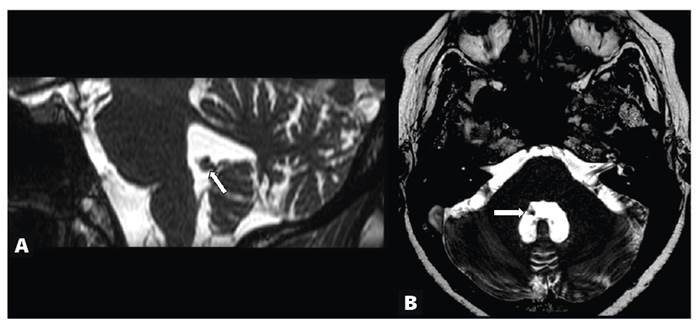
Muscular and brain cysticercosis
Affiliations
- PMID: 37878829
- PMCID: PMC10588824
- DOI: 10.1590/0037-8682-0439-2023
Item in Clipboard
Muscular and brain cysticercosis
Rev Soc Bras Med Trop.
.
No abstract available
Conflict of interest statement
Figures


References
-
- 1. Yacoub VRD, Ramos MC, Reis F. High-resolution vessel wall magnetic resonance imaging for the diagnosis of neurocysticercosis vasculitis. Rev Soc Bras Med Trop. 2022;55:e0203. - PMC - PubMed
- Yacoub VRD, Ramos MC, Reis F. High-resolution vessel wall magnetic resonance imaging for the diagnosis of neurocysticercosis vasculitis. Rev Soc Bras Med Trop. 2022;55:e0203. - PMC - PubMed
-
- 2. Garcia HH, Gonzalez AE, Gilman RH. Taenia solium cysticercosis and its impact in neurological disease. Clin Microbiol Rev. 2020;33(3):e00085-19. - PMC - PubMed
- Garcia HH, Gonzalez AE, Gilman RH. Taenia solium cysticercosis and its impact in neurological disease. Clin Microbiol Rev. 2020;33(3):e00085-19. - PMC - PubMed
-
- 3. Miyoshi IC, de Toledo AHN, Pereira FV, Villarinho LL, Dalaqua M, de Ávila Duarte J, et al. Infectious Myelitis. Semin Ultrasound CT MR. 2023;44(5):424-35. - PubMed
- Miyoshi IC, de Toledo AHN, Pereira FV, Villarinho LL, Dalaqua M, de Ávila Duarte J, et al. Infectious Myelitis. Semin Ultrasound CT MR. 2023;44(5):424–435. - PubMed
MeSH terms
LinkOut - more resources
Full Text Sources
Medical
