. 2023 Oct 25;19(1):223.
doi: 10.1186/s12917-023-03782-2.

Anti-cancer effect of palmatine through inhibition of the PI3K/AKT pathway in canine mammary gland tumor CMT-U27 cells

Affiliations

Anti-cancer effect of palmatine through inhibition of the PI3K/AKT pathway in canine mammary gland tumor CMT-U27 cells

Min-Jae Yoo et al. BMC Vet Res. .

Abstract

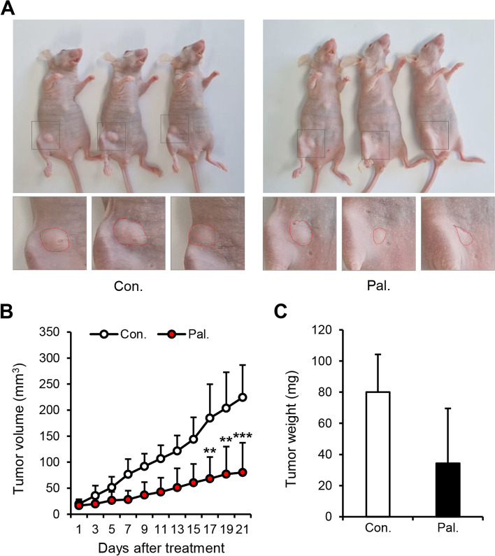
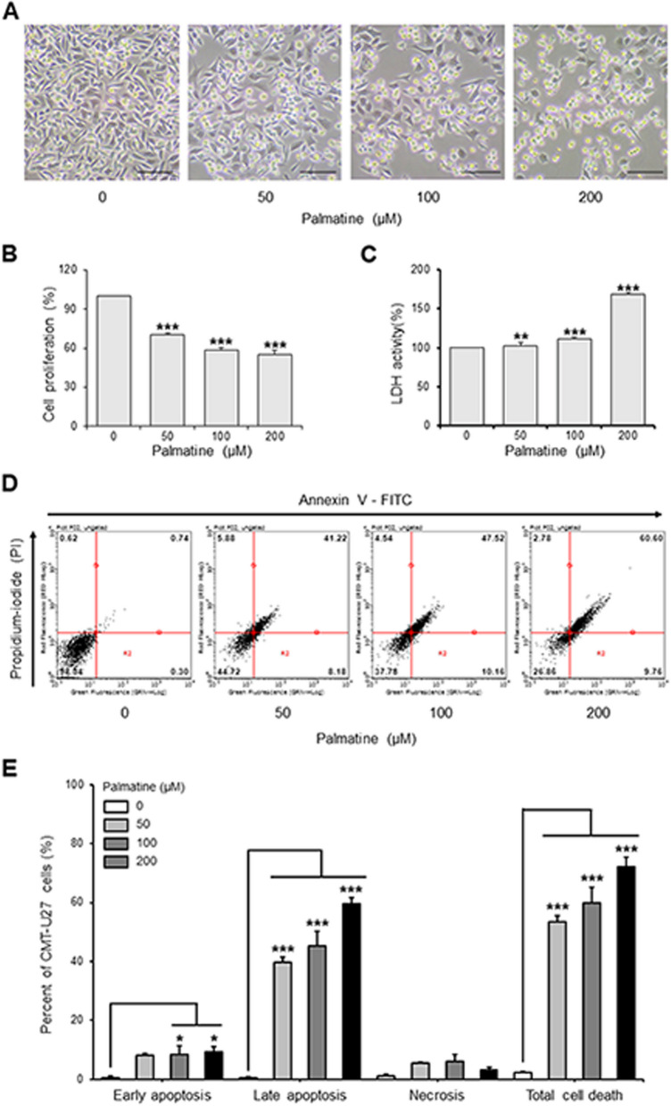
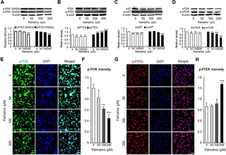
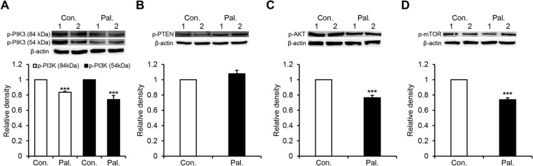
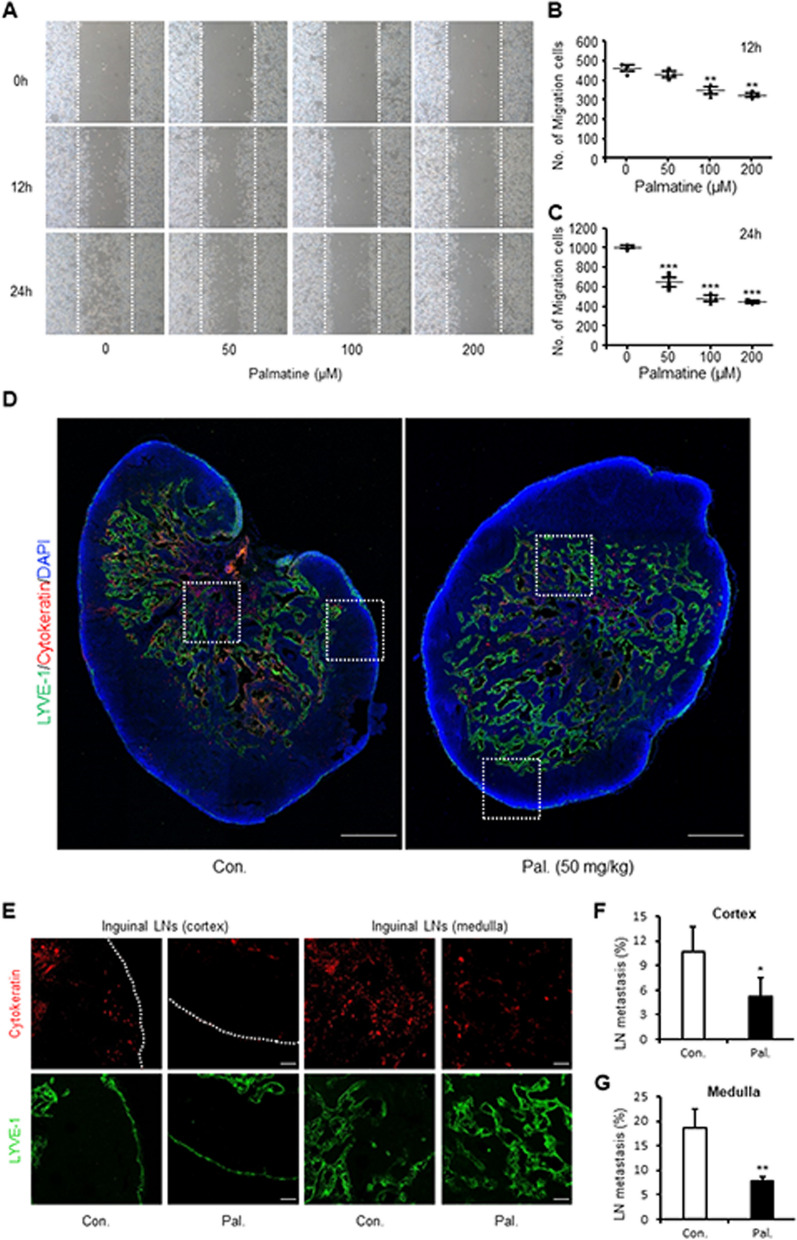
Canine mammary gland tumors (CMTs) are the most common and lethal cancers in female dogs. Dysregulated phosphoinositide 3-kinases (PI3K)/AKT pathway reportedly was involved in the growth and metastasis of CMTs. However, there are few studies on therapeutic strategies for targeting the PI3K pathway in CMTs. In this study, we aimed to determine whether palmatine, a natural isoquinoline alkaloid with anti-cancer properties, could inhibit the growth of CMTs and whether the inhibitory effect was mediated through the PI3K/AKT pathway. Our in vitro experiments on CMT-U27, a CMT cell line, showed that palmatine reduced cell proliferation and induced cell death. Western blotting results revealed that palmatine decreased the protein expression of PI3K, PTEN, AKT, and mechanistic target of rapamycin in the PI3K/AKT pathway, which was supported by the results of immunocytochemistry. Additionally, palmatine suppressed the migration and tube formation of canine aortic endothelial cells as well as the migration of CMT U27 cells. Our in vivo results showed that palmatine inhibited tumor growth in a CMT-U27 mouse xenograft model. We observed a decreased expression of proteins in the PI3K/AKT pathway in tumor tissues, similar to the in vitro results. Furthermore, palmatine significantly disrupted the tumor vasculature and inhibited metastasis to adjacent lymph nodes. In conclusion, our findings demonstrate that palmatine exerts anti-cancer effects against CMTs by inhibiting PI3K/AKT signaling pathway, suggesting that palmatine has potential as a canine-specific PI3K inhibitor for the treatment of CMTs.

Keywords: CMTs; Cancer; Canine; Canine mammary gland tumors; PI3K inhibitor; PI3K/AKT pathway; PTEN; Palmatine.


Conflict of interest statement

The authors declare no competing interests.

Figures

Fig. 1
Fig. 1
Palmatine suppresses tumor progression in an in vivo CMT-U27 mouse xenograft model. A Tumor development at 21 days after intraperitoneal injection of Palmatine (50 mg/kg) and DMSO in PBS (control) in BALB/c nude mice. The dotted lines in the magnified images demarcate CMT masses. B Comparison of tumor volume in treatment group and control group over 21 days. C Comparison of the weight of tumors harvested from treatment and control group after 21 days of injection. Unless otherwise denoted: n = 5 each group. Values are mean ± SD. ** p < 0.01; *** p < 0.001 versus control. Con., control; Pal., palmatine
Fig. 2
Fig. 2
Palmatine induces cell death of CMT-U27 cells. A Images showing morphological changes on CMT-U27 cells after palmatine-treatment. Magnification, 100 × . Scale bar, 50 µm. B, C Dose-dependent comparison of CMT-U27 cell proliferation and LDH activity. Values are mean ± SD. ** p < 0.01; *** p < 0.001 versus untreated cells by one-way ANOVA followed by Bonferroni post-hoc test. D Apoptosis in CMT-U27 cells was measured by flow cytometry with Annexin V-FITC staining. E Percentage of CMT-U27 cells in early, late apoptosis, necrosis, and total cell death when treated with palmatine by concentration. Values are mean ± SD. * p < 0.05; *** p < 0.001 versus untreated cells by one-way ANOVA followed by Bonferroni post-hoc test
Fig. 3
Fig. 3
Palmatine regulates the PI3K/AKT signaling pathway in CMT-U27 cells. A-D CMT-U27 cells grown in 6-well plates and then cultured in fresh media containing various concentrations of palmatine for 18 h at 37ºC. After treatment, the cells were harvested for western blotting to quantify the expression of p-PI3K (A), PTEN, p-PTEN (B), AKT, p-AKT (C), mTOR, p-mTOR (D), and β-actin. E–H CMT-U27 cells grown in 24-well plates and then cultured in fresh media containing various concentrations of palmatine for 18 h at 37ºC. After treatment, immunocytochemistry was performed to quantify the expression of p-PI3K (E) and p-PTEN (G). Scale bar, 50 µm. Values are mean ± SD. * p < 0.05, ** p < 0.01, *** p < 0.001 versus untreated cells by one-way ANOVA followed by Bonferroni post-hoc test
Fig. 4
Fig. 4
Palmatine inhibits PI3K/AKT pathway and increases PTEN activity in an in vivo CMT-U27 xenograft models. Tumors of CMT-U27 xenograft model injected with palmatine (treatment group) or DMSO in PBS (control group) for 21 days were collected and western blotting was performed. A-D Images and quantification of the protein expression of p-PI3K (A), p-PTEN (B), p-AKT (C), p-mTOR (D) and β-actin in tumors. Values are mean ± SD. *** p < 0.001 versus control by unpaired Student’s t-test. Con., control; Pal., palmatine
Fig. 5
Fig. 5
Palmatine suppresses angiogenesis in CMTs and CnAoECs. A-C Fluorescent images and quantification of CD31 and α-SMA expression differences between treatment and control groups. Scale bar, 50 µm. Values are mean ± SD. ** p < 0.01 versus control by unpaired Student’s t-test. Con., control; Pal., palmatine. D, E CnAOECs grown in 6-well plates and scratched by 1000 μl tip. Then, CnAOECs cultured in fresh media containing various concentrations of palmatine for 24 h at 37ºC. Images showing CnAOECs migration after palmatine treatment in a dose-dependent manner and quantification of the number of migrated cells. Values are mean ± SD. * p < 0.05 versus untreated cells by one-way ANOVA followed by Bonferroni post-hoc test. F, G CnAOECs were seeded in matrigel-coated 6-well plates and incubated for 3 h at 37ºC. Images showing CnAOECs tube formation after palmatine treatment in a dose-dependent manner and quantification of the number of tubules. Values are mean ± SD. *** p < 0.001 versus untreated cells by one-way ANOVA followed by Bonferroni post-hoc test
Fig. 6
Fig. 6
Palmatine restrains the migration and metastasis of CMT cells. A-C CMT-U27 cells grown in 6-well plates and scratched by 1000 μl tip. Then, CMT-U27 cells cultured in fresh media containing various concentrations of palmatine for 24 h at 37ºC. Images showing CMT-U27 cells migration after palmatine treatment in a dose- and time-dependent manner and quantification of the number of migrated cells over time. Magnification, 40 × . Values are mean ± SD. ** p < 0.01, *** p < 0.001 versus untreated cells by one-way ANOVA followed by Bonferroni post-hoc test. D Fluorescent images showing the lymph nodes of treatment and control groups stained for cytokeratin and LYVE-1. Scale bar, 400 μm. E–G Magnified images and quantification of lymph node metastases in the cortex and medulla regions in the control and treatment groups. The dotted lines demarcate the margin of the lymph node. Scale bar, 50 μm. Values are mean ± SD. * p < 0.05, ** p < 0.01 versus control by one-way ANOVA followed by Bonferroni post-hoc test. Con., control; Pal., palmatine
Fig. 7
Fig. 7
Schematic diagram of the mechanism of anti-cancer effect of palmatine

References

    1. Dorn CR, Taylor DO, Schneider R, Hibbard HH, Klauber MR. Survey of animal neoplasms in Alameda and contra costa counties, California. II. Cancer morbidity in dogs and cats from Alameda County. J Natl Cancer Inst. 1968;40(2):307–318. - PubMed
    1. Withrow S, Vail D, Page R. Small animal oncology. Philadelfia: Saunders; 2001.
    1. Gamlem H, Nordstoga K, Glattre E. Canine neoplasia – Introductory paper. APMIS. 2008;116(s125):5–18. - PubMed
    1. Hampe JF, Misdorp W. Tumours and dysplasias of the mammary gland. Bull World Health Organ. 1974;50(1–2):111–133. - PMC - PubMed
    1. Straw R. Treatment of mammary gland tumors and perianal neoplasia. pp. 672-675. In: The North American Veterinary Conference (NAVC) Orlando, Florida, 2005. 2005.

Substances