. 2023 Oct 25;21(1):752.
doi: 10.1186/s12967-023-04394-y.

JMJD6-BRD4 complex stimulates lncRNA HOTAIR transcription by binding to the promoter region of HOTAIR and induces radioresistance in liver cancer stem cells

Affiliations

JMJD6-BRD4 complex stimulates lncRNA HOTAIR transcription by binding to the promoter region of HOTAIR and induces radioresistance in liver cancer stem cells

Ruifeng Pei et al. J Transl Med. .

Abstract

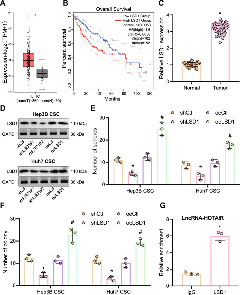
Background: Long non-coding RNA (lncRNA) HOTAIR acts importantly in liver cancer development, but its effect on radioresistance remains poorly understood. Here, our study probed into the possible impact of HOTAIR in radioresistance in liver cancer stem cells (LCSCs) and to elucidate its molecular basis.

Methods: Following sorting of stem and non-stem liver cancer cells, LCSCs were identified and subjected to RNA-seq analysis for selecting differentially expressed genes. Expression of HOTAIR was determined in liver cancer tissues and CSCs. The stemness, proliferation, apoptosis and radioresistance of LCSCs were then detected in response to altered expression of HOTAIR-LSD1-JMJD6-BRD4.

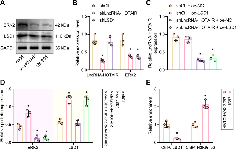
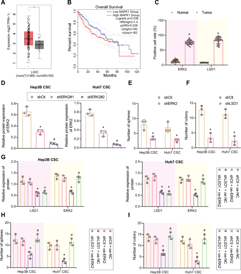
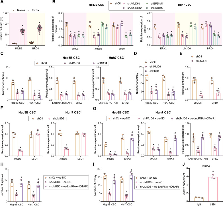
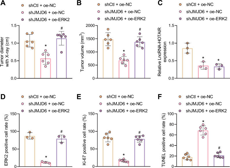
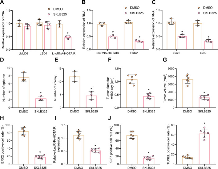
Results: Ectopic HOTAIR expression was found to promote radioresistance of LCSCs by maintaining its stemness. Mechanistic investigations indicated that HOTAIR recruited LSD1 to the MAPK1 promoter region and reduced the level of H3K9me2 in the promoter region, thus elevating ERK2 (MAPK1) expression. JMJD6-BRD4 complex promoted HOTAIR transcription by forming a complex and positively regulated ERK2 (MAPK1) expression, maintaining the stemness of LCSCs, and ultimately promoting their radioresistance in vitro and in vivo.

Conclusion: Collectively, our work highlights the promoting effect of the JMJD6-BRD4 complex on the radioresistance of LCSCs through a HOTAIR-dependent mechanism.

Keywords: CD13+CD133+; ERK2 (MAPK1); JMJD6; JMJD6 inhibitor SKLB325; LSD1; Liver cancer; Liver cancer stem cells; Long noncoding RNA HOTAIR; Radioresistance.


Conflict of interest statement

The authors declare that they have no competing interests.

Figures

Fig. 1
Fig. 1
HOTAIR expression is increased in liver cancer tissues and LCSCs, which links to stemness maintenance and radioresistance of LCSCs. A A Volcano map of the gene expression between CD13+CD133+ liver cancer cell subsets and negative liver cancer cell subsets based on the RNA-seq data. Red indicates highly expressed genes while green indicates poorly expressed genes. B The expression of HOTAIR in liver cancer and normal tissue samples in TCGA database (p = 0.03). C Correlation between the expression of HOTAIR and the progression free survival of patients with liver cancer. D The expression of HOTAIR in normal and liver cancer tissues measured by RT-qPCR, normalized to GAPDH. *p < 0.05 compared with adjacent normal tissues. E Silencing and overexpression efficiency of HOTAIR determined by RT-qPCR in Hep3B and Huh7 CSCs. *p < 0.05 compared with Hep3B and Huh7 CSCs treated with shCtl, # p < 0.05 compared with Hep3B and Huh7 CSCs treated with oeCtrl. F The effect of HOTAIR silencing or overexpression on the stemness maintenance of LCSCs, as detected by microsphere formation assay. G The colony formation ability of LCSCs after 6 Gy X-ray irradiation after HOTAIR silencing or overexpression, as examined by clonogenic assay. *p < 0.05 compared with Hep3B and Huh7 CSCs treated with shCtl, # p < 0.05 compared with Hep3B and Huh7 CSCs treated with oeCtrl. Data were represented as mean ± standard deviation. Data between cancer tissues and adjacent normal tissues were compared by paired t test, and those between the other two groups were compared by unpaired t test. The data comparison between multiple groups was performed by one-way ANOVA with Tukey’s post-hoc test. Cellular experiments were repeated in triplicate
Fig. 2
Fig. 2
LSD1 maintains the stemness of LCSCs and enhances cell radioresistance by interacting with HOTAIR. A The expression of LSD1 in liver cancer tissues was analyzed based on TCGA database. LIHC, liver hepatocellular carcinoma. The red box plots represent tumor samples and the gray box plots represent normal samples. *p < 0.01 compared with normal samples. B Correlation between the expression of LSD1 and the overall survival of patients with liver cancer. The red line indicates the liver cancer patients with high expression of LSD1, and the blue line indicates liver cancer patients with low expression of LSD1. The upper right is the significant p value and legend. C The expression of LSD1 in adjacent normal tissues and liver cancer tissues, as measured by RT-qPCR, normalized to GAPDH. *p < 0.05 compared with adjacent normal tissues. D The silencing and overexpression efficiency of LSD1 was detected by Western blot, normalized to GAPDH. E The effect of LSD1 depletion or overexpression on LCSCs stemness, as measured by microsphere formation assay. *p < 0.05 compared with shCtl-treated LCSCs, #p < 0.05 compared with oeCtl-treated LCSCs. F The colony formation ability of LCSCs with LSD1 depletion or overexpression after treatment with 6 Gy X-ray irradiation, as examined by clonogenic assay. *p < 0.05 compared with shCtl-treated LCSCs, #p < 0.05 compared with oeCtl-treated LCSCs. G Interaction between HOTAIR and LSD1 assessed by RIP assay. The relative enrichment on the Y-axis represents IP-LSD1/IgG. *p < 0.05 compared with the IgG group. The experimental data were summarized as mean ± standard deviation. Data between cancer tissues and adjacent normal tissues were compared by paired t test, and those between the other two groups were compared by unpaired t test. The data comparison between multiple groups was performed by one-way ANOVA with Tukey’s post-hoc test. Cellular experiments were repeated three times
Fig. 3
Fig. 3
HOTAIR increases ERK2 (MAPK1) expression by recruiting LSD1 to the promoter region of MAPK1. A ERK2 (MAPK1) and LSD1 expression in Hep3B and Huh7 CSCs with LSD1 silencing or HOTAIR silencing as examined by Western blot, normalized to GAPDH. B MAPK1 and HOTAIR expression in Hep3B and Huh7 CSCs with LSD1 silencing or HOTAIR silencing examined by RT-qPCR, normalized to GAPDH. *p < 0.05 compared with Hep3B and Huh7 CSCs treated with shCtl. C The expression of HOTAIR in Hep3B CSCs treated with shHOTAIR or combined with oe-LSD1 measured by RT-qPCR, normalized to GAPDH. *p < 0.05 compared with shCtl + oe-NC-treated Hep3B CSCs. D Western blot assay of ERK2 (MAPK1) and LSD1 proteins in Hep3B CSCs treated with shHOTAIR or combined with oe-LSD1, normalized to GAPDH. E The relative enrichment of LSD1 and H3K9me2 in the promoter region of MAPK1 in Hep3B CSCs with HOTAIR silencing assessed by ChIP, relative to Input. *p < 0.05 compared with shCtl-treated Hep3B CSCs. The experimental data were represented as mean ± standard deviation. Results between two groups were compared by unpaired t test. The data comparison between multiple groups was performed by one-way ANOVA with Tukey’s post-hoc test. Cellular experiments were repeated for three times
Fig. 4
Fig. 4
LSD1 increased ERK2 (MAPK1) expression, and thus maintained LCSCs stemness and further enhanced their radioresistance. A ERK2 (MAPK1) expression in normal samples and liver cancer samples, as analyzed by TCGA database. LIHC, liver hepatocellular carcinoma. Red box plots represent tumor samples, and gray box plots represent normal samples. *p < 0.01 compared with normal samples. B The correlation between ERK2 (MAPK1) expression and overall survival rate of liver cancer patients analyzed by TCGA database. The upper right is the significant p value and legend. C The positive expression of LSD1 and ERK2 (MAPK1) proteins in clinical liver cancer tissue samples, as measured by immunohistochemistry. *p < 0.05 compared with adjacent normal tissues. D The silencing efficiency of ERK2 (MAPK1) shRNAs in Hep3B and Huh7 CSCs, as detected by Western blot, normalized to GAPDH. E Microsphere formation assay was adopted to measure the stemness of LCSCs with ERK2 (MAPK1) silencing. *p < 0.05 compared with shCtl-treated cells. F The colony formation ability of LCSCs with ERK2 (MAPK1) silencing after exposure to 6 Gy X-ray, as measured by clonogenic assay. *p < 0.05 compared with shCtl-treated cells. G Quantitative analysis the relative expression of LSD1 and ERK2 (MAPK1) proteins in Hep3B and Huh7 CSCs in response to shLSD1, oe-ERK2 or in combination by Western blot, relative to GAPDH. H The LCSC stemness after treatment with shLSD1, oe-ERK2 or in combination measured by microsphere formation assay. I, After 6 Gy X-ray treatment, cell proliferation in the presence of shLSD1, oe-ERK2 or in combination, as examined by clonogenic assay. *p < 0.05 compared with shCtl + oe-NC-treated cells. #p < 0.05 compared with shLSD1 + oe-NC-treated cells. Above data were shown as mean ± standard deviation. Results between two groups were compared by unpaired t test. The data comparison between multiple groups was performed by one-way ANOVA with Tukey’s post-hoc test. Cellular experiments were repeated in triplicate
Fig. 5
Fig. 5
JMJD6 enhances HOTAIR expression to maintain the stemness of LCSCs and promotes their radioresistance. A Quantitative analysis of JMJD6 and BRD4 expression in liver cancer and adjacent normal tissues measured by immunohistochemistry. *p < 0.05 compared with adjacent normal tissues. B The expression of JMJD6, BRD4 and ERK2 (MAPK1) in Hep3B and Huh7 CSCs with JMJD6 silencing or BRD4 silencing detected by Western blot, relative to GAPDH. *p < 0.05 compared with shCtl-treated cells. C The stemness of LCSCs with JMJD6 silencing or BRD4 silencing detected by microsphere formation assay. *p < 0.05 compared with shCtl-treated cells. D After exposure to X-ray, the cell colony formation in the presence of shJMJD6 or shBRD4, *p < 0.05 compared with shCtl-treated cells. E The enrichment of JMJD6 and BRD4 in the promoter region of HOTAIR after JMJD6 silencing, as examined by ChIP assay, relative to Input. The relative enrichment on the Y-axis represents JMJD6 or BRD4/IgG. *p < 0.05 compared with shCtl-treated cells. F Detection of expression of JMJD6 and LSD1 in cells following silencing of JMJD6, *p < 0.05 compared with shCtl-treated cells. G The expression of ERK2 (MAPK1) and JMJD6 after JMJD6 depletion or in combination with HOTAIR overexpression, as examined by Western blot and RT-qPCR. *p < 0.05 compared with shCtl + oe-NC-treated cells. #p < 0.05 compared with shJMJD6 + oe-NC-treated cells. H The microsphere formation assay was adopted to measure LCSC stemness after JMJD6 depletion or in combination with HOTAIR overexpression. *p < 0.05 compared with shCtl + oe-NC-treated cells. #p < 0.05 compared with shJMJD6 + oe-NC-treated cells. I Cell colony formation after JMJD6 depletion or in combination with HOTAIR overexpression following 6 Gy X-ray treatment examined by clonogenic assay. *p < 0.05 compared with shCtl + oe-NC-treated cells. #p < 0.05 compared with shJMJD6 + oe-NC-treated cells. J, RIP assay validated the interaction of JMJD6/BRD4 complex with LSD1 (relative enrichment in Y-axis represents IP-LSD1/IgG, * p < 0.05 compared with IgG group. Measurement data were expressed as mean ± standard deviation. Results between two groups were compared by unpaired t test. The data comparison between multiple groups was performed by one-way ANOVA with Tukey’s post-hoc test. Cellular experiments were repeated three times
Fig. 6
Fig. 6
JMJD6 promotes radioresistance of LCSCs by regulating the HOTAIR-LSD1-ERK2 axis. A The diameter of xenograft tumors of nude mice in response to JMJD6 silencing alone or in combination with ERK2 (MAPK1) overexpression (n = 6). B The weight of xenografted tumors in nude mice in response to JMJD6 silencing alone or in combination with ERK2 (MAPK1) overexpression (n = 6). C The expression of HOTAIR in xenografted tumor tissues of nude mice in response to JMJD6 silencing alone or in combination with ERK2 (MAPK1) overexpression determined by RT-qPCR, normalized to GAPDH (n = 6). D The ERK2 (MAPK1) positive cells in xenograft tumor tissues of nude mice in response to JMJD6 silencing alone or in combination with ERK2 (MAPK1) overexpression, as detected by immunohistochemistry (n = 6). E Cell proliferation in xenografted tumors of mice in response to JMJD6 silencing alone or in combination with ERK2 (MAPK1) overexpression, as detected by Ki-67 staining (n = 6). F Cell apoptosis in xenografted tumors of mice in response to JMJD6 silencing alone or in combination with ERK2 (MAPK1) overexpression, as detected by TUNEL staining (n = 6). *p < 0.05 compared with shCtl + oe-NC-treated mice. #p < 0.05 compared with shJMJD6 + oe-NC-treated mice. Measurement data were represented as mean ± standard deviation. The data comparison between multiple groups was performed by one-way ANOVA with Tukey’s post-hoc test
Fig. 7
Fig. 7
JMJD6 inhibitor SKLB325 represses the LCSC stemness and relieves cell radioresistance. A The expression of HOTAIR, JMJD6 and LSD1 in Hep3B and Huh7 CSCs in response to JMJD6 inhibitor SKLB325 determined by RT-qPCR, normalized to GAPDH. B The expression of HOTAIR and MAPK1 in Hep3B and Huh7 CSCs in response to JMJD6 inhibitor SKLB325 determined by RT-qPCR, normalized to GAPDH. C The expression of Sox2 and Oct2 in Hep3B and Huh7 CSCs in response to JMJD6 inhibitor SKLB325 determined by RT-qPCR, normalized to GAPDH. D The difference of cell stemness after SKLB325 treatment, as detected by microsphere formation assay. E The impact of SKLB325 on colony formation ability in response to SKLB325 treatment, as detected by clonogenic assay. F The diameter of xenografts tumors in mice with SKLB325 treatment. G The tumor volume of xenografted tumors. H, ERK2 (MAPK1) expression in the tumor tissues of mice in response to JMJD6 inhibitor SKLB325 measured by immunohistochemistry. I The expression of HOTAIR in the tumor tissues of mice in response to JMJD6 inhibitor SKLB325 determined by RT-qPCR, normalized to GAPDH. J Ki-67 staining and TUNEL staining were adopted to examine cell proliferation and apoptosis in the tumor tissues of mice in response to JMJD6 inhibitor SKLB325. *p < 0.05 compared with DMSO-treated cells or mice. Experimental data were shown as mean ± standard deviation. Results between two groups were compared by unpaired t test
Fig. 8
Fig. 8
Mechanism graph of the regulatory network. The JMJD6–BRD4 complex elevates the expression of ERK2 (MAPK1) through the HOTAIR-LSD1 axis, thereby maintaining the stemness of LCSCs and enhancing their radioresistance

Similar articles

Cited by

References

    1. Siegel RL, Miller KD, Jemal A. Cancer statistics, 2016. CA Cancer J Clin. 2016;66(1):7–30. doi: 10.3322/caac.21332. - DOI - PubMed
    1. Torre LA, Bray F, Siegel RL, Ferlay J, Lortet-Tieulent J, Jemal A. Global cancer statistics, 2012. CA Cancer J Clin. 2015;65(2):87–108. doi: 10.3322/caac.21262. - DOI - PubMed
    1. Ferlay J, Soerjomataram I, Dikshit R, Eser S, Mathers C, Rebelo M, et al. Cancer incidence and mortality worldwide: sources, methods and major patterns in GLOBOCAN 2012. Int J Cancer. 2015;136(5):E359–E386. doi: 10.1002/ijc.29210. - DOI - PubMed
    1. Liu CY, Chen KF, Chen PJ. Treatment of liver cancer. Cold Spring Harb Perspect Med. 2015;5(9):a021535. doi: 10.1101/cshperspect.a021535. - DOI - PMC - PubMed
    1. Villanueva A, Hernandez-Gea V, Llovet JM. Medical therapies for hepatocellular carcinoma: a critical view of the evidence. Nat Rev Gastroenterol Hepatol. 2013;10(1):34–42. doi: 10.1038/nrgastro.2012.199. - DOI - PubMed

MeSH terms