. 2023 Dec;10(35):e2305042.
doi: 10.1002/advs.202305042. Epub 2023 Oct 25.

Unloading-Induced Skeletal Interoception Alters Hypothalamic Signaling to Promote Bone Loss and Fat Metabolism

Affiliations

Unloading-Induced Skeletal Interoception Alters Hypothalamic Signaling to Promote Bone Loss and Fat Metabolism

Qiaoyue Guo et al. Adv Sci (Weinh). 2023 Dec.

Abstract

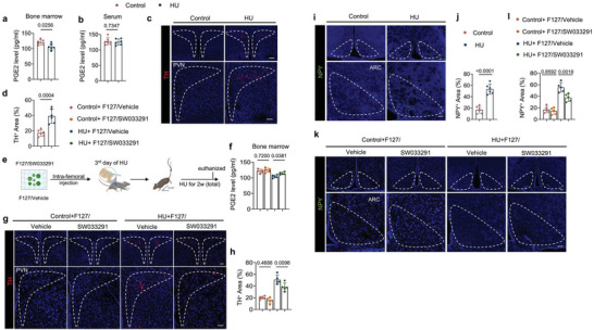
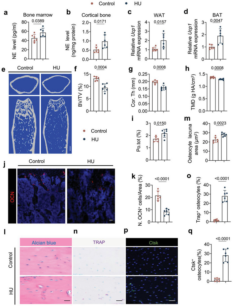
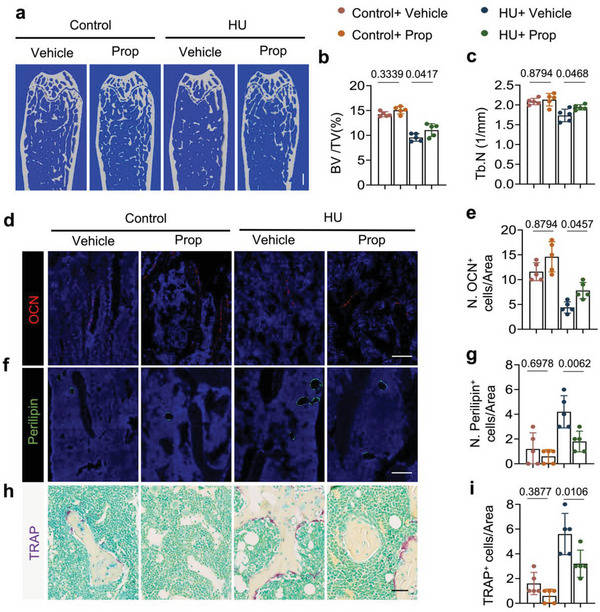
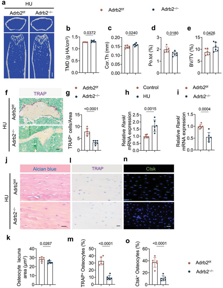
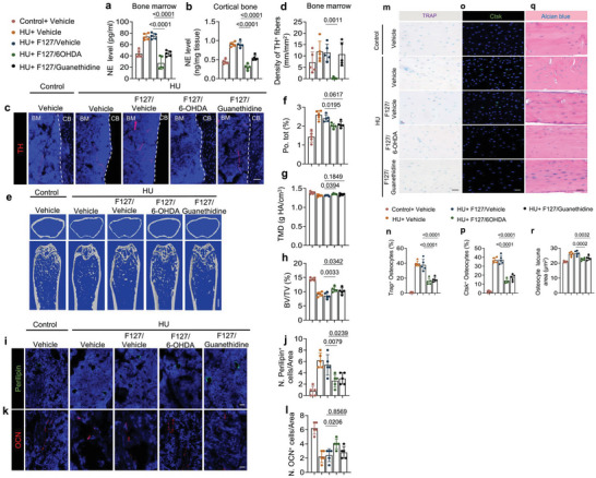
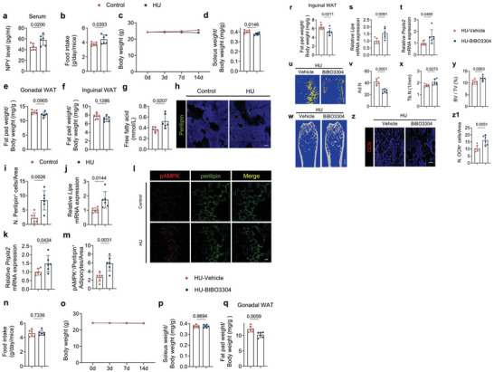
Microgravity is the primary factor that affects human physiology in spaceflight, particularly bone loss and disturbances of the central nervous system. However, little is known about the cellular and molecular mechanisms of these effects. Here, it is reported that in mice hindlimb unloading stimulates expression of neuropeptide Y (NPY) and tyrosine hydroxylase (TH) in the hypothalamus, resulting in bone loss and altered fat metabolism. Enhanced expression of TH and NPY in the hypothalamus occurs downstream of a reduced prostaglandin E2 (PGE2)-mediated ascending interoceptive signaling of the skeletal interoception. Sympathetic antagonist propranolol or deletion of Adrb2 in osteocytes rescue bone loss in the unloading model. Moreover, depletion of TH+ sympathetic nerves or inhibition of norepinephrine release ameliorated bone resorption. Stereotactic inhibition of NPY expression in the hypothalamic neurons reduces the food intake with altered energy expenditure with a limited effect on bone, indicating hypothalamic neuroendocrine factor NPY in the facilitation of bone formation by sympathetic TH activity. These findings suggest that reduced PGE2-mediated interoceptive signaling in response to microgravity or unloading has impacts on the skeletal and central nervous systems that are reciprocally regulated.

Keywords: bone metabolism; microgravity; neuropeptide Y (NPY); skeletal interoception; tyrosine hydroxylase (TH).


Conflict of interest statement

The authors declare no conflict of interest.

Figures

Figure 1
Figure 1
PGE2 ascending interoceptive signaling affects both hypothalamic tyrosine hydroxylase (TH) expression and neuropeptide Y (NPY) expression in HU mice. a,b) Enzyme‐linked immunosorbent assay (ELISA) analysis of PGE2 level in bone marrow (a) and serum (b) of control and HU mice. c) Representative immunofluorescent staining of TH‐positive nerve neurons and fibers (red) from the paraventricular nucleus (PVN) of the hypothalamus and d) their quantitative analysis in control and HU mice. Higher magnifications of selected areas are shown at the lower side of corresponding panels. Scale bar = 40 µm. N = 6. e) Schematic diagram illustrating the procedure of bone marrow injection of F127/vehicle (30% w/v F127 in PBS) and F127/SW033219 (15 mg mL−1 of SW033219 dissolved in 30% w/v F127 of PBS, the dose of SW033219 was 120 mg kg−1) in HU mice. Mice were given intra‐bone marrow injections on the 3rd day of unloading. f) PGE2 level in bone marrow determined by ELISA analysis of control and HU mice treated with F127/vehicle and F127/SW033219, respectively. N = 5. g) Representative immunofluorescent staining of TH‐positive nerve neurons and fibers (red) from PVN of hypothalamus and h) their quantitative analysis in different treatment groups. Higher magnifications of selected areas are shown at the lower side of corresponding panels. Scale bar = 40 µm. N = 5. i) Representative immunofluorescent staining of NPY (green) in the arcuate nucleus (ARC) of the hypothalamus and j) its quantitative analysis in control and HU mice. Higher magnifications of boxed areas are shown at the lower side of corresponding panels. Scale bar = 40 µm. N = 6. k) Representative immunofluorescent staining of NPY (green) in ARC of the hypothalamus and (l) its quantitative analysis in different treatment groups. Higher magnifications of selected areas are shown at the lower side of corresponding panels. Scale bar = 40 µm. N = 5. Data are presented as mean ± SEM. Statistical significance was determined by unpaired, two‐tailed Student's t‐test (a,b,d,j) and one‐way ANOVA with Dunnet post hoc test (f,h,l)
Figure 2
Figure 2
Elevated sympathetic tone induces bone loss in HU mice. a,b) ELISA analysis of norepinephrine (NE) levels in the bone marrow (a) and cortical bone (b) of control and HU mice. N = 6. c,d) mRNA expression of Ucp1 by RT‐qPCR in white fat tissue (c) and brown fat tissue (d) of control and HU mice. N = 6. e–i) Representative micro‐computed tomography (µCT) images (e) and quantitative analysis of different bone parameters, including trabecular bone fraction (BV/TV) (f), cortical thickness (Cor.Th) (g), tissue mineral density (TMD) (h) and total porosity (Po.tot) (i) of cortical bone in control and HU mice. Scale bar = 0.25 mm. N = 6. j) Representative immunofluorescent staining of osteocalcin (OCN)‐positive cells (red) and k) their quantitative analysis in femoral bone marrow area of control and HU mice. N = 6. l) Representative Alcian blue staining and m) quantification of osteocyte lacuna area of cortical bone in control and HU mice. Scale bar = 40 µm. N = 6. n) Representative tartrate‐resistant acid phosphatase (TRAP) staining (purple) images at femoral mid‐shaft of cortical bone and o) quantification of TRAP+ osteocytes on femoral cortical bone in control and HU mice. Scale bar = 40 µm. N = 6. p) Representative immunofluorescent staining of Ctsk (green) at femoral mid‐shaft of cortical bone and q) quantification of Ctsk+ osteocytes on femoral cortical bone in control and HU mice.  Scale bar = 40 µm. N = 6. Data are presented as mean ± SEM. Statistical significance was determined by unpaired, two‐tailed Student's t‐test.
Figure 3
Figure 3
Bone loss and lipolysis activity in HU mice could be attenuated by a non‐selective β‐blocker, propranolol (Prop). Control and HU mice were injected with saline or propranolol at a dose of 0.5 mg kg−1 per day for 2 weeks. a–c) Representative µCT images (a) and quantitative analysis of trabecular BV/TV (b) and Trabecular number (Tb.N) (c) in different treatment groups. Scale bar = 0.5 mm. N = 5. d) Representative immunofluorescent staining of osteocalcin (OCN)‐positive cells (red) and e) their quantitative analysis in femoral bone marrow area of different treatment groups. Scale bar = 40 µm. N = 5. f) Representative immunofluorescent staining of perilipin‐positive cells (green) and (g) their quantitative analysis in femoral bone marrow area of different treatment groups. Scale bar = 40 µm. N = 5. h) Representative TRAP staining images and i) quantification of TRAP+ cells in femoral bone marrow area of different treatment groups. Scale bar = 40 µm. N = 5. Data are presented as mean ± SEM. Statistical significance was determined by one‐way ANOVA with the Dunnet post hoc test.
Figure 4
Figure 4
Osteocyte‐specific deletion of Adr2b blunts the bone loss in HU mice. a–e) Representative µCT images (a) and quantitative analysis of different bone parameters, including TMD (b), Cor.Th (c), Po.tot (d) of cortical bone and BV/TV (e) of trabecular bone of Adrb2f/f and Adrb2−/− HU mice. Scale bar = 0.5 mm. N = 6. f) Representative TRAP staining images and g) quantification of TRAP+ cells in femoral bone marrow area of Adrb2f/f and Adrb2−/− HU mice. Scale bar = 40 µm. N = 6. h) mRNA expression of Rankl by RT‐qPCR in cortical bone of control and HU mice. N = 6. i) mRNA expression of Rankl by RT‐qPCR in cortical bone of Adrb2flox/flox and Adrb2−/− HU mice. N = 6. j) Representative Alcian blue staining and (k) quantification of osteocyte lacuna area of cortical bone in Adrb2f/f and Adrb2−/− HU mice. Scale bar = 40 µm. N = 6. l) Representative TRAP staining (purple) images at femoral mid‐shaft of cortical bone and m) quantification of TRAP+ osteocytes on femoral cortical bone in Adrb2f/f and Adrb2−/− HU mice. Scale bar = 40 µm. N = 6. n) Representative immunofluorescent staining of Ctsk (green) at femoral mid‐shaft of cortical bone and o) quantification of Ctsk+ osteocytes on femoral cortical bone in Adrb2f/f and Adrb2−/− HU mice. Scale bar = 40 µm. N = 6. Data are presented as mean ± SEM. Statistical significance was determined by unpaired, two‐tailed Student's t‐test.
Figure 5
Figure 5
Depletion of TH‐positive sympathetic nerves or inhibition of NE release blunts bone resorption in HU mice. a,b) ELISA analysis of norepinephrine (NE) level in bone marrow (a) and cortical bone (b) of mice with different treatments. N = 5. c) Representative immunofluorescent staining of TH‐positive nerve fibers (red) from femoral bone marrow and d) their quantitative analysis in different treatment groups. e–h) Representative µCT images (e) and quantitative analysis of different bone parameters, including Po.tot (f), TMD (g) of cortical bone, and BV/TV (h) of trabecular bone in different treatment groups. Scale bar = 0.5 mm. N = 5. i) Representative immunofluorescent staining of perilipin‐positive cells (green) and j) their quantitative analysis in femoral bone marrow area of different treatment groups. Scale bar = 40 µm. N = 5. k) Representative immunofluorescent staining of OCN‐positive cells (red) and l) their quantitative analysis in femoral bone marrow area of different treatment groups. Scale bar = 40 µm. N = 5. m) Representative TRAP staining (purple) images at femoral mid‐shaft of cortical bone and n) quantification of TRAP+ osteocytes on femoral cortical bone in different treatment groups. Scale bar = 40 µm. N = 5. o) Representative immunofluorescent staining of Ctsk (green) at femoral mid‐shaft of cortical bone and p) quantification of Ctsk+ osteocytes on femoral cortical bone in different treatment groups. Scale bar = 40 µm. N = 5. q) Representative Alcian blue staining and r) quantification of osteocyte lacuna area of cortical bone in different treatment groups. Scale bar = 40 µm. N = 5. The conjugates of F127/Vehicle made by 30% F127 (30% w/v F127 in PBS); the conjugates of F127/6‐OHDA made by 6‐OHDA in 30% F127, the dose of 6‐OHDA was 10 mg kg−1 per day; the conjugates of F127/guanethidine made by guanethidine in 30% F127, the dose of guanethidine was 10 mg kg−1 per day. Data are presented as mean ± SEM. Statistical significance was determined by one‐way ANOVA with the Dunnet post hoc test.
Figure 6
Figure 6
HU mice display greater NPY expression and inhibition of NPY Y1R blunts the negative effects on bone and fat metabolism of unloading. a) ELISA analysis of NPY level in serum of control and HU mice. N = 6. b) Quantitative analysis of food intake for control and HU mice. N = 6. c) Quantitative analysis of body weight for control and HU mice. N = 6. d–f) Quantitative analysis of the weight of the soleus (d), the gonadal (e), and inguinal fat pads (f) isolated from control and HU mice. N = 6. g) ELISA analysis of free fatty acid level in serum from control and HU mice. N = 6. h) Representative immunofluorescent staining of perilipin‐positive cells (green) and their quantitative analysis i) in femoral bone marrow area of control and HU mice. Scale bar = 40 µm. N = 6. j,k) mRNA expression of Lipe (j) and Pnpla2 (k) by RT‐qPCR in white adipose tissues of control and HU mice. N = 6. l,m) Representative co‐immunofluorescent staining of pAMPK (red) and perilipin (green) from gonadal white adipose tissues (l) and quantitative analysis of pAMPK+/Perilipin+ adipocytes per area (m) in control and HU mice. Scale bar = 40 µm. N = 6. n) Quantitative analysis of food intake for HU mice treated with vehicle and BIBO3304 (1 mg kg−1 per day) every day for 2 weeks. N = 6. o) Quantitative analysis of body weight for HU mice treated with vehicle and BIBO3304. N = 6. p–r) Quantitative analysis of the weight of the soleus (p) and gonadal fat pads (q), and the inguinal fat pads (r) isolated from HU mice treated with vehicle and BIBO3304. N = 6. s,t) mRNA expression of Lipe (s) and Pnpla2 (t) by RT‐qPCR in white adipose tissues of HU mice treated with vehicle and BIBO3304. N = 6. u,v) Representative µCT‐detected OsO4‐stained images of decalcified femurs (u) and quantitative analysis of the number of adipocytes (Ad.N) (v) in HU mice treated with vehicle and BIBO3304. N = 6. Scale bar = 0.5 mm. w–y) Representative µCT images (w) and quantitative analysis of different bone parameters, including trabecular number (Tb.N) (x) and trabecular BV/TV (y) in HU mice treated with vehicle and BIBO3304, respectively. Scale bar = 1 mm. N = 6. z,z1) Representative immunofluorescent staining of OCN (red) (z) and quantification of OCN‐positive cells (z1) in femoral bone marrow area of HU mice treated with vehicle and BIBO3304, respectively. N = 6. Scale bar = 40 µm. Data are presented as mean ± SEM. Statistical significance was determined by unpaired, two‐tailed Student's t‐test.
Figure 7
Figure 7
Knockdown of NPY mRNA expression in the ARC leads to alterations in energy expenditure with limited effects on TH expression and bone resorption. a) Representative immunofluorescent staining of NPY (green) in ARC of the hypothalamus and b) its quantitative analysis in HU mice treated with AAV‐Control and AAV‐shNPY, respectively. Higher magnifications of selected areas are shown at the lower side of corresponding panels. N = 6. Scale bar = 40 µm. c) mRNA expression of Npy by RT‐qPCR in the hypothalamus of HU mice treated with AAV‐Control and AAV‐shNPY. N = 6. d) ELISA analysis of NPY level in serum of HU mice treated with AAV‐Control and AAV‐shNPY. N = 6. e) Representative immunofluorescent staining of TH‐positive neurons and nerve fibers (red) from PVN of hypothalamus and f) their quantitative analysis in HU mice treated with AAV‐Control and AAV‐shNPY. Scale bar = 40 µm. N = 6. g) ELISA analysis of NE level in serum of HU mice treated with AAV‐Control and AAV‐shNPY. N = 6. h) Quantitative analysis of food intake for HU mice treated with AAV‐Control and AAV‐shNPY, respectively. N = 6. i) Quantitative analysis of body weight for HU mice treated with AAV‐Control and AAV‐shNPY, respectively. N = 6. j–l) Quantitative analysis of the weight of the soleus (j), the gonadal (k), and inguinal fat pads (l) isolated from HU mice treated with AAV‐Control and AAV‐shNPY, respectively. N = 6. m) ELISA analysis of free fatty acid level in serum from HU mice treated with AAV‐Control and AAV‐shNPY. N = 6. n,o) mRNA expression of Lipe (n) and Pnpla2 (o) by RT‐qPCR in white adipose tissues of HU mice treated with AAV‐Control and AAV‐shNPY. N = 6. p) Representative co‐immunofluorescent staining and (q) quantitative analysis of pAMPK (red) and perilipin (green) from gonadal white adipose tissues of HU mice treated with AAV‐Control and AAV‐shNPY. Scale bar = 40 µm. N = 6. r) Representative µCT‐detected OsO4‐stained images of decalcified femurs and s)quantitative analysis of Ad.N in HU mice treated with AAV‐Control and AAV‐shNPY. Scale bar = 1 mm. N = 6. t–x) Representative µCT images (t) and quantitative analysis of different bone parameters, including Po.tot (u) and Cor.Th (v) of cortical bone, BV/TV (w), and Tb.N (x) of trabecular bone in HU mice treated with AAV‐Control and AAV‐shNPY. Scale bar = 0.5 mm. N = 6. y) Representative immunofluorescent staining of OCN (red) and (z) quantification of perilipin‐positive and OCN‐positive cells in femoral bone marrow area of HU mice treated with AAV‐Control and AAV‐shNPY. Scale bar = 40 µm. N = 6. z1) Diagram showing that decreased skeletal PGE2/EP4 ascending signal increased the expression of both sympathetic tone and NPY level in the hypothalamus as the independent descending interoceptive signal for bone and fat metabolism in the HU model. Data are presented as mean ± SEM. Statistical significance was determined by unpaired, two‐tailed Student's t‐test.

References

    1. Garrett‐Bakelman F. E., Darshi M., Green S. J., Gur R. C., Lin L., Macias B. R., Mckenna M. J., Meydan C., Mishra T., Nasrini J., Piening B. D., Rizzardi L. F., Sharma K., Siamwala J. H., Taylor L., Vitaterna M. H., Afkarian M., Afshinnekoo E., Ahadi S., Ambati A., Arya M., Bezdan D., Callahan C. M., Chen S., Choi A. M. K., Chlipala G. E., Contrepois K., Covington M., Crucian B. E., De Vivo I., et al., Science 2019, 364, eaau8650. - PMC - PubMed
    1. Williams D., Kuipers A., Mukai C., Thirsk R., Cmaj 2009, 180, 1317. - PMC - PubMed
    1. Vico L., Collet P., Guignandon A., Lafage‐Proust M.‐H., Thomas T., Rehailia M., Alexandre C., Lancet 2000, 355, 1607. - PubMed
    1. Fitts R. H., Riley D. R., Widrick J. J., J. Appl. Physiol. 2000, 89, 823. - PubMed
    1. Aubert A. E., Beckers F., Verheyden B., Acta Cardiologica 2005, 60, 129. - PubMed

Publication types

LinkOut - more resources